Shaping Epigenetic Memory via Genomic Bookmarking: Supplementary Information
Abstract
Reconciling the stability of epigenetic patterns with the rapid turnover of histone modifications and their adaptability to external stimuli is an outstanding challenge. Here, we propose a new biophysical mechanism that can establish and maintain robust yet plastic epigenetic domains via genomic bookmarking (GBM). We model chromatin as a recolourable polymer whose segments bear non-permanent histone marks (or colours) which can be modified by “writer” proteins. The three-dimensional chromatin organisation is mediated by protein bridges, or “readers”, such as Polycomb Repressive Complexes and Transcription Factors. The coupling between readers and writers drives spreading of biochemical marks and sustains the memory of local chromatin states across replication and mitosis. In contrast, GBM-targeted perturbations destabilise the epigenetic patterns. Strikingly, we demonstrate that GBM alone can explain the full distribution of Polycomb marks in a whole Drosophila chromosome. We finally suggest that our model provides a starting point for an understanding of the biophysics of cellular differentiation and reprogramming.
I Introduction
Cells belonging to distinct tissues in a multi-cellular organism possess exactly the same genome, yet the DNA sequence is expressed differently. This is made possible by the establishment of lineage-specific epigenetic patterns (or “landscapes”) – the heritable marking of post-translational modifications (PTM) on histones and of methylation on DNA Waddington1942 ; Alberts2014 ; Strahl2000 ; Kakutani2001 ; Cavalli2013 ; Pal2013 ; Probst2009 ; Ng2008 . Epigenetic patterns are robust, as they can be remembered across many rounds of cell division Alberts2014 ; Waddington1942 ; Angel2011 ; Waddington1942 ; Kouskouti2005a ; Ciabrelli2017 ; Probst2009 . At the same time, they are plastic and dynamic. They can adapt in response to external stimuli Waddington1942 ; Stern2012 ; Wood2014 ; Angel2011 ; Feuerborn2015 , and they are affected by disease and ageing Pal2016 ; Heard2014 . Additionally, many biochemical marks encoding the epigenetic information can turn over rapidly and are lost during DNA replication Zentner2013 ; Klosin2017 . For example, acetyl groups on histones have half-lives minutes Zentner2013 ; Barth2010 , methyl groups on histones change during the period of one cell cycle Kheir2010 ; Alabert2015 ; Zentner2013 and DNA methylation is modified during development Heard2014 . The turnover may originate from histone replacement/displacement during transcription Zentner2013 ; Skene2013 ; Probst2009 ; Festuccia2017 , replication Scharf2009 ; Klosin2017 ; Probst2009 or from stochastic PTM deposition and removal Arnold2013 ; Dodd2007 ; Berry2017 .
Our goal is to develop a biophysical model that can reconcile the reproducible and robust formation of heritable yet plastic epigenetic landscapes across cell populations in the face of the rapid turnover of the underlying histone marks. In particular we will be interested in models which can yield “epigenetic domains”, by which we mean 1D stretches of similarly-marked histones which tend to be co-localised in 3D and co-regulated Sexton2012 ; Dixon2012 ; Jost2014B ; Rao2014 ; Dixon2015 . [Note that in the context of our model, the terms histone marks, chromatin states and PTM will be used interchangeably.]
Existing models describe changes of PTMs in one-dimension (1D) or through effective long-range contacts; they yield smooth transitions between stable states and weak (transient) bistability Dodd2007 ; Micheelsen2010 ; Dodd2011 ; Anink-Groenen2014 ; Obersriebnig2016 ; Jost2014B ; Arnold2013 ; Erdel2016 ; Erdel2013 ; Erdel2017 . In contrast, our model explicitly takes into account the realistic structure and dynamics of the chromatin fibre in 3D (Fig. 1) – crucial elements for the spreading of histone marks in vivo Talbert2006 ; Lanzuolo2007 ; Engreitz2013 ; Pinter2012 ; Schauer2017 ; Ciabrelli2017 ; Deng2014 .
From the physical perspective, accounting for realistic 3D interactions (e.g., the formation of loops and trans-contacts driven by the binding of bi- and multi-valent transcription factors) triggers “epigenetic memory” Probst2009 ; Ng2008 , i.e., stability of the epigenetic patterns against extensive perturbations such as DNA replication Michieletto2016prx . Within this framework, the possible “epigenetic phases” of the system are either disordered (no macroscopic epigenetic domain is formed) or homogeneous (only one histone mark spreads over the whole chromosome). Thus, no existing biophysical model can currently predict the spontaneous emergence of multiple heritable epigenetic domains starting from a “blank” chromatin canvas Michieletto2016prx .
Here, we propose a model for the de novo formation, spreading and inheritance of epigenetic domains that relies solely on three elements. First, we assume a positive feedback between multivalent PTM-binding proteins (“readers”) and other proteins which replace such marks (“writers”). This captures the well-known observations that, for instance, HP1 (a reader binding to heterochromatin) recruits SUV39h1 (a writer for H3K9me3 Hathaway2012 ), and that the Polycomb-Repressive-Complex PRC2 (a reader) contains the enhancer-of-zeste EZH2 (a writer) that spreads H3K27me3 Angel2011 ; Zentner2013 ; Hauri2016 ; Collinson2016 ; Michieletto2016prx . Second, we assume the presence of genomic bookmarking (GBM) factors, typically transcription factors that can bind to their cognate sites and remain dynamically associated with chromatin through mitosis Teves2016 . Examples of such GBMs include Polycomb-Group-Proteins (PcG) Kassis2013 ; Schuettengruber2014 ; Ciabrelli2017 ; Laprell2017 , and Posterior-Sex-Combs (PSC) Follmer2012 bound to Polycomb-Response-Elements (PREs) in Drosophila Lanzuolo2007 ; Laprell2017 ; Follmer2012 ; Ciabrelli2017 , GATA Kadauke2012 ; Kadauke2013a and UBF Grob2014 in humans and Esrbb Festuccia2016 ; Festuccia2017 and Sox2 Teves2016 ; Deluz2016 in mouse. Here, we will use the term transcription factor (TF) to include both activators and repressors. Third, we assume that the recruitment of read-write machineries is coupled to specific GBM binding. These three assumptions allow our model to reconcile short-term turnover of PTM with long-term epigenetic memory and plasticity. Finally, we show that our model can quantitatively recapitulate the distribution of H3K27me3 mark seen in Drosophila cells in vivo.
II Material and Methods
II.1 A Polymer Model for Dynamic Epigenetic Patterns
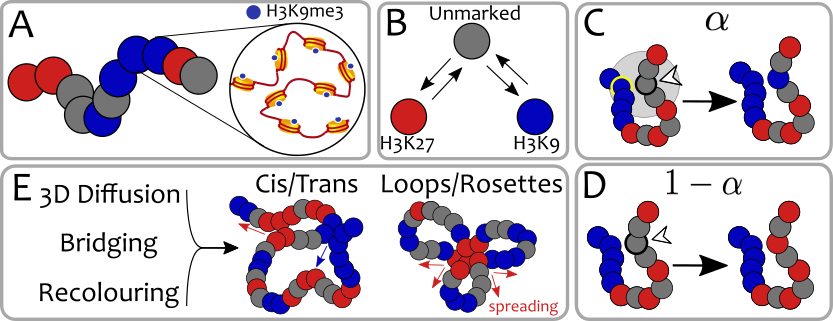
To capture the dynamic nature of epigenetic landscape due to PTM turnover and histone displacement Festuccia2016 ; Zentner2013 , we enhance the (semi-flexible) bead-spring polymer model for chromatin Rosa2008 ; Fudenberg2016 ; Mirny2011 ; Brackley2016nar ; Brackley2016nucleus ; Brackley2013pnas ; Cheng2015 ; Sanborn2015 ; Rosa2010 by adding a further degree of freedom to each bead. Specifically, each bead – corresponding to one or few nucleosomes (choosing a different coarse-graining leaves our result qualitatively unaffected) – bears a “colour” representing the instantaneous local chromatin state (e.g., H3K9me3, H3K27me3, H3K27ac, etc., see Fig. 1(A)), which can dynamically change in time according to realistic biophysical rules Michieletto2016prx ; Dodd2007 ; Arnold2013 (see Fig. 1(B)). This is in contrast with previous works that only accounted for static epigenetic patterns via co-polymer modelling Jost2014B ; DiPierro2016 ; Brackley2016nar ; Barbieri2012 .
We first consider a toy model in which beads may be found in one of three possible states: grey (unmarked), red (e.g., Polycomb-rich) and blue (e.g., heterochromatin-rich). [A more realistic model will be discussed later]. Beads bearing the same histone mark are mutually “sticky”, indicating the presence of implicit bridging proteins Zentner2013 ; Brackley2016nar ; Brackley2013pnas , and can thus bind to each other with interaction energy (see Fig. 1(E)). All other interactions are purely repulsive. The natural time-scale for our simulations is the Brownian time which is the typical diffusion time for a bead of size . As discussed in the SM, this time can be estimated as ms which is equivalent to considering a nucleoplasm viscosity of cP and a bead of size nm Michieletto2016prx .
The action of writer proteins is modelled through “recolouring” moves occurring at rate ; here, we set which is close to typical timescales for acetylation marks Barth2010 . In selected cases, we have also employed a faster recolouring rate of to ensure faster convergence to steady state (see SM for details on simulations and time-mapping).
Our model couples reading and writing as follows. First, a bead is selected randomly. Next, with probability , it recruits a neighbour from spatially-proximate beads (within , where is bead size). The colour of the first bead is then shifted one step “closer” to the colour of the second (Fig. 1B-C). Otherwise (with probability 1-), the bead undergoes a noisy conversion to one of the other colours (see Fig. 1D and SM for further details).
This re-colouring scheme encodes a purely non-equilibrium process and it is akin to a “voter” or “infection-type” model Dodd2007 ; Arnold2013 . In SM, we describe a “Potts” recolouring scheme which can be arbitrarily tuned either in- or out-of-equilibrium Michieletto2016prx . Both schemes couple 1D epigenetic information along the chromatin strand to 3D folding. Both drive a positive feedback loop between readers (which bind and bridge chromatin segments) and writers (which can change the underlying epigenetic pattern). Strikingly, both strategies lead to qualitatively similar behaviours, in which cis/trans contacts, globules and rosettes (Fig. 1E) spontaneously emerge and drive the spreading of histone modifications. To simplify the presentation of our results, and because the observed behaviours are similar, we choose to report in the main text the finding obtained via the “infection-type” model. This model may better capture the one-to-one nature of the chemical reactions required for the deposition (or writing) of histone marks (see SM for more details).
III Results
III.1 The Phase Diagram of the System Entails Epigenetic Memory
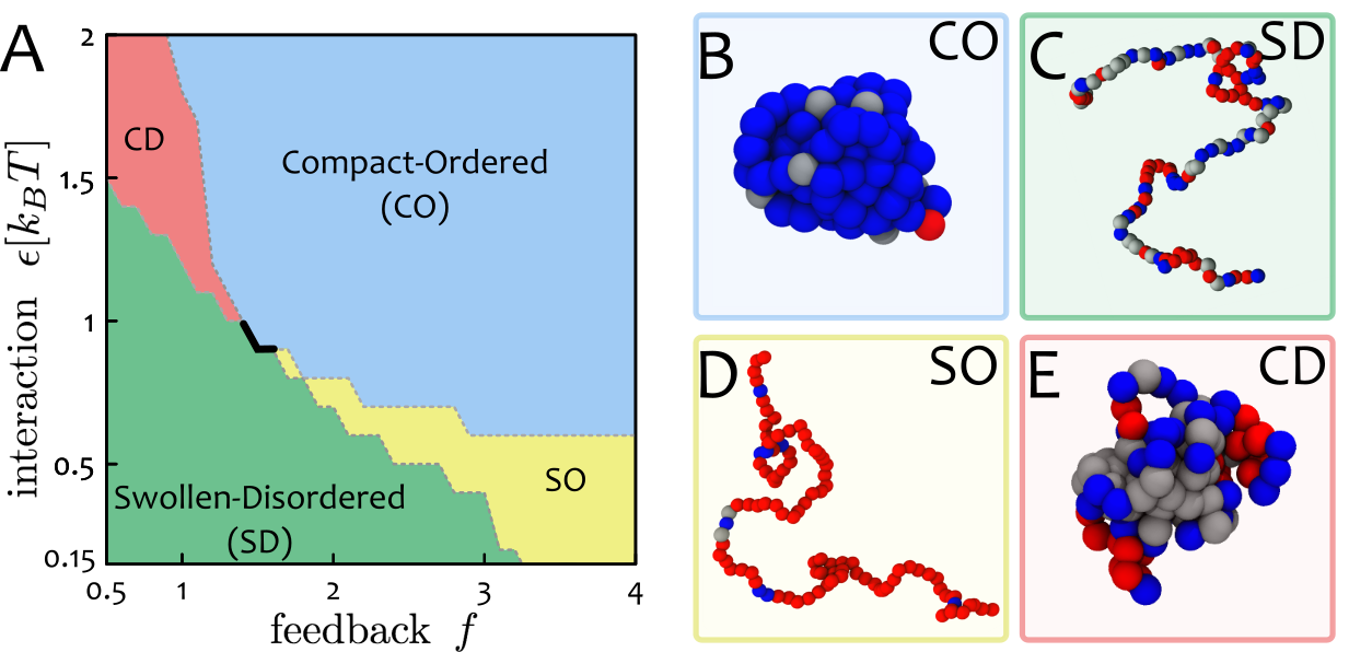
We first map the phase diagram obtained by varying the “feedback” parameter and the attraction energy between any two like-coloured beads. A more realistic model accounting for different attractions between “Polycomb-rich” and “heterochromatin-rich” beads is considered later.
Figure 2A shows that there are four distinct phases predicted by our minimal model. First, at small and , the fibre is swollen and epigenetically disordered (SD). At large and , the system is in the compact epigenetically ordered (CO) phase. These two states are separated by a discontinuous transition, signalled by the presence of hysteresis and coexistence (see SM). The discontinuous nature of the transition is important because it confers metastability to the two phases with respect to perturbations in the parameter space. In addition, perturbing a compact heterochromatin-rich state by extensively erasing PTM marks (e.g. during replication) fails to drive the system out of that epigenetic state Michieletto2016prx ; in other words, the global epigenetic state is remembered across genome-wide re-organisation Michieletto2016prx ; Angel2011 .
The two remaining regions of the phase diagram (Fig. 2A) are (i) an ordered-swollen phase (SO), observed at large but small or moderate , and (ii) a compact-disordered phase (CD), found at small and large . Our simulations suggest that the transitions from, or to, these states are smooth and unlike that between the SD and CO phases.
We highlight that the first order line (black thick line in Fig. 2A) entails hysteresis (see SM, Fig. S3) and robustness of the states against small perturbations in the parameter space. On the other hand, a pathway that brings, for instance, a CO state into a SD one passing through the SO region, crosses continuous lines. Such a pathway in the parameter space may be a valid model to describe a change of identity of a cell, for instance during reprogramming. While this is an appealing avenue, we leave its exploration for future work as it requires a more detailed mapping between the recolouring rules of real systems and our parameter space.
III.2 Polymer Simulations of the Minimal Model Capture Realistic Chromatin Conformations
Intriguingly, some of the phases in the phase diagram in Fig. 2 correspond to structures seen in eukaryotic chromosomes. Most notably, the compact-ordered phase provides a primitive model for the structure of the inactive copy of the X chromosome in female mammals; this is almost entirely transcriptionally silent, and this state is inherited through many cell divisions Alberts2014 .
The compact-disordered phase is reminiscent of “gene deserts” (or black chromatin Sexton2012 ; Kharchenko2011 ). This is a state without a coherent epigenetic mark which tends to co-localise in 3D, possibly due to the linker histone H1 Sexton2012 ; Kharchenko2011 ; Filion2010 . Finally, the swollen-ordered phase is reminiscent of open and transcriptionally-active chromatin Gilbert2004 ; Gilbert2006 ; Nozawa2017 .
In this simplified model, feedback between readers and writers leads to unlimited spreading of a single histone mark in both ordered phases (CO and SO, see Fig. 2) Michieletto2016prx ; Michieletto2017scirep . Although near-unlimited spreading of silencing marks is seen in telomere position effects in yeast Talbert2006 and position-effect variegation in Drosophila Schotta2002 ), this minimal model cannot recapitulate the existence of multiple epigenetic domains, or “heterogeneous” epigenetic patterns.
III.3 A Biophysical Model for Genomic Bookmarking
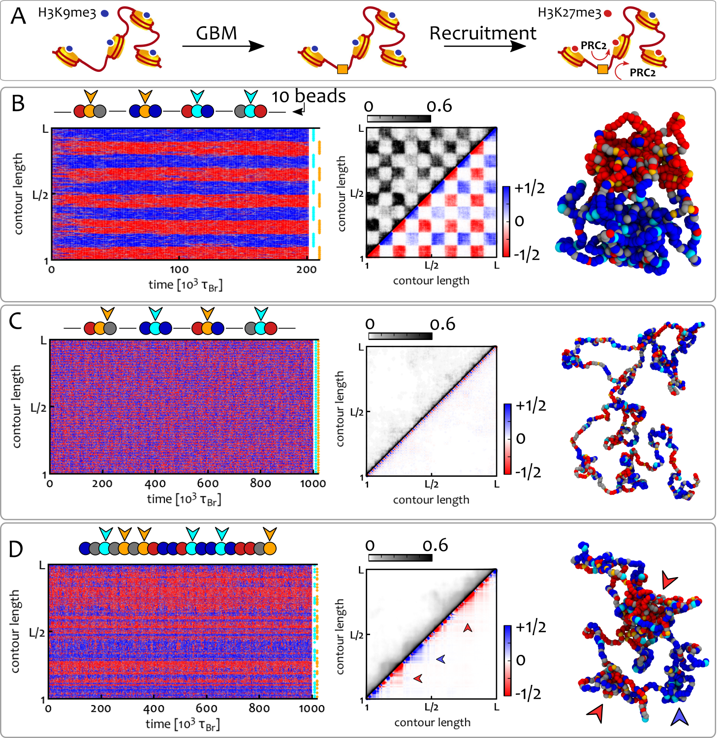
We now introduce genomic bookmarking (GBM) to account for heterogeneous epigenetic patterns, coexistence of heritable epigenetic domains and active/inactive (A/B) compartments Rao2014 ; Dixon2015 . A bookmark is here considered as a TF (activator or repressor) that binds to a cognate site and recruits appropriate readers or writers (see Fig. 3A).
A mechanistic model of how bookmarks might guide the re-establishment of the previous epigenetic patterns after mitosis remains elusive Heard2014 ; Kadauke2013a ; Sarge2005 ; Teves2016 . Here, we assume that GBMs are expressed in a tissue-specific manner and remain (dynamically) associated to chromatin during mitosis Teves2016 ; Follmer2012 . Then, on re-entering into inter-phase, they can recruit appropriate read/write machineries and re-set the previous transcriptional programme.
In our polymer model, we account for bookmarks by postulating that some of the beads cannot change their chromatin state (Fig. 3A). Thus, a red (blue) bookmark is a red (blue) bead that cannot change its colour, and otherwise behaves like other red (blue) beads. In Figure 3A, a bookmark is indicated by an orange square that binds to DNA (rather than a PTM) and recruits read/write machineries (e.g., PRC2), which then spread a histone mark (e.g., H3K27me3) to the neighbours Alberts2014 ; Cheutin2012 ; Zentner2013 ; Cavalli2013 .
It is important to stress that, in these polymer simulations, spreading of a colour is driven by the local increase in the density of that color. Indeed, bridging drives like-colour attractions and increases the probability that a random bead will be “infected” by a 3D-proximal bead bearing that mark. The choice of which mark dominates the local spreading is decided via symmetry breaking and we thus bias the local concentration of marks by introducing DNA-bound enzymes, i.e. bookmarks (see Supplementary Movie 1).
III.4 GBM Drives Stable Coexistence of 1D Epigenetic Domains and Shapes the 3D Chromatin Organisation
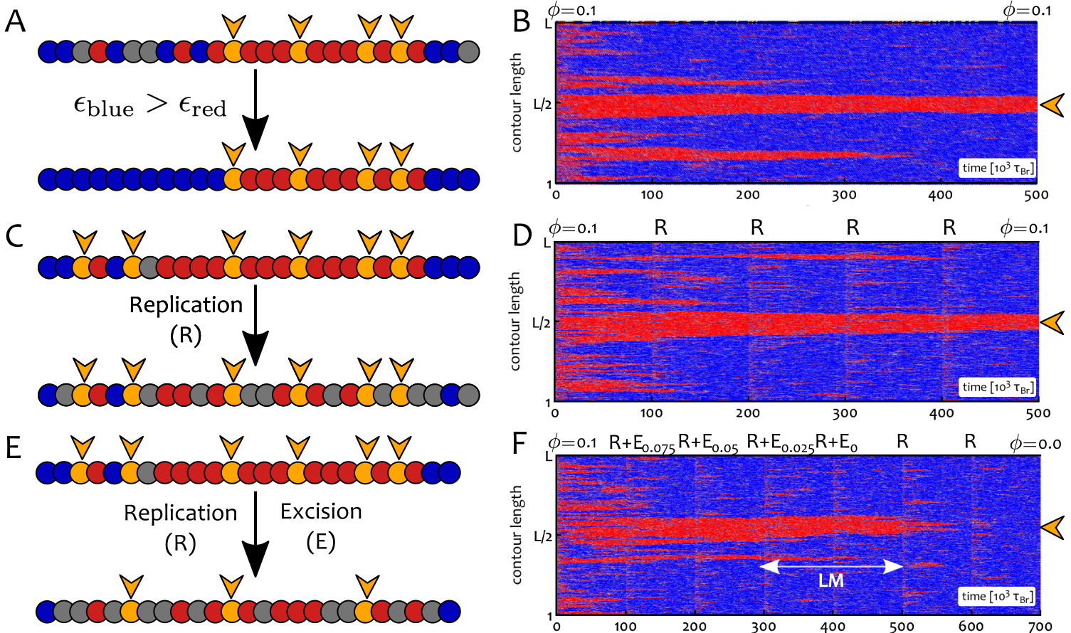
We now consider a chromatin fibre where a fraction of beads are “bookmarks” and analyse how their spatial distribution affects the epigenetic patterns in steady state. We consider three possible GBM distributions as follows: (i) Clustered: bookmarks are equally spaced along the fibre; the colour alternates after every consecutive bookmarks ( defines the cluster size). (ii) Mixed: same as clustered, but now colours alternate every other bookmark (). (iii) Random: random bookmarks are placed along the fibre while the fraction is kept constant.
Figures 3B-D show the results for and a chromatin fibre beads long. This correspond to about Mbp, or nucleosomes, for a coarse graining of kbp per bead, i.e., a fibre with approximately one bookmark every nucleosomes. Simulations are initialised with the chromatin fibre in the swollen-disordered phase and non-bookmarked regions contain equal numbers of red, blue and grey beads.
The clustered distribution of bookmarks (Fig. 3B) reaches a stable epigenetic pattern with blocks of alternating colours (domains). On the contrary, the mixed bookmark distribution hinders domain formation, and the fibre remains in the SD state (Fig. 3C). Remarkably, random bookmarks also yield domains in 1D (Fig. 3D), even in the absence of any correlation between the location of bookmarks.
Importantly, we highlight that the bookmarking pattern affects 3D structure. Thus, in Figure 3C-D, both the random and mixed patterns yield swollen or partially-collapsed fibres, even though the parameters used normally drive the system to a collapsed phase. [Note that our parameter choice accounts for the fact that the critical marking the SD-CO transition decreases with .]
For the random distribution, the contact map exhibits locally compact structures with coherent epigenetic marks (see arrowhead in Fig. 3D) while long-range interactions between like-coloured domains are supressed. This result is in marked contrast with equilibrium models with static epigenetic pattern Jost2014B ; Barbieri2012 ). On the other hand, for clustered bookmarks, red and blue domains separately coalesce in 3D (macro-phase-separation), to give a checker-board appearance of the contact map (Fig. 3B) reminiscent of the pattern formaed by A/B compartments in Hi-C maps after suitable normalisation Lieberman-Aiden2009 ; Rao2014 .
We highlight that these patterns are achieved independently of the chosen initial configuration. As shown in the SM (Fig. S4), a system initialised from deep into the collapsed-disordered phase (reminiscent of condensed mitotic chromosomes) leads to the same 1D pattern of marks and 3D organisation found in Fig. 3 at large times.
III.5 A Critical Density of Bookmarks is Required to Form Stable Domains
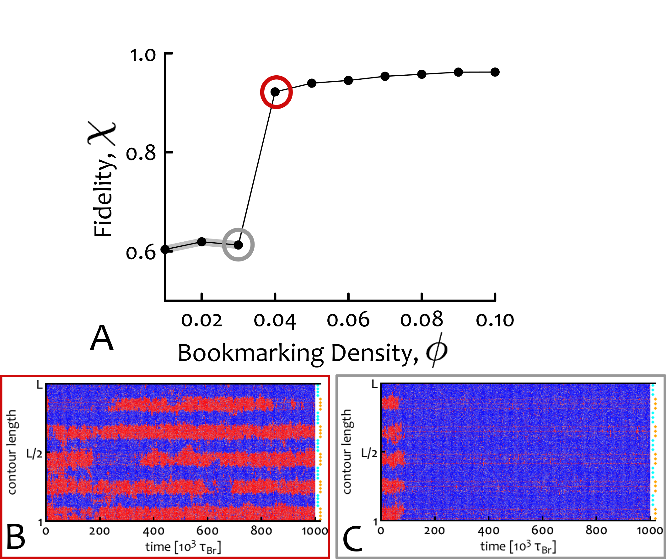
We now ask what is the minimum density of like-coloured bookmarks needed to form stable domains. To address this question we systematically vary bookmark density and perform simulations with clustered patterns (Fig. 3B) as these are the most effective way to create domains. Here, varies from to for a chain with . To facilitate the analysis, we fix the domain size at beads ( kbp), which is in the range of typical HiC domains Dixon2015 ; Lieberman-Aiden2009 ; Rao2014 . We set the system to be in the collapsed-ordered phase, i.e. and , and quantify the efficiency of domain formation by measuring the probability that bead () is in a “red” state, . If ideal regular domains are formed along the fibre (i.e., if all beads have the intended colour, that of the closest bookmarks) then would be a perfect square wave (Fig. 4, caption). The fidelity of domain formation can then be estimated as , where is the mean square deviation (variance) between , measured in simulations, and , i.e. . The fidelity parameter is , when the epigenetic pattern is far from the ordered block-like state and is dominated by a single colour, whereas for ideal block-like domain formation.
We observe (Fig. 4A) that the system displays a phase transition near the critical density . For , stable domains are seen in kymographs and . For instead, a single mark takes over the whole fibre. Near there is a sharp transition between these two regimes in which domains appear and disappear throughout the simulation (see kymograph in Fig. 4B).
The critical density corresponds to about or nucleosomes in about as not all nucleosomes coarse-grained in a “bookmark bead” need to be bookmarked. We argue that, crucially, not all the genome must have this critical density of bookmarks, but only regions required to robustly develop a specific domain of coherent histone marks in a given cell-line.
III.6 Biasing Epigenetic Landscapes with Asymmetric Interactions
Thus far, we have considered symmetric interactions between like-coloured beads. In other words, red-red and blue-blue interaction strengths were equal. However, such binding energies may differ if mediated by distinct proteins. Consider the case where red and blue marks encode Polycomb repression and constitutive heterochromatin, respectively. If the blue-blue interaction is larger than the red-red one, the thermodynamic symmetry of the system is broken and the blue mark eventually takes over all non-bookmarked regions (Fig. 5A). However, if there are bookmarks for the red mark, they locally favour the red state, whereas the stronger attraction globally favours the blue mark. This competition creates an additional route to form stable domains as exemplified in Figure 5A,B. Here, red bookmarks (identified by orange beads) are concentrated in the central segment of a chromatin fibre. Starting from a swollen and epigenetically disordered fibre, where red, blue and grey beads are equal in number, we observe that blue marks quickly invade non-bookmarked regions and convert red beads into blue ones (a process mimicking heterochromatic spreading in vivo Hathaway2012 ). However, the central segment containing the bookmarks displays a stable red domain (Fig. 5A,B).
III.7 Bookmark Excision but not DNA Replication Destabilises the Epigenetic Landscape
We next asked whether the epigenetic pattern established through GBM is also stable against extensive perturbations such as DNA replication. In order to investigate this scenario we simulated semi-conservative replication of the chromatin fibre by replacing half of the (non-bookmarked) beads with new randomly coloured beads Berry2017 . In Figure 5C-D we show that our model can “remember” the established epigenetic pattern through multiple rounds of cell division. Importantly, the combination of “memory” and local epigenetic order (via bookmarks) may allow cells to display “epialleles”, i.e., alleles with different transcriptional behaviours thus explaining local (or “cis-”) memory Berry2015 ; Berry2017 .
We next considered a set-up relevant in light of recent experiments in Drosophila Laprell2017 ; Coleman2017 , where the role of Polycomb-Response-Elements (PREs) in epigenetic memory was investigated. In these works, polycomb-mediated gene repression was perturbed as a consequence of artificial insertion or deletion of PREs. In Figure 5 we thus performed a simulated dynamic experiment where replication was accompanied by random excision of bookmarks Laprell2017 (Fig. 5E,F); in practice, we remove of the initial number of bookmarks at each replication event. Then each “cell cycle” successively dilutes the bookmarks which at some point can no longer sustain the local red state and the region is consequently flooded with blue marks.
Importantly, the system does not display immediate loss of the red domain as soon as ; on the contrary, this domain is temporarily retained through local memory (see Fig. 5F, LM) Angel2011 ; Berry2015 ; Berry2017 . This originates from an enhanced local density of marks together with the positive read/write feedback (see SM). [The persistence of the local memory can be tuned via the parameters of our polymer model.] These results are again consistent with experiments, as regions of the Drosophila genome marked with H3K27me3 are only gradually lost after PRE excision Laprell2017 . Similarly, epialleles have been observed to be temporarily remembered across cell division Berry2015 .
We finally highlight that the results presented in Fig. 5 are independent on the chosen initial configuration. In SM (Figs.S4-S5) we show that starting from a collapsed and epigenetically disordered chromatin (CD phase), resembling heavily condensed and sparsely marked mitotic structures, leads to the same behaviour and strongly supports the robustness of our findings.
III.8 Chromosome-Wide Simulations Predict the Epigenetic Landscape in Drosophila
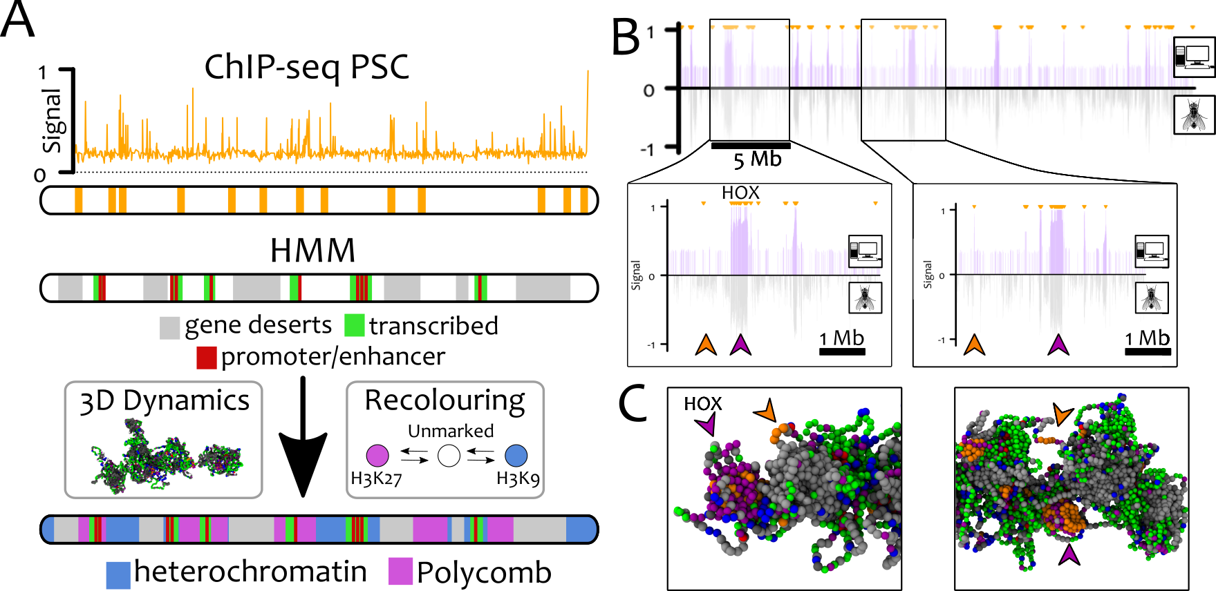
Simplified models considered thus far are useful to identify generic mechanisms; we now aim to test our model in a realistic scenario. To do so, we perform polymer simulations of the whole right arm of chromosome 3 in Drosophila S2 cells.
Bookmarks (orange, in Fig. 6) are located on the chromosome using PSC ChIP-Seq data Follmer2012 , as PSC binds to PREs during inter-phase and mitosis Follmer2012 as well as recruiting PRC2 (via molecular bridging). Some other beads are permanently coloured according to the “9-state” Hidden Markov Model (HMM, Kharchenko2011 ). If they correspond to gene deserts (state 9), promoter/enhancers (state 1) or transcriptionally active regions (states 2-4) they are coloured grey, red and green, respectively. We further introduce an interaction between promoter and enhancer beads to favour looping, plus, an attractive interaction between gene desert (grey) beads mimicking their compaction by H1 linker histone Sexton2012 (see SM for full list of parameters). The remaining 20% of the polymer is left blank and these “unmarked” beads are allowed to dynamically change their chromatin state into heterochromatin (blue) or polycomb (purple) according to our recolouring scheme.
We evolve the system to steady state and we evaluate the probability of finding a Polycomb mark at a certain genomic position. [To determine these probability, a bookmarked bead is counted as bearing the H3K27me3 mark when it is near beads with polycomb marks, or within large stretches of bookmarked beads]. This provides us with an in silico ChIP-seq track for Polycomb marks which can be compared with in vivo ChIP-Seq data Kharchenko2011 (see Fig. 6B). The two are in excellent agreement (Pearson correlation coefficient , against for a random dataset).
Remarkably, not all bookmarked segments (orange) are populated by Polycomb marks; instead we observe that H3K27me3 spreading requires appropriate 3D folding (Fig. 6B-C, insets). Bookmarks which do not contact other bookmarks due to the local epigenetic landscape do not nucleate H3K27me3 spreading. Again, this is consistent with 3D chromatin conformation being crucial for the spreading and establishment of epigenetic patterns Ciabrelli2017 ; Engreitz2013 ; Deng2014 .
IV Discussion
We proposed and investigated a new biophysical mechanism for the de novo establishment of epigenetic domains and their maintenance through interphase and mitosis. Our simplest model requires only one element: a positive feedback between readers (e.g., binding proteins HP1, PRC2, etc.) and writers (e.g., methyltransferases SUV39, EzH2, etc.).
We performed large-scale simulations in which chromatin is modelled as a semi-flexible bead-and-spring polymer chain overlaid with a further degree of freedom representing a dynamic epigenetic patterning. Specifically, each bead is assigned a colour corresponding to the local instantaneous epigenetic state. Readers are implicitly included by setting an attraction between like-coloured beads Brackley2016nar ; Barbieri2012 , whereas writers are modelled by performing re-colouring moves according to realistic and out-of-equilibrium rules Dodd2007 ; Dodd2011 (see Fig. 1).
We find that, if read-write positive feedback is sufficiently strong, a single histone mark can spread over the whole fibre and drives a discontinuous transition to a collapsed-ordered state (see Fig. 2). This state is stable and robust against extensive perturbations such as those occurring during replication Talbert2006 ; Zentner2013 ; Cavalli2013 , when most histones are removed or displaced Alberts2014 ; Zentner2013 ; Festuccia2017 . In other words, our model displays “epigenetic memory”.
The main limitation of this simple model is that epigenetic order in real chromosomes is local, rather than global. Distinct epigenetic domains coexist on a chromosome, thereby forming an “heterogeneous” epigenetic pattern. Our main result is that this feature of real chromosomes can be reproduced by our model when we include genomic bookmarking (GBM).
Here, we envisage bookmarks which can perform functions typical of many TFs: they recruit read/write machineries, and hence nucleate the spreading of epigenetic marks and the establishment of epigenetic domains. We also assumed that bookmarking TFs are permanently bound to DNA, however our conclusions should hold even for dynamic bookmarks that switch between bound and unbound state Brackley2016ephe ; Teves2016 .
We find that stable domains can be formed with only one type of bookmark when the competing epigenetic mark is thermodynamically favoured (Fig. 5). This result rationalises the common understanding that heterochromatin can spread at lengths (blue mark in Fig. 5A,B) and it is stopped by actively transcribed (bookmarked) regions. Further, it is in agreement with recent genome editing experiments in Drosophila: when PRE is inserted into the genome, it provides a bookmark for H3K27me3 which leads to spreading of that mark Laprell2017 , whereas PRE excision leads to (gradual) loss of the mark Laprell2017 (Fig. 5). Additionally, the expression of HOX and other Polycomb-regulated genes (which contain multiple PREs) is predicted by our model to be less sensitive to deletion of single PREs De2016 . We suggest that this is because domains remain stable if bookmark density is kept above the critical threshold (Fig. 4).
Our results strongly suggest that bookmarks can establish specific epigenetic domains by exploiting the local diffusion of chromatin and thereby “infecting” 3D-proximal chromatin segments. The local increase in the density of a mark is then stopped either by thermodynamics (Fig. 5A) or competition with other bookmarks (Fig. 3B). Crucially, our model does not require any boundary element to stop the spreading of marks, which is instead self-regulated.
Losing bookmarks (via artificial excision or DNA mutation) will thus impair the ability of cells to inherit the cell-line-specific epigenetic patterns. In addition, we argue that newly activated bookmarks (for instance subsequently to inflammation response or external stimuli Kirmes2015 ; Feuerborn2015 ; Wood2014 ) may drive the de novo formation of transient epigenetic domains which allow the plastic epigenetic response to environmental changes.
We show that our model can recreate the pattern of H3K27me3 in Drosophila S2 cells starting solely from the position of PSC proteins acting as Polycomb bookmarks Intriguingly, our simulations show that not all bookmarks end up in H3K27me3 domains; whether or not they do, depends on their network of chromatin contacts in 3D. This is agreement with recent experiments Ciabrelli2017 ; Engreitz2013 ; Deng2014 and it is also reminiscent of the well-known position effect according to which the activity of a gene depends on its local environment Feuerborn2015 .
While our framework can be directly applied to model competition between repressive epigenetic marks, the deposition of active marks may be better modelled as resulting from a co-transcriptional positive feedback loop. In light of this, in the SM we show that a model with thermodynamically favoured heterochromatin competing with local recolouring due to transcription leads to results that are qualitatively similar to those presented in the previous sections, as long as promoters are seen as bookmarks for active marks (see SM for more details).
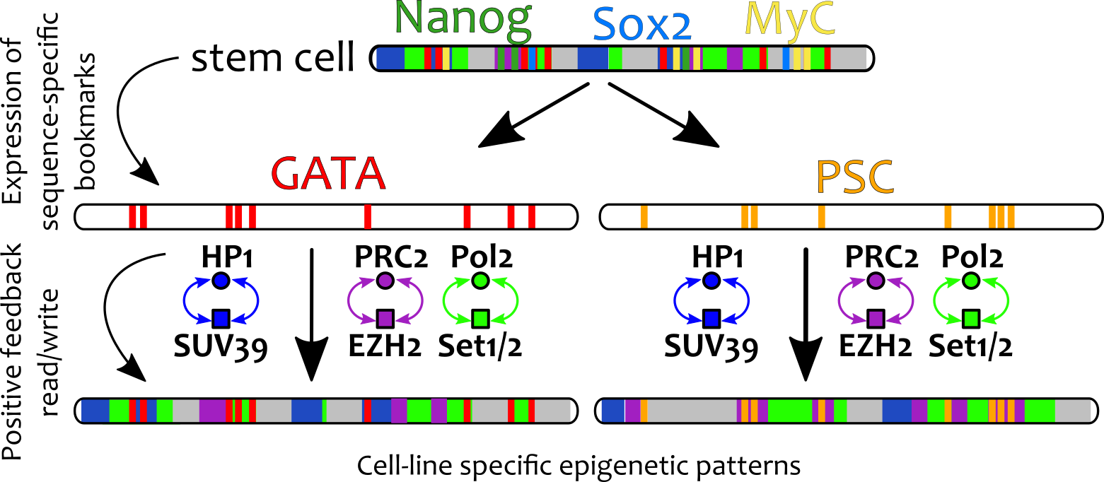
Our results also prompt several further questions. First, starting from a stem cell, how might different cell lineages be established? We suggest that environmental and morphological cues trigger production of lineage-specific bookmarks such as GATA Kadauke2013a and PSC Follmer2012 , which nucleate the positive feedback between readers and writers to generate and sustain new cell-line specific epigenetic patterns (Fig. 7). Thus, bookmarks are here envisaged as key elements that should be targeted in order to understand, and manipulate, cellular differentiation. Second, how might reprogramming factors like Nanog work? We argue that their binding can “mask” the action of pre-existing bookmarks, thereby allowing the establishment of new epigenetic patterns Festuccia2016 (see also BioRxiv: https://doi.org/10.1101/127522).
In conclusion, we have extended the existing notion of GBM to include the ability of nucleating the spreading of epigenetic marks by triggering local read/write feedback loops. This model predicts the de novo establishment of heterogeneous epigenetic patterns which can be remembered across replication and can adapt in response to GBM-targeted perturbations.
Within our framework, architectural elements such as CTCF Alberts2014 , Cohesins Fudenberg2016 and SAF-A Nozawa2017 may provide the initial 3D chromatin conformation upon which the GBM-driven establishment of epigenetic landscape takes place.
V ACKNOWLEDGEMENTS
We acknowledge the European Research Council for funding (Consolidator Grant THREEDCELLPHYSICS, Ref. 648050). Work in the Papantonis lab is supported by CMMC core funding. The authors thank C. A. Brackley, A. Buckle, N. Gilbert, J. Allan and G. Cavalli for insightful remarks on the manuscript.
References
- (1) Waddington,C.H. (1942) Canalization of Development and the Inheritance of Acquired Characters. Nature, 150(3811), 563–565.
- (2) Alberts,B., Johnson,A., Lewis,J., Morgan,D., and Raff,M. (2014) Molecular Biology of the Cell, Taylor & Francis, .
- (3) Strahl,B. and Allis,C. (2000) The language of covalent histone modifications. Nature, 403(January), 41–45.
- (4) Jenuwein,T. and Allis,C.D. (2001) Translating the Histone Code. Science, 293(August), 1074–1081.
- (5) Cavalli,G. and Misteli,T. (mar, 2013) Functional implications of genome topology.. Nat. Struct. Mol. Biol., 20(3), 290–9.
- (6) Pal,B., Bouras,T., Shi,W., Vaillant,F., Sheridan,J.M., Fu,N., Breslin,K., Jiang,K., Ritchie,M.E., Young,M., Lindeman,G.J., Smyth,G.K., and Visvader,J.E. (2013) Global Changes in the Mammary Epigenome Are Induced by Hormonal Cues and Coordinated by Ezh2. Cell Rep., 3(2), 411–426.
- (7) Probst,A.V., Dunleavy,E., and Almouzni,G. (mar, 2009) Epigenetic inheritance during the cell cycle.. Nat. Rev. Mol. Cell. Biol., 10(3), 192–206.
- (8) Ng,R.K. and Gurdon,J.B. (2008) Epigenetic memory of an active gene state depends on histone H3.3 incorporation into chromatin in the absence of transcription.. Nat. Cell Biol., 10(1), 102–9.
- (9) Angel,A., Song,J., Dean,C., and Howard,M. (2011) A Polycomb-based switch underlying quantitative epigenetic memory. Nature, 476(7358), 105–108.
- (10) Kouskouti,A. and Talianidis,I. (2005) Histone modifications defining active genes persist after transcriptional and mitotic inactivation.. EMBO J., 24(2), 347–357.
- (11) Ciabrelli,F., Comoglio,F., Fellous,S., Bonev,B., Ninova,M., Szabo,Q., Xuéreb,A., Klopp,C., Aravin,A., Paro,R., Bantignies,F., and Cavalli,G. (2017) Stable Polycomb-dependent transgenerational inheritance of chromatin states in Drosophila. Nat. Genet., (March).
- (12) Stern,S., Fridmann-Sirkis,Y., Braun,E., and Soen,Y. (2012) Epigenetically Heritable Alteration of Fly Development in Response to Toxic Challenge. Cell Rep., 1(5), 528–542.
- (13) Wood,S. and Loudon,A. (2014) Clocks for all seasons: Unwinding the roles and mechanisms of circadian and interval timers in the hypothalamus and pituitary. J. Endocrinol., 222(2).
- (14) Feuerborn,A. and Cook,P.R. (2015) Why the activity of a gene depends on its neighbors. Trends Genet., 31(9), 483–490.
- (15) Pal,S. and Tyler,J. (2016) Epigenetics and aging. Sci. Adv., 2(e1600584), 253–254.
- (16) Heard,E. and Martienssen,R.A. (2014) Transgenerational epigenetic inheritance: Myths and mechanisms. Cell, 157(1), 95–109.
- (17) Zentner,G.E. and Henikoff,S. (2013) Regulation of nucleosome dynamics by histone modifications.. Nat. Struct. Mol. Biol., 20(3), 259–66.
- (18) Klosin,A., Reis,K., Hidalgo-Carcedo,C., Casas,E., Vavouri,T., and Lehner,B. (2017) Impaired DNA replication derepresses chromatin and generates a transgenerationally inherited epigenetic memory. Sci. Adv., 3(8).
- (19) Barth,T.K. and Imhof,A. (2010) Fast signals and slow marks: the dynamics of histone modifications. Trends Biochem. Sci., 35(11), 618–626.
- (20) Kheir,T.B. and Lund,A.H. (2010) Epigenetic dynamics across the cell cycle. Essays Biochem., 48, 107–120.
- (21) Alabert,C., Barth,T.K., Reverón-Gómez,N., Sidoli,S., Schmidt,A., Jensen,O., Imhof,A., and Groth,A. (2015) Two distinct modes for propagation of histone PTMs across the cell cycle. Genes Dev., 29(6), 585–590.
- (22) Skene,P.J. and Henikoff,S. (2013) Histone variants in pluripotency and disease.. Development, 140, 2513–24.
- (23) Festuccia,N., Gonzalez,I., and Navarro,P. (2017) The Epigenetic Paradox of Pluripotent ES Cells. J. Mol. Biol., 429(10), 1476–1503.
- (24) Scharf,A.N.D., Barth,T.K., and Imhof,A. (2009) Establishment of histone modifications after chromatin assembly. Nucleic Acids Res., 37(15), 5032–5040.
- (25) Arnold,C., Stadler,P.F., and Prohaska,S.J. (2013) Chromatin computation: Epigenetic inheritance as a pattern reconstruction problem. J. Theor. Biol., 336, 61–74.
- (26) Dodd,I.B., Micheelsen,M.A., Sneppen,K., and Thon,G. (2007) Theoretical Analysis of Epigenetic Cell Memory by Nucleosome Modification. Cell, 129(4), 813–822.
- (27) Berry,S., Dean,C., and Howard,M. (2017) Slow Chromatin Dynamics Allow Polycomb Target Genes to Filter Fluctuations in Transcription Factor Activity. Cell Syst., 4(4), 445–457.e8.
- (28) Sexton,T., Yaffe,E., Kenigsberg,E., Bantignies,F., Leblanc,B., Hoichman,M., Parrinello,H., Tanay,A., and Cavalli,G. (feb, 2012) Three-Dimensional Folding and Functional Organization Principles of the Drosophila Genome. Cell, 148(3), 458–472.
- (29) Dixon,J.R., Selvaraj,S., Yue,F., Kim,A., Li,Y., Shen,Y., Hu,M., Liu,J.S., and Ren,B. (2012) Topological domains in mammalian genomes identified by analysis of chromatin interactions. Nature, 485(7398), 376–380.
- (30) Jost,D., Carrivain,P., Cavalli,G., and Vaillant,C. (2014) Modeling epigenome folding: formation and dynamics of topologically associated chromatin domains. Nucleic Acids Research, 42(15), 1–9.
- (31) Rao,S.S.P., Huntley,M.H., Durand,N.C., Stamenova,E.K., Bochkov,I.D., Robinson,J.T., Sanborn,A.L., Machol,I., Omer,A.D., Lander,E.S., and Aiden,E.L. (2014) A 3D map of the human genome at kilobase resolution reveals principles of chromatin looping. Cell, 159(7), 1665–1680.
- (32) Dixon,J.R., Jung,I., Selvaraj,S., Shen,Y., Antosiewicz-Bourget,J.E., Lee,A.Y., Ye,Z., Kim,A., Rajagopal,N., Xie,W., Diao,Y., Liang,J., Zhao,H., Lobanenkov,V.V., Ecker,J.R., Thomson,J.A., and Ren,B. (2015) Chromatin architecture reorganization during stem cell differentiation. Nature, 518(7539), 331–336.
- (33) Micheelsen,M.A., Mitarai,N., Sneppen,K., and Dodd,I.B. (2010) Theory for the stability and regulation of epigenetic landscapes.. Phys. Biol., 7(2), 026010.
- (34) Dodd,I.B. and Sneppen,K. (2011) Barriers and silencers: A theoretical toolkit for control and containment of nucleosome-based epigenetic states. J. Mol. Biol., 414(4), 624–637.
- (35) Anink-Groenen,L.C.M., Maarleveld,T.R., Verschure,P.J., and Bruggeman,F.J. (2014) Mechanistic stochastic model of histone modification pattern formation.. Epigenetics chromatin, 7(1), 30.
- (36) Obersriebnig,M.J., Pallesen,E.M.H., Sneppen,K., Trusina,A., and Thon,G. (2016) Nucleation and spreading of a heterochromatic domain in fission yeast.. Nat. Commun., 7(May), 11518.
- (37) Erdel,F. and Greene,E.C. (2016) Generalized nucleation and looping model for epigenetic memory of histone modifications. Proc. Nat. Acad. Sci. USA, 113(29), E4180–E4189.
- (38) Erdel,F., Müller-Ott,K., and Rippe,K. (dec, 2013) Establishing epigenetic domains via chromatin-bound histone modifiers. Ann. N. Y. Acad. Sci., 1305(1), 29–43.
- (39) Erdel,F. (2017) How Communication Between Nucleosomes Enables Spreading and Epigenetic Memory of Histone Modifications. BioEssays, 1700053, 1700053.
- (40) Talbert,P.B. and Henikoff,S. (2006) Spreading of silent chromatin: inaction at a distance.. Nat. Rev. Genet., 7(10), 793–803.
- (41) Lanzuolo,C., Roure,V., Dekker,J., Bantignies,F., and Orlando,V. (2007) Polycomb response elements mediate the formation of chromosome higher-order structures in the bithorax complex.. Nat. Cell Biol., 9(10), 1167–1174.
- (42) Engreitz,J.M., Pandya-jones,A., Mcdonel,P., Shishkin,A., Sirokman,K., Surka,C., Kadri,S., Xing,J., Goren,A., Lander,E.S., Plath,K., and Guttman,M. (2013) The Xist lncRNA Exploits Three-Dimensional Genome Architecture to Spread Across the X Chromosome. Science, 341(August), 1–9.
- (43) Pinter,S.F., Sadreyev,R.I., Yildirim,E., Jeon,Y., Ohsumi,T.K., Borowsky,M., and Lee,J.T. (2012) Spreading of X chromosome inactivation via a hierarchy of defined Polycomb stations. Genome Res., 22, 1864–1876.
- (44) Schauer,T., Ghavi-Helm,Y., Sexton,T., Albig,C., Regnard,C., Cavalli,G., Furlong,E.E., and Becker,P.B. (2017) Chromosome topology guides the Drosophila Dosage Compensation Complex for target gene activation. EMBO reports,.
- (45) Deng,W., Rupon,J.W., Krivega,I., Breda,L., Motta,I., Jahn,K.S., Reik,A., Gregory,P.D., Rivella,S., Dean,A., and Blobel,G.A. (2014) Reactivation of developmentally silenced globin genes by forced chromatin looping. Cell, 158(4), 849–860.
- (46) Michieletto,D., Orlandini,E., and Marenduzzo,D. (2016) Polymer Model with Epigenetic Recolouring Reveals a Pathway for the de novo Establishment and 3D Organisation of Chromatin Domains. Phys. Rev. X, 6, 041047.
- (47) Hathaway,N.A., Bell,O., Hodges,C., Miller,E.L., Neel,D.S., and Crabtree,G.R. (2012) Dynamics and memory of heterochromatin in living cells. Cell, 149(7), 1447–1460.
- (48) Hauri,S., Comoglio,F., Seimiya,M., Gerstung,M., Glatter,T., Hansen,K., Aebersold,R., Paro,R., Gstaiger,M., and Beisel,C. (2016) A High-Density Map for Navigating the Human Polycomb Complexome. Cell Rep., 17(2), 583–595.
- (49) Collinson,A., Collier,A.J., Morgan,N.P., Sienerth,A.R., Chandra,T., Andrews,S., and Rugg-Gunn,P.J. (2016) Deletion of the Polycomb-Group Protein EZH2 Leads to Compromised Self-Renewal and Differentiation Defects in Human Embryonic Stem Cells. Cell Rep., 17(10), 2700–2714.
- (50) Teves,S.S., An,L., Hansen,A.S., Xie,L., Darzacq,X., and Tjian,R. (2016) A dynamic mode of mitotic bookmarking by transcription factors. Elife, 5(NOVEMBER2016), 1–24.
- (51) Kassis,J.A. and Brown,J.L. (2013) Polycomb group response elements in Drosophila and vertebrates. Advances in genetics, 81, 83.
- (52) Schuettengruber,B., Oded Elkayam,N., Sexton,T., Entrevan,M., Stern,S., Thomas,A., Yaffe,E., Parrinello,H., Tanay,A., and Cavalli,G. (2014) Cooperativity, specificity, and evolutionary stability of polycomb targeting in Drosophila. Cell Rep., 9(1), 219–233.
- (53) Laprell,F., Finkl,K., and Müller,J. (2017) Propagation of Polycomb-repressed chromatin requires sequence-specific recruitment to DNA. Science, 8266, eaai8266.
- (54) Follmer,N.E., Wani,A.H., and Francis,N.J. (2012) A Polycomb Group Protein Is Retained at Specific Sites on Chromatin in Mitosis. PLoS Genet., 8(12).
- (55) Kadauke,S., Udugama,M.I., Pawlicki,J.M., Achtman,J.C., Jain,D.P., Cheng,Y., Hardison,R.C., and Blobel,G.A. (2012) Tissue-specific mitotic bookmarking by hematopoietic transcription factor GATA1. Cell, 150(4), 725–737.
- (56) Kadauke,S. and Blobel,G.A. (2013) Mitotic bookmarking by transcription factors.. Epigenetics chromatin, 6(1), 6.
- (57) Grob,A., Colleran,C., and McStay,B. (2014) Construction of synthetic nucleoli in human cells reveals how a major functional nuclear domain is formed and propagated through cell division. Genes Dev., 28(3), 220–230.
- (58) Festuccia,N., Dubois,A., Vandormael-Pournin,S., Tejeda,E.G., Mouren,A., Bessonnard,S., Mueller,F., Proux,C., Cohen-Tannoudji,M., and Navarro,P. (2016) Mitotic binding of Esrrb marks key regulatory regions of the pluripotency network. Nat. Cell Biol., 18(11), 1139–1148.
- (59) Deluz,C., Friman,E.T., Strebinger,D., Benke,A., Raccaud,M., Callegari,A., Leleu,M., Manley,S., and Suter,D.M. (2016) A role for mitotic bookmarking of SOX2 in pluripotency and differentiation. Genes Dev., 30(22), 2538–2550.
- (60) Kharchenko,P.V., Alekseyenko,A.A., Schwartz,Y.B., Minoda,A., Riddle,N.C., Ernst,J., Sabo,P.J., Larschan,E., Gorchakov,A.A., Gu,T., Linder-Basso,D., Plachetka,A., Shanower,G., Tolstorukov,M.Y., Luquette,L.J., Xi,R., Jung,Y.L., Park,R.W., Bishop,E.P., Canfield,T.K., Sandstrom,R., Thurman,R.E., MacAlpine,D.M., Stamatoyannopoulos,J.A., Kellis,M., Elgin,S.C.R., Kuroda,M.I., Pirrotta,V., Karpen,G.H., and Park,P.J. (2011) Comprehensive analysis of the chromatin landscape in Drosophila melanogaster.. Nature, 471(7339), 480–5.
- (61) Gilbert,N. and Bickmore,W.A. (2006) Structure and Transcription. Biochem. Soc. Symp., 73, 59–66.
- (62) Rosa,A. and Everaers,R. (jan, 2008) Structure and dynamics of interphase chromosomes. PLoS Comp. Biol., 4(8), 1.
- (63) Fudenberg,G., Imakaev,M., Lu,C., Goloborodko,A., Abdennur,N., and Mirny,L.A. (2016) Formation of Chromosomal Domains by Loop Extrusion. Cell Rep., 15, 2038–2049.
- (64) Mirny,L.A. (jan, 2011) The fractal globule as a model of chromatin architecture in the cell.. Chromosom. Res., 19(1), 37–51.
- (65) Brackley,C.A., Johnson,J., Kelly,S., Cook,P.R., and Marenduzzo,D. (2016) Simulated binding of transcription factors to active and inactive regions folds human chromosomes into loops, rosettes and topological domains. Nucleic Acids Res., 44(8), 3503–3512.
- (66) Brackley,C.A., Michieletto,D., Mouvet,F., Johnson,J., Kelly,S., Cook,P.R., and Marenduzzo,D. (2016) Simulating topological domains in human chromosomes with a fitting-free model. Nucleus, 7(5), 453–461.
- (67) Brackley,C.A., Taylor,S., Papantonis,A., Cook,P.R., and Marenduzzo,D. (sep, 2013) Nonspecific bridging-induced attraction drives clustering of DNA-binding proteins and genome organization.. Proc. Natl. Acad. Sci. USA, 110(38), E3605–11.
- (68) Cheng,T.M., Heeger,S., Chaleil,R.A., Matthews,N., Stewart,A., Wright,J., Lim,C., Bates,P.A., and Uhlmann,F. (2015) A simple biophysical model emulates budding yeast chromosome condensation. Elife, 4, 1–22.
- (69) Sanborn,A.L., Rao,S.S.P., Huang,S.C., Durand,N.C., Huntley,M.H., Jewett,A.I., Bochkov,I.D., Chinnappan,D., Cutkosky,A., Li,J., Geeting,K.P., Gnirke,A., Melnikov,A., McKenna,D., Stamenova,E.K., Lander,E.S., and Aiden,E.L. (2015) Chromatin extrusion explains key features of loop and domain formation in wild-type and engineered genomes. Proc. Natl. Acad. Sci. USA, 112(47), 201518552.
- (70) Rosa,A., Becker,N.B., and Everaers,R. (2010) Looping probabilities in model interphase chromosomes. Biophys. J., 98(11), 2410–2419.
- (71) Di Pierro,M., Zhang,B., Aiden,E.L., Wolynes,P.G., and Onuchic,J.N. (2016) Transferable model for chromosome architecture. Proc. Natl. Acad. Sci. USA, 113(43), 201613607.
- (72) Barbieri,M., Chotalia,M., Fraser,J., Lavitas,L.M., Dostie,J., Pombo,A., and Nicodemi,M. (oct, 2012) Complexity of chromatin folding is captured by the strings and binders switch model.. Proc. Natl. Acad. Sci. USA, 109(40), 16173–8.
- (73) Filion,G.J., van Bemmel,J.G., Braunschweig,U., Talhout,W., Kind,J., Ward,L.D., Brugman,W., de Castro,I.J., Kerkhoven,R.M., Bussemaker,H.J., and van Steensel,B. (oct, 2010) Systematic protein location mapping reveals five principal chromatin types in Drosophila cells.. Cell, 143(2), 212–24.
- (74) Gilbert,N., Gilchrist,S., and Bickmore,W.A. (2004) Chromatin Organization in the Mammalian Nucleus. Int. Rev. Cytol., 242, 283–336.
- (75) Nozawa,R.S., Boteva,L., Soares,D.C., Naughton,C., Dun,A.R., Ramsahoye,B., Bruton,P.C., Saleeb,R.S., Arnedo,M., Hill,B., Duncan,R., Maciver,S.K., and Gilbert,N. (2017) SAF-A regulates interphase chromosome structure through oligomerisation with chromatin- associated RNAs. Cell,.
- (76) Michieletto,D., Orlandini,E., and Marenduzzo,D. (2017) Epigenetic Transitions and Knotted Solitons in Stretched Chromatin. Sci. Rep., 7, 14642.
- (77) Schotta,G., Ebert,A., Krauss,V., Fischer,A., Hoffmann,J., Rea,S., Jenuwein,T., Dorn,R., and Reuter,G. (2002) Central role of Drosophila SU(VAR)3-9 in histone H3-K9 methylation and heterochromatic gene silencing. EMBO J., 21(5), 1121–1131.
- (78) Sarge,K.D. and Park-Sarge,O.K. (2005) Gene bookmarking: Keeping the pages open. Trends Biochem. Sci., 30(11), 605–610.
- (79) Cheutin,T. and Cavalli,G. (2012) Progressive polycomb assembly on H3K27me3 compartments generates Polycomb bodies with developmentally regulated motion. PLoS Genet., 8(1).
- (80) Lieberman-Aiden,E., van Berkum,N.L., Williams,L., Imakaev,M., Ragoczy,T., Telling,A., Amit,I., Lajoie,B.R., Sabo,P.J., Dorschner,M.O., Sandstrom,R., Bernstein,B., Bender,M.A., Groudine,M., Gnirke,A., Stamatoyannopoulos,J., Mirny,L.A., Lander,E.S., and Dekker,J. (oct, 2009) Comprehensive mapping of long-range interactions reveals folding principles of the human genome.. Science, 326(october), 289–93.
- (81) Berry,S., Hartley,M., Olsson,T.S.G., Dean,C., and Howard,M. (2015) Local chromatin environment of a Polycomb target gene instructs its own epigenetic inheritance. Elife, 4(MAY), 1–11.
- (82) Coleman,R.T. and Struhl,G. (2017) Causal role for inheritance of H3K27me3 in maintaining the OFF state of a Drosophila HOX gene. Science, 8236, 10.1126/science.aai8236.
- (83) Brackley,C.A., Liebchen,B., Michieletto,D., Mouvet,F., Cook,P.R., and Marenduzzo,D. (2017) Ephemeral Protein Binding to DNA Shapes Stable Nuclear Bodies and Chromatin Domains. Biophys J., 112(6), 1085–1093.
- (84) De,S., Mitra,A., Cheng,Y., Pfeifer,K., and Kassis,J.A. (2016) Formation of a Polycomb-Domain in the Absence of Strong Polycomb Response Elements. PLoS Genet., 12(7), 1–22.
- (85) Kirmes,I., Szczurek,A., Prakash,K., Charapitsa,I., Heiser,C., Musheev,M., Schock,F., Fornalczyk,K., Ma,D., Birk,U., Cremer,C., and Reid,G. (2015) A transient ischemic environment induces reversible compaction of chromatin.. Genome Biol., 16(1), 246.