Stability and roughness of interfaces in mechanically-regulated tissues
Abstract
Cell division and death can be regulated by the mechanical forces within a tissue. We study the consequences for the stability and roughness of a propagating interface, by analysing a model of mechanically-regulated tissue growth in the regime of small driving forces. For an interface driven by homeostatic pressure imbalance or leader-cell motility, long and intermediate-wavelength instabilities arise, depending respectively on an effective viscosity of cell number change, and on substrate friction. A further mechanism depends on the strength of directed motility forces acting in the bulk. We analyse the fluctuations of a stable interface subjected to cell-level stochasticity, and find that mechanical feedback can help preserve reproducibility at the tissue scale. Our results elucidate mechanisms that could be important for orderly interface motion in developing tissues.
Interfaces are ubiquitous in tissue biology, between a tissue and its environment Hong et al. (2016); Ravasio et al. (2015); Basan et al. (2013) or between cell populations Baker (2011); Ninov and Martin-Blanco (2007); Vincent et al. (2013); Gogna et al. (2015); Podewitz et al. (2016). There is great interest in how interfaces propagate smoothly or maintain their shape in the face of cell proliferation and renewal Hong et al. (2016); Marianes and Spradling (2013); Ninov and Martin-Blanco (2007); Landsberg et al. (2009); de la Loza and Thompson (2017); Curtius et al. (2017), for example by line tension acting at tissue boundaries Sussman et al. (2018); Cayuso et al. (2015); Landsberg et al. (2009); Bielmeier et al. (2016).
Theoretical efforts have focused on contour instabilities in cancer Tracqui (2009); Cristini et al. (2005); Poplawski et al. (2010); Ciarletta et al. (2011); Ben Amar et al. (2011), branching Lubkin and Murray (1995); Miura (2015) or folding Bayly et al. (2013), and wound healing Zimmermann et al. (2014); Tarle et al. (2015); Basan et al. (2013). In models that include nutrient diffusion, protruding regions access more nutrient, triggering further growth Castro et al. (2005); Poplawski et al. (2010); Cristini et al. (2005), reminiscent of the Mullins-Sekerka instability in non-living systems Mullins and Sekerka (1963). An epithelium-stroma interface could form undulations due to mechanical stresses from cell turnover Basan et al. (2011); Risler and Basan (2013), while a Saffman-Taylor-like instability based on viscosity contrast has been proposed to underlie branching in the developing lung Lubkin and Murray (1995). A recent cell-based simulation of imbalanced mechanically-regulated growth between two epithelia observed a stable interface, and quantified its roughness Podewitz et al. (2016). A related simulation of cells in an inert medium found finger-like protrusions, arising for higher friction in the medium relative to the cells Drasdo and Hoehme (2012); Lorenzi et al. (2017). Ref. Risler et al. (2015) calculated the steady-state surface fluctuations of a non-growing tissue maintained in its homeostatic state.
In tissue replacement, such as in the developing Drosophila abdominal epidermis Ninov et al. (2010); Bischoff (2012), interface propagation occurs. This may be driven by imbalances in pressure associated with cell division, and/or directed cell motility, which cause the expansion of one tissue at the other’s expense.
In this Letter, we ask whether factors that drive an interface’s propagation can also affect its stability and roughness. We are particularly interested in the consequences of mechanically-regulated cell division and death for the behaviour of interfaces. If cell number change is sensitive to mechanical forces Montel et al. (2011); Streichan et al. (2014); Puliafito et al. (2012); Benham-Pyle et al. (2015); Aegerter-Wilmsen et al. (2007); Pan et al. (2016); Levayer et al. (2016); Gudipaty et al. (2017); de la Loza and Thompson (2017); Fletcher et al. (2018); LeGoff and Lecuit (2016) it leads to a “homeostatic pressure” Basan et al. (2009); Ranft et al. (2014); Recho et al. (2016); Podewitz et al. (2016) which can drive interface propagation without coherently-directed cell motility forces. Alternatively, active, directed migration is proposed to drive interface motion in wound healing Cochet-Escartin et al. (2014); Ravasio et al. (2015); Poujade et al. (2007) or tissue replacement Bischoff (2012).
We study a model of competing epithelial tissues, with an interface driven by homeostatic pressure or by directed motility, acting either at the interface (a “leader-cell” limit) or in bulk 111See Supplemental Material for further theoretical calculations, estimates of parameters, and Refs. Saffman and Taylor (1958); Edwards and Wilkinson (1982); Blanch-Mercader et al. (2017); Bonnet et al. (2012); Bambardekar et al. (2015); Sepúlveda et al. (2013); Curtain and Morris (2009); Trächtler (2016); Ans . Farooqui and Fenteany (2004); Trepat et al. (2009); Scarpa and Mayor (2016). Our results encompass also the cases of stationary interfaces maintained under constant cell renewal Marianes and Spradling (2013), and of a single growing tissue Hong et al. (2016).
We find a Saffman-Taylor-like instability involving substrate friction, and a long-wavelength instability dependent on an effective viscosity of cell number change. Bulk motile forces induce an instability depending on their strength and direction in each tissue. The free boundary of a growing single tissue is generally stabilised by the mechanisms studied here. Adding a driving noise to represent, e.g., stochastic cell division, we calculate the roughness and centre-of-mass diffusion of a stably-propagating interface, and find that mechanical feedback can help preserve reproducibility at the tissue scale Hong et al. (2016).
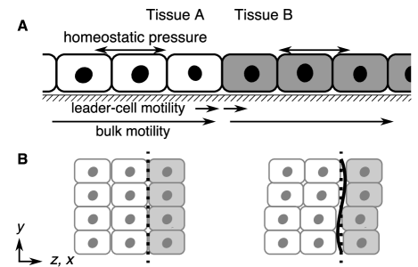
We use a 2D hydrodynamic description in terms of cell density and velocity fields. Tissues and cover an infinite domain, meeting at a flat interface (Fig. 1A). Since we assume a sharp interface, we state equations for a general tissue, unless decorated with or .
We begin with continuity of the areal cell density, ,
| (1) |
where is the velocity field and
| (2) |
is an expansion of the net division/death rate about the homeostatic density , with a characteristic timescale Basan et al. (2009). The units of each tissue are independent, so we set . We consider a linearised, isotropic elastic stress,
| (3) |
where is a tissue’s homeostatic stress, its elastic modulus and . The homeostatic pressure imbalance is . The quantity is an effective bulk viscosity for cell number change Lubkin and Murray (1995); on a timescale , a tissue loses its elastic character as cells are lost or created in response to elastic stress Ranft et al. (2010); Risler et al. (2015). Based on parameter estimates (see supplement Note (1)), we neglect viscous stresses , anticipating Recho et al. (2016); Note (1) . Force balance expresses the tissue velocity as
| (4) |
for substrate friction and a density of active motility forces directed normal to the interface. “Leader-cell” motility at the interface gives an effective contribution to Note (1). We thus take in Eq. 4 as uniform in a given tissue, to account for “bulk” directed motility forces, as may arise from cryptic lamellipodia away from tissue edges Farooqui and Fenteany (2004); Trepat et al. (2009); Scarpa and Mayor (2016).
Moving steady state.
We first solve for the steady propagation of a flat interface (cf. Refs. Recho et al. (2016); Podewitz et al. (2016)). The comoving coordinate is , with the velocity of the interface at , propagating in . Assuming driving forces small enough that nonlinear terms in , can be neglected in Eq. 1, we write
| (5) |
where . The resulting propagating steady state is derived in the supplement Note (1) by matching the tissues’ stress and velocity at the interface. Density perturbations decay from the interface (Fig. 2) governed by each tissue’s hydrodynamic length . Their sign (see Eq. S2 Note (1)) depends on , and on , a difference in “bare” velocities associated to the bulk directed motilities. In Fig. 2A, the growing tissue has decreased density so, by Eq. 2, is proliferative near the interface, while the shrinking tissue has increased density so undergoes net apoptosis near the interface. In Fig. 2B, both tissues are apoptotic near the interface. The steady interface velocity,
| (6) |
is, for and , that found in Ref. Podewitz et al. (2016). To justify ignoring nonlinear terms, we require that stresses from homeostatic pressure, motility and interface line tension are small relative to the tissues’ elastic moduli: , , . Much stronger stresses would lead to nonlinear responses and, eventually, tissue rupture Recho et al. (2016); Harris et al. (2012); Note (1).

Interface stability.
In the supplement Note (1), starting from Fourier and Laplace transforms and of Eq. 5, we perturb the propagating steady state calculated above, to find the fate of an interface fluctuation (Fig. 1B) , where . For line tension (e.g., increased myosin at heterotypic junctions Landsberg et al. (2009) or a supracellular actin cable Hayes and Solon (2017)),
| (7) |
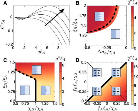
where is the deviation from stress of the propagating steady state. The dominant growth rate is denoted , with , indicating stability or instability (in the applicable parameter regime we do not find complex poles, so treat as real Note (1)). Dispersion relations (e.g., Fig. 3A) maximised over yield phase diagrams (Fig. 3B,C,D) of the most-unstable wavenumber . We approximate the dispersion relation in limits of Note (1) to find the analytic criteria discussed below.
Interface driven by homeostatic pressure or leader-cell motility.
We first discuss growth of tissue driven by (Fig. 3A,B,C), without bulk directed motility (). Analytic dispersion relations Note (1) show that, for strong enough , the interface is unstable if tissue has greater friction or effective viscosity . Instability criteria (given ) are
| (8) |
and
| (9) |
where Eq. Interface driven by homeostatic pressure or leader-cell motility. is approximate Note (1). Two types of transition arise. Fig. 3A and Eq. Interface driven by homeostatic pressure or leader-cell motility. show a “type Is” transition in the Cross-Hohenberg classification Cross and Hohenberg (1993), where an intermediate band becomes unstable (“s” indicates that the instabilities found are stationary, not oscillatory). Fig. S2A Note (1) and Eq. 9 show a “type IIs” transition, with unstable band and onset at . Then, one expects near threshold that the characteristic wavelength scales with system size. Eqs. Interface driven by homeostatic pressure or leader-cell motility., 9 are combined with phase diagrams of in Fig. 3B,C.
Interface driven by bulk directed motility.
We now consider the case , with bulk directed motility forces . Instability occurs when Note (1) satisfies
| (10) |
Fig. S2B Note (1) shows dispersion relations crossing the type IIs transition of Eq. 10. The phase diagram in Fig. 3D shows that a static interface (), marginally stable for Note (1), can be stable or unstable depending on the direction of , .
Single tissue.
The free boundary of a growing single tissue (e.g., epithelium invading empty substrate Ravasio et al. (2015)), is stabilised by the mechanisms studied here (Eqs. S20–S22 Note (1)). Protrusion formation is often observed in wound-healing, via a number of proposed mechanisms we have not included Zimmermann et al. (2014); Tarle et al. (2015); Basan et al. (2013); Mark et al. (2010).

A stable interface subject to noise.
Interface propagation and maintenance takes place in the presence of stochasticity in cell divisions, motilities, material parameters, etc. In the supplement Note (1) we model this with i) a driving noise in Eq. 5, where , corresponding to a contribution of random cell division; or (ii) a noisy motile force contribution to Eq. 4, . Noisy motility could arise, e.g., from ‘swirling’ patterns Basan et al. (2013), provided that the correlation length of these patterns is small compared to other length scales discussed. We focus here on cell division noise, but find qualitatively similar results for noise on the motile force Note (1).
Excluding the mode (discussed below), we calculate the correlation function of an interface in the stable parameter regime. The saturation (late-time) roughness as the system size in , , becomes large, is
| (11) |
The dependence on is as in 1-dimensional Edwards-Wilkinson deposition Antal and Rácz (1996). The positive denominator is expanded in the applicable regime of small . Roughness can be reduced by three now-familiar mechanisms: line tension ; stabilising effective viscosity contrast , ; stabilising bulk motilities . For identical tissues without line tension, the “interface” is an arbitrary line in the tissue: Eq. A stable interface subject to noise. then diverges, i.e., the roughness grows indefinitely. Identical tissues with line tension yield , so that mechanical regulation (i.e., smaller ) reduces boundary roughness. This is true also for a single tissue,
| (12) |
where if the tissue is growing () the roughness is decreased. Fig. 4A shows this behaviour quantitatively for estimated physical values of the parameters Note (1).
The mode leads to an effective diffusion coefficient for the interface centre-of-mass,
| (13) |
and for a single tissue,
| (14) |
The behaviour of Eq. 14 for varying friction and elastic modulus is shown in Fig. 4B. These equations control the accumulating uncertainty in tissue size as the interface centre-of-mass progressively diffuses away from its noise-free trajectory. A larger friction coefficient leads to more precise growth (Fig. 4B) but decreases the velocity (Eq. 6), which suggests trade-offs might be necessary to optimise the speed and precision of growth.
Discussion.
Given experimental evidence of mechanically-regulated cell number change Montel et al. (2011); Streichan et al. (2014); Puliafito et al. (2012); Benham-Pyle et al. (2015); Aegerter-Wilmsen et al. (2007); Pan et al. (2016); Levayer et al. (2016); Gudipaty et al. (2017); de la Loza and Thompson (2017); Fletcher et al. (2018), models of the type used here are widely studied Ranft et al. (2014); Basan et al. (2009); Recho et al. (2016); Podewitz et al. (2016); Lorenzi et al. (2017). There is much interest in mechanisms of boundary maintenance between cell populations Landsberg et al. (2009); Hayes and Solon (2017); Taylor et al. (2017). Recent simulations showed how the topology of cellular interactions can stabilise anomalously smooth interfaces Sussman et al. (2018), while experiments suggest that interface maintenance is not only local but is connected to mechanical waves and jamming processes deep within neighbouring tissues Rodríguez-Franco et al. (2017). Our results add to this picture, showing that mechanically-regulated cell number change within in the tissue bulk can exert an important influence on the properties of interfaces. We have shown how the forces driving overall interface propagation can also generate instabilities, and affect the response of interfaces to cell-level stochasticity.
A Saffman-Taylor-like instability based on substrate friction Drasdo and Hoehme (2012); Lorenzi et al. (2017) (Eqs. Interface driven by homeostatic pressure or leader-cell motility., S17 Note (1)) accords with the tumour literature, where tissues with weaker cell-matrix adhesions tend to be more invasive Frieboes (2006). A longer-wavelength instability (Eq. 9) occurs if the effective bulk viscosity for cell number change is smallest in the growing tissue. Cell-based simulations Podewitz et al. (2016) could explore our predictions, which could in turn be extended to include, e.g., cell growth anisotropy, proposed to play a role in the stable interfaces found in Ref. Podewitz et al. (2016).
The effects of bulk directed motility depend on (Fig. 3D). Repulsive migration, known to occur due to Eph and ephrin signalling Cayuso et al. (2015); Taylor et al. (2017), should yield , favouring stability. In Drosophila abdominal epidermis, larval epithelial cells being replaced by histoblasts are proposed to actively migrate away from the propagating interface Bischoff (2012). This motility force would promote stability, presumably desirable to ensure reproducible, well-controlled tissue replacement. This Drosophila system, or model experiments Rodríguez-Franco et al. (2017), could be used to test our theory by perturbing, e.g., motility, substrate friction, or cell division, and observing the effect on interfaces.
We found that mechanical feedback can help to smooth a stable interface in the presence of noise, as well as determining how quickly the interface centre-of-mass diffuses away from its noise-free position. These findings are relevant to the question of how tissue-level reproducibility is achieved despite cell-level stochasticity Hong et al. (2016).
Acknowledgements.
We thank Anna Ainslie, John Robert Davis, Federica Mangione and Nic Tapon for discussions. J. J. W. acknowledges support by a Wellcome Trust Investigator award to Dr Nic Tapon (107885/Z/15/Z), and acknowledges discussions with Ruth Curtain, Claire McIlroy, Pasha Tabatabai and Ansgar Trächtler. G. S. and J. J. W. acknowledge support by the Francis Crick Institute which receives its core funding from Cancer Research UK (FC001317, FC001175), the UK Medical Research Council (FC001317, FC001175), and the Wellcome Trust (FC001317, FC001175).References
- Hong et al. (2016) L. Hong, M. Dumond, S. Tsugawa, A. Sapala, A.-L. Routier-Kierzkowska, Y. Zhou, C. Chen, A. Kiss, M. Zhu, O. Hamant, R. S. Smith, T. Komatsuzaki, C.-B. Li, A. Boudaoud, and A. H. K. Roeder, Developmental Cell 38, 15 (2016).
- Ravasio et al. (2015) A. Ravasio, I. Cheddadi, T. Chen, T. Pereira, H. T. Ong, C. Bertocchi, A. Brugues, A. Jacinto, A. J. Kabla, Y. Toyama, X. Trepat, N. Gov, L. Neves de Almeida, and B. Ladoux, Nature Communications 6, 7683 EP (2015).
- Basan et al. (2013) M. Basan, J. Elgeti, E. Hannezo, W. J. Rappel, and H. Levine, Proceedings of the National Academy of Sciences 110, 2452 (2013).
- Baker (2011) N. E. Baker, Curr. Biol. 21, R11 (2011).
- Ninov and Martin-Blanco (2007) N. Ninov and E. Martin-Blanco, Nature Protocols 2, 3074 (2007).
- Vincent et al. (2013) J.-P. Vincent, A. G. Fletcher, and L. A. Baena-Lopez, Nature Reviews Molecular Cell Biology 14, 581 (2013).
- Gogna et al. (2015) R. Gogna, K. Shee, and E. Moreno, Annual review of genetics 49, 697 (2015).
- Podewitz et al. (2016) N. Podewitz, F. Jülicher, G. Gompper, and J. Elgeti, New Journal of Physics 18, 083020 (2016).
- Marianes and Spradling (2013) A. Marianes and A. C. Spradling, eLife 2, e00886 (2013).
- Landsberg et al. (2009) K. P. Landsberg, R. Farhadifar, J. Ranft, D. Umetsu, T. J. Widmann, T. Bittig, A. Said, F. Julicher, and C. Dahmann, Current Biology 19, 1950 (2009).
- de la Loza and Thompson (2017) M. D. de la Loza and B. Thompson, Mechanisms of Development 144, Part A, 23 (2017).
- Curtius et al. (2017) K. Curtius, N. A. Wright, and T. A. Graham, Nature Reviews Cancer 18, 19 (2017).
- Sussman et al. (2018) D. M. Sussman, J. M. Schwarz, M. C. Marchetti, and M. L. Manning, Phys. Rev. Lett. 120, 058001 (2018).
- Cayuso et al. (2015) J. Cayuso, Q. Xu, and D. G. Wilkinson, Developmental Biology 401, 122 (2015).
- Bielmeier et al. (2016) C. Bielmeier, S. Alt, V. Weichselberger, M. La Fortezza, H. Harz, F. Jülicher, G. Salbreux, and A.-K. Classen, Current Biology 26, 563 (2016).
- Tracqui (2009) P. Tracqui, Reports on Progress in Physics 72, 056701 (2009).
- Cristini et al. (2005) V. Cristini, H. B. Frieboes, R. Gatenby, S. Caserta, M. Ferrari, and J. Sinek, Clinical Cancer Research 11, 6772 (2005).
- Poplawski et al. (2010) N. J. Poplawski, A. Shirinifard, U. Agero, J. S. Gens, M. Swat, and J. A. Glazier, PLOS ONE 5, 1 (2010).
- Ciarletta et al. (2011) P. Ciarletta, L. Foret, and M. Ben Amar, Journal of Royal Society Interface 8, 345 (2011).
- Ben Amar et al. (2011) M. Ben Amar, C. Chatelain, and P. Ciarletta, Phys. Rev. Lett. 106, 148101 (2011).
- Lubkin and Murray (1995) S. R. Lubkin and J. D. Murray, Journal of Mathematical Biology 34, 77 (1995).
- Miura (2015) T. Miura, The Journal of Biochemistry 157, 121 (2015).
- Bayly et al. (2013) P. V. Bayly, R. J. Okamoto, G. Xu, Y. Shi, and L. A. Taber, Physical Biology 10, 016005 (2013).
- Zimmermann et al. (2014) J. Zimmermann, M. Basan, and H. Levine, The European Physical Journal Special Topics 223, 1259 (2014).
- Tarle et al. (2015) V. Tarle, A. Ravasio, V. Hakim, and N. S. Gov, Integr. Biol. 7, 1218 (2015).
- Castro et al. (2005) M. Castro, C. Molina-París, and T. S. Deisboeck, Phys. Rev. E 72, 041907 (2005).
- Mullins and Sekerka (1963) W. W. Mullins and R. F. Sekerka, Journal of Applied Physics 34, 323 (1963).
- Basan et al. (2011) M. Basan, J.-F. Joanny, J. Prost, and T. Risler, Phys. Rev. Lett. 106, 158101 (2011).
- Risler and Basan (2013) T. Risler and M. Basan, New Journal of Physics 15, 065011 (2013).
- Drasdo and Hoehme (2012) D. Drasdo and S. Hoehme, New Journal of Physics (2012).
- Lorenzi et al. (2017) T. Lorenzi, A. Lorz, and B. Perthame, Kinetic and Related Models 10, 299 (2017).
- Risler et al. (2015) T. Risler, A. Peilloux, and J. Prost, Phys. Rev. Lett. 115, 258104 (2015).
- Ninov et al. (2010) N. Ninov, S. Menezes-Cabral, C. Prat-Rojo, C. Manjón, A. Weiss, G. Pyrowolakis, M. Affolter, and E. MartIn-Blanco, Current Biology 20, 513 (2010).
- Bischoff (2012) M. Bischoff, Developmental Biology 363, 179 (2012).
- Montel et al. (2011) F. Montel, M. Delarue, J. Elgeti, L. Malaquin, M. Basan, T. Risler, B. Cabane, D. Vignjevic, J. Prost, G. Cappello, and J.-F. Joanny, Phys. Rev. Lett. 107, 188102 (2011).
- Streichan et al. (2014) S. J. Streichan, C. R. Hoerner, T. Schneidt, D. Holzer, and L. Hufnagel, Proceedings of the National Academy of Sciences 111, 5586 (2014).
- Puliafito et al. (2012) A. Puliafito, L. Hufnagel, P. Neveu, S. Streichan, A. Sigal, D. K. Fygenson, and B. I. Shraiman, Proceedings of the National Academy of Sciences 109, 739 (2012).
- Benham-Pyle et al. (2015) B. W. Benham-Pyle, B. L. Pruitt, and W. J. Nelson, Science 348, 1024 (2015).
- Aegerter-Wilmsen et al. (2007) T. Aegerter-Wilmsen, C. M. Aegerter, E. Hafen, and K. Basler, Mechanisms of Development 124, 318 (2007).
- Pan et al. (2016) Y. Pan, I. Heemskerk, C. Ibar, B. I. Shraiman, and K. D. Irvine, Proceedings of the National Academy of Sciences 113, E6974 (2016).
- Levayer et al. (2016) R. Levayer, C. Dupont, and E. Moreno, Current Biology 26, 670 (2016).
- Gudipaty et al. (2017) S. A. Gudipaty, J. Lindblom, P. D. Loftus, M. J. Redd, K. Edes, C. F. Davey, V. Krishnegowda, and J. Rosenblatt, Nature (2017).
- Fletcher et al. (2018) G. C. Fletcher, M.-d.-C. Diaz-de-la Loza, N. Borreguero-Muñoz, M. Holder, M. Aguilar-Aragon, and B. J. Thompson, Development (2018), 10.1242/dev.159467.
- LeGoff and Lecuit (2016) L. LeGoff and T. Lecuit, Cold Spring Harbor perspectives in biology 8, a019232 (2016).
- Basan et al. (2009) M. Basan, T. Risler, J.-F. Joanny, X. Sastre-Garau, and J. Prost, HFSP Journal 3, 265 (2009).
- Ranft et al. (2014) J. Ranft, M. Aliee, J. Prost, F. Jülicher, and J.-F. Joanny, New Journal of Physics , 1 (2014).
- Recho et al. (2016) P. Recho, J. Ranft, and P. Marcq, Soft Matter 12, 2381 (2016).
- Cochet-Escartin et al. (2014) O. Cochet-Escartin, J. Ranft, P. Silberzan, and P. Marcq, Biophys. J. 106, 65 (2014).
- Poujade et al. (2007) M. Poujade, E. Grasland-Mongrain, A. Hertzog, J. Jouanneau, P. Chavrier, B. Ladoux, A. Buguin, and P. Silberzan, Proceedings of the National Academy of Sciences 104, 15988 (2007).
- Note (1) See Supplemental Material for further theoretical calculations, estimates of parameters, and Refs. Saffman and Taylor (1958); Edwards and Wilkinson (1982); Blanch-Mercader et al. (2017); Bonnet et al. (2012); Bambardekar et al. (2015); Sepúlveda et al. (2013); Curtain and Morris (2009); Trächtler (2016); Ans .
- Farooqui and Fenteany (2004) R. Farooqui and G. Fenteany, J. Cell Sci. 118, 51 (2004).
- Trepat et al. (2009) X. Trepat, M. R. Wasserman, T. E. Angelini, E. Millet, D. A. Weitz, J. P. Butler, and J. J. Fredberg, Nature Physics 5, 426 (2009).
- Scarpa and Mayor (2016) E. Scarpa and R. Mayor, The Journal of Cell Biology 212, 143 (2016).
- Ranft et al. (2010) J. Ranft, M. Basan, J. Elgeti, J.-F. Joanny, J. Prost, and F. Jülicher, Proc. Natl. Acad. Sci. 107, 20863 (2010).
- Harris et al. (2012) A. R. Harris, L. Peter, J. Bellis, B. Baum, A. J. Kabla, and G. T. Charras, PNAS 109, 16449 (2012).
- Hayes and Solon (2017) P. Hayes and J. Solon, Mechanisms of Development 144, 2 (2017).
- Cross and Hohenberg (1993) M. C. Cross and P. C. Hohenberg, Rev. Mod. Phys. 65, 851 (1993).
- Mark et al. (2010) S. Mark, R. Shlomovitz, N. S. Gov, M. Poujade, E. Grasland-Mongrain, and P. Silberzan, Biophysical Journal 98, 361 (2010).
- Antal and Rácz (1996) T. Antal and Z. Rácz, Physical Review E (Statistical Physics 54, 2256 (1996).
- Taylor et al. (2017) H. B. Taylor, A. Khuong, Z. Wu, Q. Xu, R. Morley, L. Gregory, A. Poliakov, W. R. Taylor, and D. G. Wilkinson, Journal of The Royal Society Interface 14, 20170338 (2017).
- Rodríguez-Franco et al. (2017) P. Rodríguez-Franco, A. Brugues, A. Marín-Llauradó, V. Conte, G. Solanas, E. Batlle, J. J. Fredberg, P. Roca-Cusachs, R. Sunyer, and X. Trepat, Nature Materials 16, 1029 (2017).
- Frieboes (2006) H. B. Frieboes, Cancer Research 66, 1597 (2006).
- Saffman and Taylor (1958) P. G. Saffman and G. Taylor, Proceedings of the Royal Society of London A: Mathematical, Physical and Engineering Sciences 245, 312 (1958).
- Edwards and Wilkinson (1982) S. F. Edwards and D. R. Wilkinson, Proceedings of the Royal Society of London A: Mathematical, Physical and Engineering Sciences 381, 17 (1982).
- Blanch-Mercader et al. (2017) C. Blanch-Mercader, R. Vincent, E. Bazellières, X. Serra-Picamal, X. Trepat, and J. Casademunt, Soft Matter 13, 1235 (2017).
- Bonnet et al. (2012) I. Bonnet, P. Marcq, F. Bosveld, L. Fetler, Y. Bellaiche, and F. Graner, Journal of The Royal Society Interface 9, 2614 (2012).
- Bambardekar et al. (2015) K. Bambardekar, R. Clément, O. Blanc, C. Chard s, and P.-F. Lenne, Proceedings of the National Academy of Sciences 112, 1416 (2015).
- Sepúlveda et al. (2013) N. Sepúlveda, L. Petitjean, O. Cochet, E. Grasland-Mongrain, P. Silberzan, and V. Hakim, PLoS Computational Biology 9, e1002944 (2013).
- Curtain and Morris (2009) R. Curtain and K. Morris, Automatica 45, 1101 (2009).
- Trächtler (2016) A. Trächtler, ArXiv e-prints (2016), arXiv:1603.01059 [math.DS] .
- (71) A. Trächtler, private communication.