Reorientations, relaxations, metastabilities, and multidomains of skyrmion lattices
Abstract
Magnetic skyrmions are nano-sized topologically protected spin textures with particle-like properties. They can form lattices perpendicular to the magnetic field and the orientation of these skyrmion lattices with respect to the crystallographic lattice is governed by spin-orbit coupling. By performing small angle neutron scattering measurements, we investigate the coupling between the crystallographic and skyrmion lattices in both Cu2OSeO3 and the archetype chiral magnet MnSi. The results reveal that the orientation of the skyrmion lattice is primarily determined by the magnetic field direction with respect to the crystallographic lattice. In addition, it is also influenced by the magnetic history of the sample which can induce metastable lattices. Kinetic measurements show that these metastable skyrmion lattices may or may not relax to their equilibrium positions under macroscopic relaxation times. Furthermore, multidomain lattices may form when two or more equivalent crystallographic directions are favored by spin-orbit coupling and oriented perpendicular to the magnetic field.
I Introduction
Magnetic skyrmions are nano-sized topologically protected spin textures with particle-like properties which may form lattices oriented perpendicular to the magnetic field.(Mühlbauer et al., 2009; Fert et al., 2013; Nagaosa and Tokura, 2013; Milde et al., 2013; Romming et al., 2013; Seki and Mochizuki, 2015; Bauer and Pfleiderer, 2016) These Skyrmion Lattices (SkL) were first identified in cubic chiral magnets by Small Angle Neutron Scattering (SANS) inside the A-phase, which is a pocket in the magnetic field () - temperature () phase diagram just below the critical temperature . Soon after their first observation in MnSi,(Mühlbauer et al., 2009) skyrmion lattices were found in other cubic chiral magnets including Fe1-xCoxSi,(Münzer et al., 2010) FeGe,(Wilhelm et al., 2011) Cu2OSeO3(Seki et al., 2012a, b; Adams et al., 2012) and Co-Zn-Mn alloys.(Tokunaga et al., 2015; Karube et al., 2016) More recently, skyrmions and their lattices have been observed in polar magnets,Kézsmárki et al. (2015) in thin films,Tonomura et al. (2012); Wilson et al. (2014) and at surfaces and interfaces of different atomic layersHeinze et al. (2011). Skyrmions are topologically stable and can be controlled with extremely small electric currents which frees the path for successful applications in novel spintronic and information storage devices.(Yu et al., 2012; Nagaosa and Tokura, 2013; Fert et al., 2013; White et al., 2014)
In chiral magnets, the Dzyaloshinskii-Moriya interaction Dzyaloshinski (1958); Moriya (1960) plays a crucial role in stabilizing the helimagnetic order and skyrmion lattices.Bak and Jensen (1980) However, this interaction in itself cannot explain the thermodynamic stability of skyrmion lattices, which has been attributed to additional terms including thermal fluctuations,(Mühlbauer et al., 2009; Buhrandt and Fritz, 2013; Bauer and Pfleiderer, 2016) spin exchange stiffness and/or uniaxial anisotropy.(Bogdanov and Yablonskii, 1989; Bogdanov and Hubert, 1994; Rößler et al., 2006; Butenko et al., 2010; Rößler et al., 2011) The specific orientation of the skyrmion lattice with respect to the crystallographic lattice can be accounted for by higher order spin-orbit coupling terms.Mühlbauer et al. (2009) In order to describe this orientation dependence, it is convenient to consider the skyrmion lattice as a single domain, long-range ordered state resulting from the superposition of three helical vectors separated by 60∘.Adams et al. (2011, 2012) These helices propagate in the plane perpendicular to the magnetic field and lead to the characteristic six-fold symmetric SANS patterns.Mühlbauer et al. (2009) In MnSi one of the three helices preferentially aligns along the crystallographic direction. For Cu2OSeO3 this orientational preference is along ,Adams et al. (2012); Zhang et al. (2016a, b) although some SANS measurements seem to indicate that for specific fields and temperatures the direction is preferred.Seki et al. (2012b); Makino et al. (2017)
Besides this orientational preference, patterns with twelve or more peaks have been observed by SANS and resonant x-ray scattering on Cu2OSeO3.Langner et al. (2014); Zhang et al. (2016a, b); Makino et al. (2017) These patterns indicate the coexistence of multiple skyrmion lattice domains with different orientations. In fact, such multidomain states have also been seen by Lorentz transmission electron microscopy in thin films of MnSi and Cu2OSeO3(Mochizuki et al., 2014; Pöllath et al., 2017) and by SANS in single crystals of Fe1-xCoxSi.Münzer et al. (2010); Bannenberg et al. (2016a) The occurrence of these multidomain states appears for bulk Cu2OSeO3 to be related to the thermal history.Makino et al. (2017); Reim et al. (2017) In addition, it was proposed that multidomains are stabilized by magnetic fields applied along directions deviating from the major cubic axes.(Zhang et al., 2016b, a) However, none of these previous studies provides a holistic view on the interrelation between the orientation of skyrmion lattices and the crystallographic lattice, the occurrence of multidomain lattices and the influence of meta-stabilities induced by different magnetic field histories for all cubic chiral magnets.
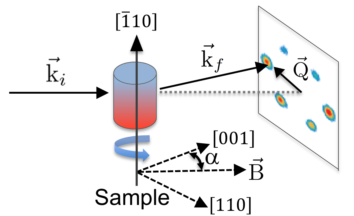
In the following we fill this gap by studying the skyrmion lattice orientation with respect to the crystallographic lattice and the occurrence of multidomain lattices in both the insulator Cu2OSeO3 and the archetype chiral magnet MnSi. For this purpose, we performed SANS measurements in a way so far not considered. As schematically shown in Fig. 1, the measurements were performed by rotating the sample around its vertical axis, and thus having the magnetic field applied along different crystallographic directions. By rotating the sample both in zero and under field, we study the effect of the magnetic field history on the orientation of the skyrmion lattice with respect to the crystallographic lattice. The results show unambiguously that the orientation of the skyrmion lattice and the occurrence of multidomain states is primarily governed by the magnetic field direction with respect to the crystallographic lattice. The orientation of the skyrmion lattice with respect to the crystallographic lattice is also influenced by the magnetic history of the sample. The latter can induce metastable orientations of the skyrmion lattice with respect to the crystallographic one that may or may not relax to their equilibrium orientation under macroscopic relaxation times. Furthermore, multidomain lattices may form when two or more equivalent crystallographic directions are favored by spin-orbit coupling and oriented perpendicular to the magnetic field.
II Experimental
The SANS measurements on Cu2OSeO3 were performed on a single crystal with dimensions of about mm3 grown by chemical vapor transport. The sample was aligned with the [10] crystallographic direction vertical within 3∘. The monochromatic SANS instrument PA20 of the Laboratoire Léon Brillouin was used with a wavelength of = 0.6 nm, / = 0.12 and the detector placed 12.7 m from the sample. The 3He XY multidetector is made of 128128 pixels of 55 mm2. The magnetic field was applied parallel to the incoming neutron beam designated by its wave-vector using an Oxford Instruments horizontal field cryomagnet.
The MnSi sample is a cubic single crystal with dimensions of about mm3 and was already used in a previous experiment.Pappas et al. (2017) The MnSi sample was also aligned with the [10] crystallographic direction vertical within 5∘. The SANS measurements were performed at the time-of-flight instrument LARMOR of the ISIS neutron source using neutrons with wavelengths of 0.09 nm1.25 nm. The sample was placed 4.4 m from the detector that consists of eighty 8 mm wide 3He tubes. The magnetic field was applied with a three-dimensional (3D) 2T vector cryomagnet along the incoming neutron beam.111The field homogeneity of the 3D cryomagnet, defined as ‘the maximum field error over a 10 mm diameter spherical volume’, is determined by the manufacturer as 0.11 % of the applied magnetic field. In particular, we eliminated the residual field of the cryomagnet by warming up the entire cryomagnet before the experiment.222We mounted a field probe on the window of the cryostat to check directly the residual field taking into account its decay with distance. This field probe confirmed the absence of a sizeable residual field. In addition, we made magnetic field scans from negative to positive fields to evaluate the residual field. As the plot of the integrated scattered intensity vs magnetic field was symmetric and centered around zero, a sizable residual field was absent.
All SANS patterns are normalized to standard monitor counts and background corrected using a high temperature measurement at 60 K for MnSi and 70 K for Cu2OSeO3.
III Results
III.1 Cu2OSeO3
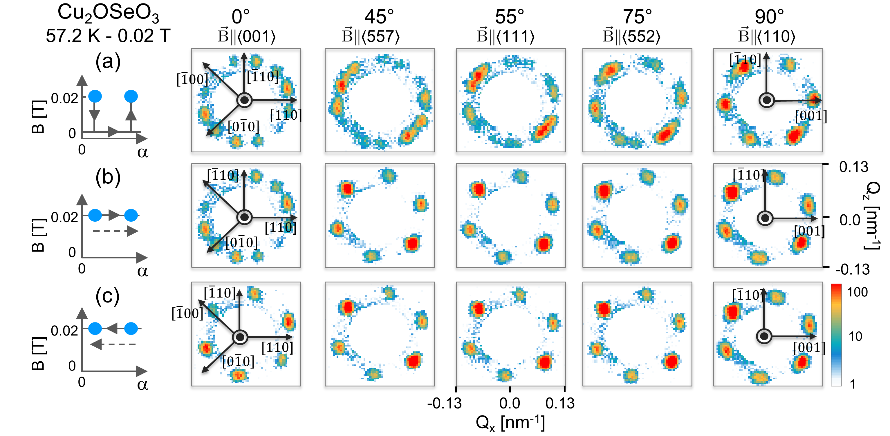
We first consider the case of Cu2OSeO3 and display in Fig. 2(a) and Supplemental Movie 1foo a series of SANS patterns measured at = 57.4 K and = 0.02 T. The field was applied during the measurements and switched off when the sample was rotated in steps of 5∘ from = 0∘ to 90∘. The angle is defined as the angle between the magnetic field and the [001] crystallographic axis in the horizontal plane, implying that = 0∘ corresponds to [001] and = 90∘ corresponds to .
At = 0∘, twelve Bragg peaks are found corresponding to two six-peak patterns rotated 30∘ from each other. This indicates the stabilization of two distinct skyrmion lattice domains with different orientations with respect to the crystallographic lattice. One of these domains has one set of two peaks aligned along [100] while the other domain has two peaks along the [010] direction. The intensity differences between the peaks are most likely due to a small misalignment of the single crystal and/or demagnetization effects that could also cause small differences in domain populations.
When the sample is rotated and increases, the [100] and [010] directions are no longer perpendicular to the field and the skyrmion lattices can no longer orient along either of these directions. For small values of , the twelve-peak patterns persist implying that the coexistence of the two skyrmion domains remains energetically favorable. For larger values of around 45∘, the patterns seem to indicate 18 peaks. Unfortunately, the resolution of the measurement is not sufficient to unambiguously confirm the existence of these 18 peaks, which would suggest the coexistence of three skyrmion lattice domains as reported elsewhere.Zhang et al. (2016a)
When is further increased, the third [001] direction approaches the direction perpendicular to the field and the domains gradually merge into one. At = 90∘ only six peaks are visible and among them two are aligned along the [001] direction. At this position, the lattice has rotated by 15∘ with respect to each of the two lattices seen at = 0∘.333The scattering patterns of this rotation scan are equivalent to those obtained after zero field cooling the sample.
A similar rotation scan was performed but this time with the field on during the rotation of the sample. The results, displayed in Fig. 2(b) and Supplemental Movie 2,foo show two important differences with respect to the previous case. During this rotation, the scattering from one of the two skyrmion lattice domains is suppressed, whereas the other one is enhanced. Furthermore, the SkL does not reorient for any of the field directions including at = 90∘. At this angle the favorable [001] direction is perpendicular to the field, but the skyrmion lattice is oriented 15∘ away from it and is pinned by the field to the position it had at = 0∘. This six fold symmetry persists when the sample is rotated back from = 90∘ to 0∘ under magnetic field. As shown in Fig. 2(c), the twelve fold symmetry is still not recovered at = 0∘ where only one skyrmion lattice domain is found. These measurements show that the orientation of skyrmion lattices and the stabilization of multiple domains are strongly influenced by the history of the applied magnetic field direction within the equilibrium skyrmion phase.
III.2 MnSi
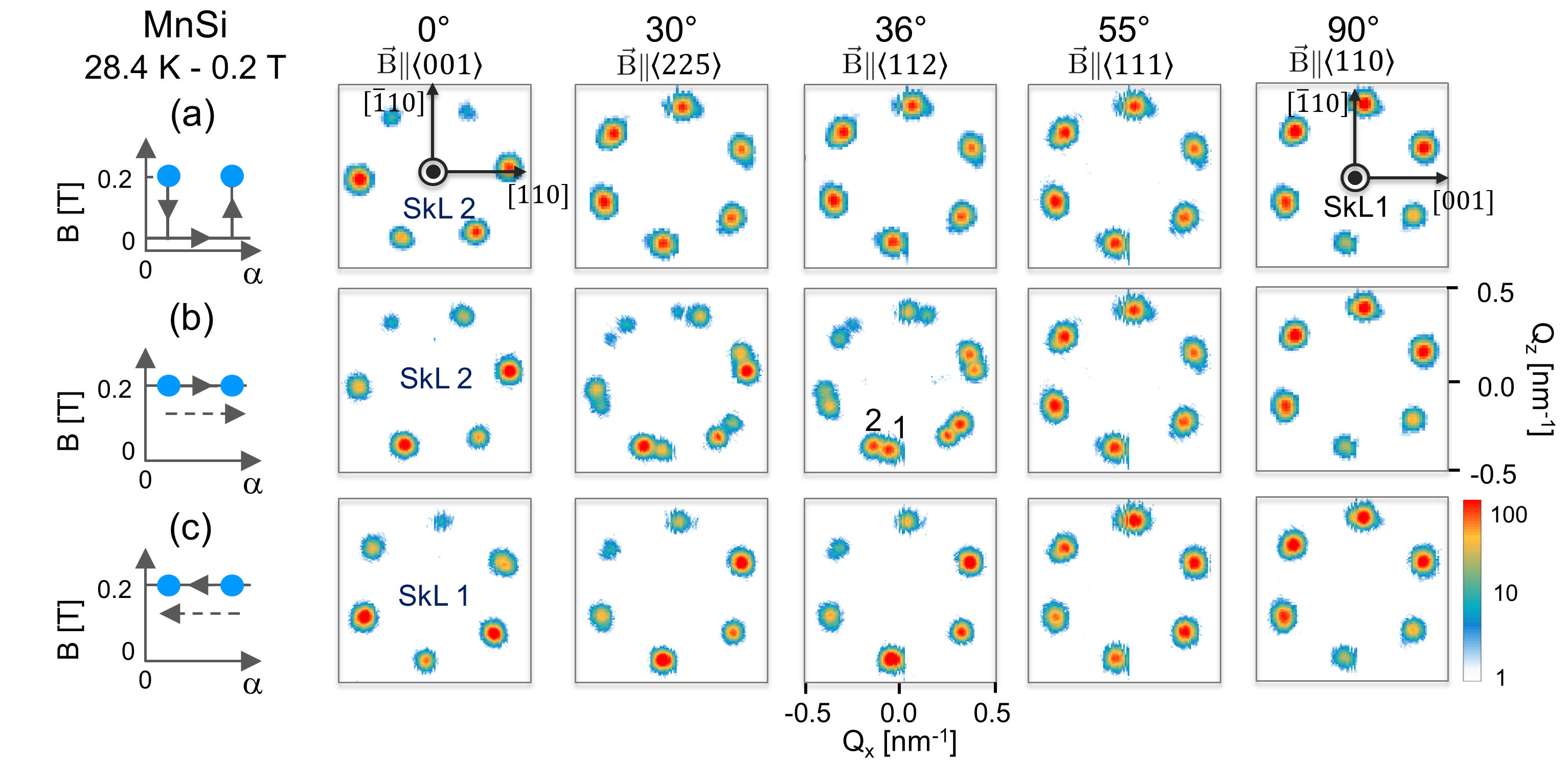
We now consider the case of MnSi and display in Fig. 3(a) a series of SANS patterns measured at = 28.4 K and = 0.2 T after zero-field cooling. For = 0∘, i.e. for , two directions ([110] and ), which are in MnSi preferred by spin-orbit coupling, are perpendicular to the magnetic field. In contrast to Cu2OSeO3 we do not observe twelve peaks, but only six, originating from a single SkL orientation along [110]. Six peaks are also seen for 0∘ but this time the SkL aligns along the [] direction. This orientation, which we name SkL 1, has a scattering pattern that is 30∘ rotated with respect to the one at = 0∘ which we name SkL 2.
The MnSi sample was also rotated from = 0∘ to 90∘ with a magnetic field of 0.2 T on during the measurements and the rotation of the sample. The SANS patterns were recorded every 2∘ and are presented in Supplemental Movie 3foo with a selection given in Fig. 3(b). For 0 28∘, the patterns correspond to SkL 2. The patterns qualitatively change for 28∘ and show a superposition of SkL 2 and SkL 1. By further increasing , SkL 2 decreases in intensity and totally vanishes at 45∘, while at the same time SkL 1 becomes more prominent. As for Cu2OSeO3, the patterns remain the same when the sample is rotated under field back to = 0∘. SkL 2 is still not recovered after waiting 30 min at = 0∘. These results indicate that in both Cu2OSeO3 and MnSi the orientation of the SkL is determined by the magnetic field orientation with respect to crystallographic lattice as well as by the history of the magnetic field (direction) of the sample. They also demonstrate that it is possible to induce metastable skyrmion states in both Cu2OSeO3 and MnSi within the thermodynamic equilibrium skyrmion phase.
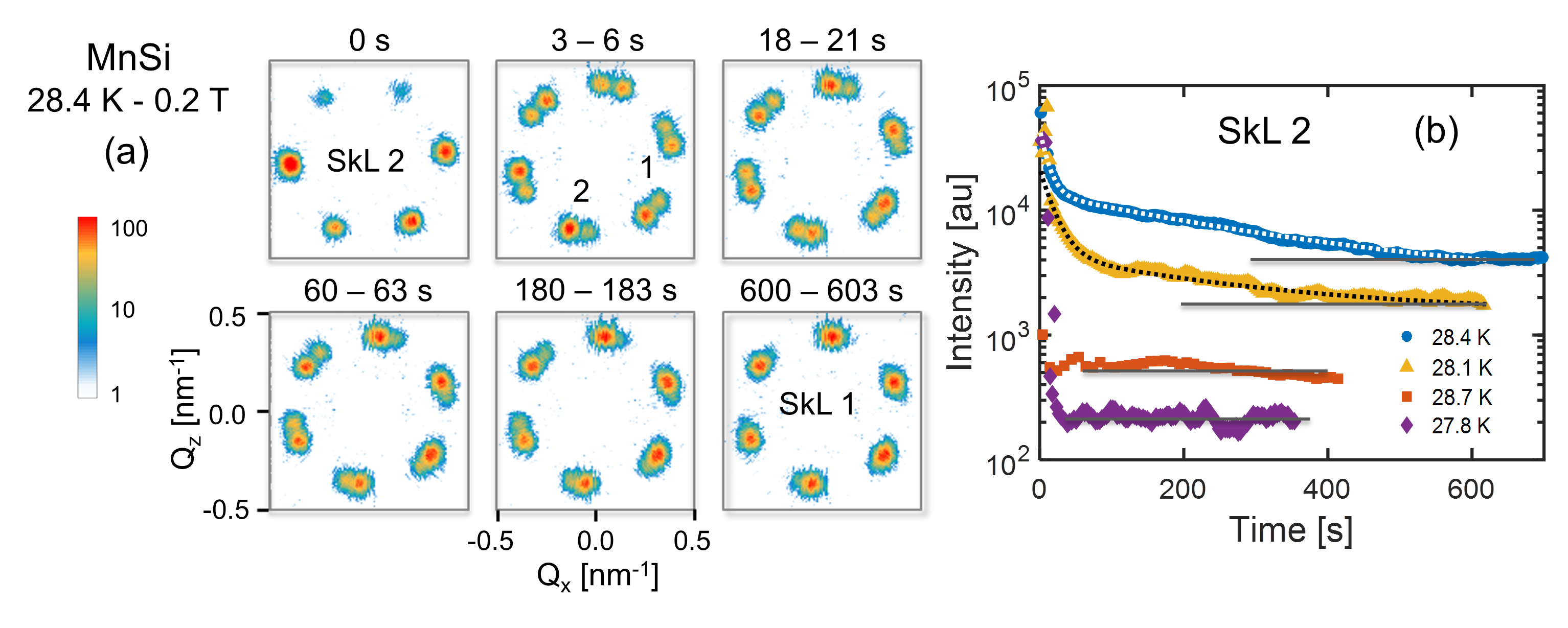
In order to follow the re-orientation from SkL 2 to SkL 1 as a function of time, we performed fast rotations from = 0∘ () to = 55∘ () while keeping the field on. The SANS patterns where recorded continuously using event mode data acquisition. Subsequently, they were processed such that a pattern was obtained for every 3 s. All patterns are displayed in Supplemental Movie 4foo and a selection of them is shown in Fig. 4(a). During the first 3 seconds, SkL 1 forms and coexists with SkL 2. Afterwards, the intensity of SkL 2 drops gradually whereas the intensity of SkL 1 increases. After 25 s, the rate at which the intensity of SkL 2 decreases slows down considerably, and it takes almost ten minutes for SkL 2 to completely disappear.
A quantitative analysis of this reorientation is provided by considering the time dependence of the total intensity of all six Bragg peaks of SkL 2. The results are displayed in Fig. 4(b) and show a fast decrease in intensity within the first 25 seconds followed by a slow decay. Therefore, we fitted the data to a superposition of two exponentials:
| (1) |
The fit at = 28.4 K provides = 3.1 0.1 104, = 1.1 0.1 104, = 2.5 0.2 104 and the two time constants = 11.2 0.6 s and = 3.0 0.3 102 s. As such, it shows that the relaxation is governed by two separate processes occurring on the seconds and minutes time-scales, respectively. These processes possibly reflect the movement of (topological) domain walls and/or their pinning to defects of the crystallographic lattice.Pöllath et al. (2017) Slow relaxations that originate from multiple processes have also been observed around the A-phase by ac magnetic susceptibility measurements in Fe1-xCoxSi.Bannenberg et al. (2016b)
The relaxation of skyrmion lattices does not change dramatically in the center of the A-phase. At a lower temperature of 28.1 K, the estimated values are = 18.9 0.4 s and = 2.6 0.2 102 s. Thus, the characteristic time of the slow relaxation remains almost unchanged, whereas that of the faster process is almost doubled. The relaxation times are very different near the low- and high temperature borders as shown for 27.8 and 28.7 K in Fig. 4(b). The acceleration of the relaxation is not surprising for the high temperature border where it can be attributed to increased thermal fluctuations. The acceleration at the low temperature limit of = 27.8 K may be due to the fact that at this temperature the SkL is stable only for .Bauer et al. (2010) Indeed, we observe that the skyrmion lattice relaxes to the conical phase at this temperature and the SkL is thus not stable for . This highlights the importance of anisotropy terms.
IV Discussion and Conclusion
The results presented above show that the skyrmion lattice orients along the crystallographic directions expected from spin-orbit coupling when the samples are zero field cooled. Indeed, based on a Ginzburg-Landau analysis,Mühlbauer et al. (2009); Münzer et al. (2010); Seki et al. (2012b) one expects from the relevant fourth and sixth order spin-orbit coupling terms and , with being the Fourier transform of the local magnetization , an alignment of the skyrmion lattice with one of the helical vectors as for MnSi, or as for Cu2OSeO3.
Multidomain lattices may form when several equivalent crystallographic directions, as preferred by spin-orbit coupling, are simultaneously perpendicular to the magnetic field. This is exemplified by the schematic drawings in Fig. 5. Figures 5(a) and 5(c) show that two domains of skyrmion lattices are expected for both Cu2OSeO3 and MnSi for = 0∘ and , where there are two favorable and crystallographic directions perpendicular to the field. In the other configuration of = 90∘ and shown in Fig. 5(b) and 5(d), only one and crystallographic direction is perpendicular to the field, and hence, only one SkL domain is expected for both chiral magnets. A comparison with experiment shows, however, that multidomain SkLs are nucleated only in Cu2OSeO3. This is in agreement with the literature, where, to our knowledge, no stable multidomain lattice has been reported for bulk single-crystal MnSi so far.
This important difference between Cu2OSeO3 and MnSi may be attributed to substantial differences in the most relevant terms in the free energy Landau-Ginzburg functional that contains the Ferromagnetic exchange, Dzyaloshinskii-Moriya interaction, Zeeman energy and anisotropy/spin-orbit coupling terms.Mühlbauer et al. (2009) The most obvious difference is in the coupling to the external field and the Zeeman energy, which is almost an order of magnitude stronger for MnSi than Cu2OSeO3. Indeed, the magnetic fields required to stabilize the skyrmion lattice phase are almost an order of magnitude stronger for MnSi than for Cu2OSeO3 although the volume magnetizations of both systems are very similar to each other.Bauer et al. (2010); Qian et al. (2016) An additional more subtle but most relevant difference is in the spin-orbit coupling that pins the skyrmion lattice to the crystallographic one. The higher order spin-orbit coupling terms seem to be significantly stronger for MnSi than for Cu2OSeO3. In the later, both the fourth and sixth order terms responsible for this coupling are very weak as pointed out by a previous study.Seki et al. (2012b) Consequently, multidomain lattices are stabilized in Cu2OSeO3 for a wide range of field directions with respect to the crystallographic lattice. A similar case has also been documented for Fe1-xCoxSi where the coupling of the skyrmion lattice to the crystallographic lattice is likely even weaker.Münzer et al. (2010); Bannenberg et al. (2016a) If the spin-orbit coupling is stronger, as is likely the case for MnSi, a different field orientation with respect to the crystallographic lattice has a larger impact on the energy levels of the energy minima. Thus, in MnSi both the stronger Zeeman and spin orbit coupling terms in conjunction with even small sample misalignments, imperfections, or demagnetizing fields would raise the degeneracy between different and equivalent skyrmion lattice domains and thus favor a single domain configuration. We therefore conjecture that multidomain lattices should also exist in MnSi, but only within a very narrow region of field orientations with respect to the crystallographic lattice that has not been realized experimentally until now.
Our results also show that the specific history of the magnetic field (direction) has a significant impact on the SkL orientation. When rotations are performed under field, the multidomain SkL stabilized for in Cu2OSeO3 evolves to a single-domain SkL. Upon further rotation, this single domain does not reorient to its zero-field cooled configuration, which one may assume to be the most energetically favorable one. On the other hand, the skyrmion lattice may reorient under certain conditions for MnSi involving macroscopic relaxation times and metastable multidomain lattices. Thus, for both systems relatively large energy barriers prevent SkLs from reorienting to their thermodynamically most favorable state. The existence of such high energy barriers is not surprising as such a reorientation of the skyrmion lattice involves a rearrangement of the magnetic configuration over very large (macroscopic) volumes. These results thus show that it is possible to induce metastable skyrmion states in Cu2OSeO3 and MnSi within the thermodynamic equilibrium skyrmion phase.
The stabilization of the multidomain SkL in Cu2OSeO3 has previously been attributed to magnetic field directions deviating from the major cubic axesZhang et al. (2016a, b) or to the thermal and magnetic history.Seki et al. (2012b) Our results show that specific magnetic field histories can indeed suppress multidomain lattices. However, in contrast to previous work,Zhang et al. (2016a, b) we find that multidomain SkLs can also be stabilized when the field is applied along a major cubic axis such as the crystallographic direction. The occurrence of multidomain lattices can thus be understood on the basis of symmetry arguments, as illustrated in Fig. 5.
In summary, the results presented above show that the orientation of the skyrmion lattice is governed primarily by the magnetic field direction with respect to the crystallographic lattice, but is also influenced by the magnetic history of the sample. Multidomain lattices may form when two or more equivalent crystallographic directions are favored by spin-orbit coupling and oriented perpendicular to the magnetic field. These results provide new insights into the factors that stabilize skyrmion lattices and influence their orientation. They shed new light on the puzzle of the occurrence of multiple skyrmion lattice domains, an issue that is of general relevance to chiral magnetism.
Acknowledgements.
The authors wish to express their gratitude to the ISIS and LLB technical support staff for their assistance and are grateful for the kind help of C. Decorse with aligning the single crystals. The experiments at the ISIS Pulsed Neutron and Muon Source were supported by a beamtime allocation from the Science and Technology Facilities Council and the Netherlands Organization for Scientific Research (NWO). The work of L.J.B and C.P. is financially supported by The Netherlands Organization for Scientific Research through Project No. 721.012.102. F.Q. thanks the China Scholarship Council for financial support. D.L.S. and T.A.L. acknowledge support from the U.S. Department of Energy, Office of Basic Energy Sciences, Division of Materials Sciences and Engineering under Contract No DE-AC02-07CH11358.References
- Mühlbauer et al. (2009) S. Mühlbauer, B. Binz, F. Jonietz, C. Pfleiderer, A. Rosch, A. Neubauer, R. Georgii, and P. Böni, Science 323, 915 (2009).
- Fert et al. (2013) A. Fert, V. Cros, and J. Sampaio, Nature Nanotechnology 8, 152 (2013).
- Nagaosa and Tokura (2013) N. Nagaosa and Y. Tokura, Nature Nanotechnology 8, 899 (2013).
- Milde et al. (2013) P. Milde, D. Köhler, J. Seidel, L. Eng, A. Bauer, A. Chacon, J. Kindervater, S. Mühlbauer, C. Pfleiderer, S. Buhrandt, et al., Science 340, 1076 (2013).
- Romming et al. (2013) N. Romming, C. Hanneken, M. Menzel, J. E. Bickel, B. Wolter, K. von Bergmann, A. Kubetzka, and R. Wiesendanger, Science 341, 636 (2013).
- Seki and Mochizuki (2015) S. Seki and M. Mochizuki, Skyrmions in Magnetic Materials (Springer, 2015).
- Bauer and Pfleiderer (2016) A. Bauer and C. Pfleiderer, in Topological Structures in Ferroic Materials (Springer, 2016) pp. 1–28.
- Münzer et al. (2010) W. Münzer, A. Neubauer, T. Adams, S. Mühlbauer, C. Franz, F. Jonietz, R. Georgii, P. Böni, B. Pedersen, M. Schmidt, et al., Physical Review B 81, 041203 (2010).
- Wilhelm et al. (2011) H. Wilhelm, M. Baenitz, M. Schmidt, U. K. Rößler, A. A. Leonov, and A. N. Bogdanov, Physical Review Letters 107, 127203 (2011).
- Seki et al. (2012a) S. Seki, X. Yu, S. Ishiwata, and Y. Tokura, Science 336, 198 (2012a).
- Seki et al. (2012b) S. Seki, J.-H. Kim, D. S. Inosov, R. Georgii, B. Keimer, S. Ishiwata, and Y. Tokura, Physical Review B 85, 220406 (2012b).
- Adams et al. (2012) T. Adams, A. Chacon, M. Wagner, A. Bauer, G. Brandl, B. Pedersen, H. Berger, P. Lemmens, and C. Pfleiderer, Physical Review Letters 108, 237204 (2012).
- Tokunaga et al. (2015) Y. Tokunaga, X. Z. Yu, J. S. White, H. M. Rønnow, D. Morikawa, Y. Taguchi, and Y. Tokura, Nature Communications 6, 7638 (2015).
- Karube et al. (2016) K. Karube, J. S. White, N. Reynolds, J. L. Gavilano, H. Oike, A. Kikkawa, F. Kagawa, Y. Tokunaga, H. M. Rønnow, and Y. e. a. Tokura, Nature Materials 14, 1116 (2016).
- Kézsmárki et al. (2015) I. Kézsmárki, S. Bordács, P. Milde, E. Neuber, L. Eng, J. White, H. M. Rønnow, C. Dewhurst, M. Mochizuki, K. Yanai, et al., Nature Materials 14, 1116 (2015).
- Tonomura et al. (2012) A. Tonomura, X. Yu, K. Yanagisawa, T. Matsuda, Y. Onose, N. Kanazawa, H. S. Park, and Y. Tokura, Nano Letters 12, 1673 (2012).
- Wilson et al. (2014) M. N. Wilson, A. B. Butenko, A. N. Bogdanov, and T. L. Monchesky, Physical Review B 89, 094411 (2014).
- Heinze et al. (2011) S. Heinze, K. Von Bergmann, M. Menzel, J. Brede, A. Kubetzka, R. Wiesendanger, G. Bihlmayer, and S. Blügel, Nature Physics 7, 713 (2011).
- Yu et al. (2012) X. Z. Yu, N. Kanazawa, W. Z. Zhang, T. Nagai, T. Hara, K. Kimoto, Y. Matsui, Y. Onose, and Y. Tokura, Nature Communications 3, 988 (2012).
- White et al. (2014) J. White, K. Prša, P. Huang, A. A. Omrani, I. Živković, M. Bartkowiak, H. Berger, A. Magrez, J. Gavilano, G. Nagy, et al., Physical Review Letters 113, 107203 (2014).
- Dzyaloshinski (1958) I. E. Dzyaloshinski, J. Phys. Chem. Solids 4, 241 (1958).
- Moriya (1960) T. Moriya, Phys. Rev 120, 91 (1960).
- Bak and Jensen (1980) P. Bak and M. H. Jensen, Journal of Physics C: Solid State Physics 13, L881 (1980).
- Buhrandt and Fritz (2013) S. Buhrandt and L. Fritz, Physical Review B 88, 195137 (2013).
- Bogdanov and Yablonskii (1989) A. N. Bogdanov and D. A. Yablonskii, Zh. Eksp. Teor. Fiz 95, 182 (1989).
- Bogdanov and Hubert (1994) A. N. Bogdanov and A. Hubert, Journal of Magnetism and Magnetic Materials 138, 255 (1994).
- Rößler et al. (2006) U. K. Rößler, A. N. Bogdanov, and C. Pfleiderer, Nature 442, 797 (2006).
- Butenko et al. (2010) A. B. Butenko, A. A. Leonov, U. K. Rößler, and A. N. Bogdanov, Physical Review B 82, 052403 (2010).
- Rößler et al. (2011) U. K. Rößler, A. A. Leonov, and A. N. Bogdanov, in Journal of Physics: Conference Series, Vol. 303 (IOP Publishing, 2011) p. 012105.
- Adams et al. (2011) T. Adams, S. Mühlbauer, C. Pfleiderer, F. Jonietz, A. Bauer, A. Neubauer, R. Georgii, P. Böni, U. Keiderling, K. Everschor, et al., Physical Review Letters 107, 217206 (2011).
- Zhang et al. (2016a) S. Zhang, A. Bauer, D. M. Burn, P. Milde, E. Neuber, L. M. Eng, H. Berger, C. Pfleiderer, G. V. V. van der Laan, and T. Hesjedal, Nano Letters 16, 3285 (2016a).
- Zhang et al. (2016b) S. L. Zhang, A. Bauer, H. Berger, C. Pfleiderer, G. V. V. Van der Laan, and T. Hesjedal, Physical Review B 93, 214420 (2016b).
- Makino et al. (2017) K. Makino, J. D. Reim, D. Higashi, D. Okuyama, T. J. Sato, Y. Nambu, E. P. Gilbert, N. Booth, S. Seki, and Y. Tokura, Physical Review B 95, 134412 (2017).
- Langner et al. (2014) M. C. Langner, S. Roy, S. K. Mishra, J. C. T. Lee, X. W. Shi, M. Hossain, Y. D. Chuang, S. Seki, Y. Tokura, S. D. Kevan, et al., Physical Review Letters 112, 167202 (2014).
- Mochizuki et al. (2014) M. Mochizuki, X. Z. Yu, S. Seki, N. Kanazawa, W. Koshibae, J. Zang, M. Mostovoy, Y. Tokura, and N. Nagaosa, Nature Materials 13, 241 (2014).
- Pöllath et al. (2017) S. Pöllath, J. Wild, L. Heinen, T. N. Meier, M. Kronseder, L. Tutsch, A. Bauer, H. Berger, C. Pfleiderer, J. Zweck, et al., Physical Review Letters 118, 207205 (2017).
- Bannenberg et al. (2016a) L. J. Bannenberg, K. Kakurai, F. Qian, E. Lelièvre-Berna, C. D. Dewhurst, Y. Onose, Y. Endoh, Y. Tokura, and C. Pappas, Physical Review B 94, 104406 (2016a).
- Reim et al. (2017) J. D. Reim, K. Makino, D. Higashi, Y. Nambu, D. Okuyama, T. J. Sato, E. P. Gilbert, N. Booth, and S. Seki, in Journal of Physics: Conference Series, Vol. 828 (IOP Publishing, 2017) p. 012004.
- Pappas et al. (2017) C. Pappas, L. J. Bannenberg, E. Lelièvre-Berna, F. Qian, C. D. Dewhurst, R. M. Dalgliesh, D. L. Schlagel, T. A. Lograsso, and P. Falus, Physical Review Letters 119, 047203 (2017).
- Note (1) The field homogeneity of the 3D cryomagnet, defined as ‘the maximum field error over a 10 mm diameter spherical volume’, is determined by the manufacturer as 0.11 % of the applied magnetic field.
- Note (2) We mounted a field probe on the window of the cryostat to check directly the residual field taking into account its decay with distance. This field probe confirmed the absence of a sizeable residual field. In addition, we made magnetic field scans from negative to positive fields to evaluate the residual field. As the plot of the integrated scattered intensity vs magnetic field was symmetric and centered around zero, a sizable residual field was absent.
- (42) See Supplemental Material for the supplemental movies. .
- Note (3) The scattering patterns of this rotation scan are equivalent to those obtained after zero field cooling the sample.
- Bannenberg et al. (2016b) L. J. Bannenberg, A. J. E. Lefering, K. Kakurai, Y. Onose, Y. Endoh, Y. Tokura, and C. Pappas, Physical Review B 94, 134433 (2016b).
- Bauer et al. (2010) A. Bauer, A. Neubauer, C. Franz, W. Münzer, M. Garst, and C. Pfleiderer, Physical Review B 82, 064404 (2010).
- Qian et al. (2016) F. Qian, H. Wilhelm, A. Aqeel, T. T. M. Palstra, A. J. E. Lefering, E. H. Brück, and C. Pappas, Physical Review B 94, 064418 (2016).