CO2 packing polymorphism under pressure: mechanism and thermodynamics of the I-III polymorphic transition
Abstract
In this work we describe the thermodynamics and mechanism of CO2 polymorphic transitions under pressure from form I to form III combining standard molecular dynamics, well-tempered metadynamics and committor analysis. We find that the phase transformation takes place through a concerted rearrangement of CO2 molecules, which unfolds via an anisotropic expansion of the CO2 supercell. Furthermore, at high pressures we find that defected form I configurations are thermodynamically more stable with respect to form I without structural defects. Our computational approach shows the capability of simultaneously providing an extensive sampling of the configurational space, estimates of the thermodynamic stability and a suitable description of a complex, collective polymorphic transition mechanism.
I Introduction
Polymorphism, namely the possibility that molecular crystals assemble in the solid phase in different crystal lattices, is ubiquitous in nature. The spatial arrangement of molecules is key in defining mechanical, physical, chemical, and functional properties of materials. Understanding the molecular details of the thermodynamics and mechanisms underlying polymorphism is therefore key to develop detailed, rational descriptions of many natural and industrial processes Cruz-Cabeza2015 ; Cruz-Cabeza2014 ; Price2008 ; Price2015 ; Zykova-Timan2008 ; Valdes-Aguilera1989 ; Vishweshwar2005 ; Bauer2001 .
In this direction, a notable effort is put in developing both ab initio and enhanced sampling techniques to predict polymorphs of a molecule (in particular, CSP techniques Price2008 ; Bond2016 ; Reilly2016 ; Dunitz2004 ), to evaluate their relative stability at finite temperature and pressure, i.e. at conditions relevant for the life-cycle of a solid product, and to study transition mechanism and kinetics.
Among enhanced sampling techniques, metadynamicsLaio2002 ; Martonak2003 ; Martonak2005 ; Martonak2007 ; Ceriani2004 ; Zipoli2004 ; Karamertzanis2008 ; Raiteri2005 ; Zykova-Timan2008 ; Lukinov2015 (MetaD) and adiabatic free energy dynamicsYu2011 ; Yu2014 ; Schneider2016 (AFED) are employed in literature to study polymorphism.
Indeed, over the years these techniques have been tested, developed and compared on benchmark systems and combined with CSP methods. Such works made a successful step towards the characterisation of solid phase transition, proving these tools to be powerful in the prediction of new structures and transition pathways as well as of the phase diagram without any a priori knowledge.
However, a complete and systematic investigation of polymorphic transitions is still challenging.
In this work our aim is to exploit state of the art enhanced sampling simulations to investigate the thermodynamics and transition mechanisms at play in polymorphic transitions. To this aim, we combine well-tempered MetaD and committor analysis in order to identify a suitable low dimensional description of the transition between two polymorphic phases in collective variable space. To do so, here we focus our attention on solid CO2, more precisely on the - polymorphic transition that characterises CO2 packing polymorphism. Packing polymorphism arises when two solid phases differ in the packing of molecules, which have all the same molecular structure, as opposite to conformational polymorphismCruz-Cabeza2014 .
In molecular solid phases, CO2 molecules maintain their gas phase conformation. To a first, crude, approximation, each molecule can in fact be described as rigid and the bending of the O-C-O 180∘ angle can be reasonably neglected. Thanks to such limited conformational flexibility, it is as easy as spontaneous to identify each CO2 molecule with a vector passing through its axis; moreover, the centre of mass corresponds to the carbon atom at every simulation time. As a result, the state of each molecule can be completely characterised by the position of its centre of mass and the vector representing its orientation in space.
Despite its simple molecular structure, CO2 has a rather complex solid-state phase diagramDatchi2009 (partially reported in Figure 1 (a)). Indeed, at high temperature and pressure, seven different crystal structures have been detected so far, among which many are still debatedDatchi2009 ; Datchi2012 ; Santoro2006 ; Yoo1999 ; Yoo2002 ; Park2003 ; Yoo2013 ; Giordano2007 ; Iota2001 ; Datchi2016 ; Iota2007 ; Shieh2013 ; Aoki1994 ; Bonev2003 ; Kuchta1988 ; Etters1989 ; Kuchta1993 ; Li2013 ; Hirata2014 . The first form detected was molecular phase I, also called dry ice, crystallised directly from the melt; phase III followed, obtained through the compression of dry ice; the discovery of a polymeric structure, classified as phase V, attracted more interest to the study of this system, resulting in the identification of two more phases, II and IV. Phases II and IV are currently object of discussion as different groups hold contrasting views on their nature and role in the transition between molecular to non-molecular phasesYoo2002 ; Bonev2003 ; Datchi2016 ; Park2003 ; Yoo2013 ; Santoro2006 ; Gorelli2004 ; Datchi2009 . Furthermore, an amorphous phase (VI) is also identified, and the existence of molecular form VII as a phase itself is still under investigation (see Figure 1 (a)).
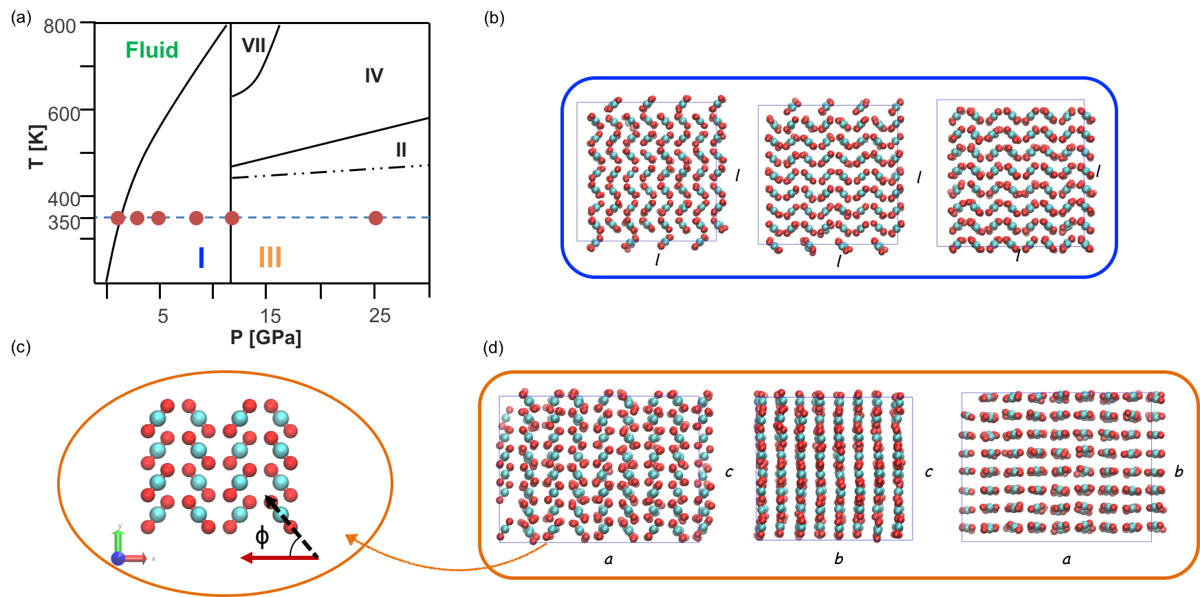
In this work we study the transition between phases I and III, which are largely accepted and well characterised in the literature (Figure 1 (b) and (d)). Their structural arrangements appear to hold several similarities. Both polymorphs, indeed, are face centred with four molecules in the unit cell, but while polymorph I’s lattice is cubic Pa, III’s is orthorombic Cmca. A major difference is the orientation of the CO2 particles: in phase I the molecular axis is in fact aligned with the diagonal of the cell, while in phase III they are arranged in parallel layers in which molecules describe a characteristic 52∘ angle, , with the side of the lattice (Figure 1 (c)).
The transformation takes place at around 11-12 (11.8) GPa Datchi2009 independently of temperature (); nevertheless defining the transition conditions is a difficult task, and the pressure transition range is suggested to be wider (7 - 15 GPa)Shieh2013 , while Olijnyk et al.Olijnyk1988 observe transition III to I under unloading at around 2.5 - 4.5 GPa at 80 K.
There is good agreement on the occurrence of a hysteresis of the specific volume, which decreases of about 2% from I to III Aoki1994 ; Bonev2003 . It is also generally accepted that the transition takes place through a concerted rotation of the molecules together with a deformation of the cubic structure to a parallelepiped, thanks to the peculiar geometrical features of the two phases.
The early works by Kuchta and Etters Kuchta1988 ; Etters1989 ; Kuchta1993 study such transition through NPT Monte Carlo (MC) simulations coupled with equalization of the Gibbs free energy in the phases under investigation, not without uncertaintiesKuchta1993 . Moreover, the authors identify the orientation of the molecules in the lattice as the most relevant feature changing in the transition and thus they employ it as a transition coordinate to estimate the free energy profile associated to the transformation; their calculations both at 0 KKuchta1988 and room temperatureKuchta1993 locate the transition pressure at 4.3 GPa.
Li et al.Li2013 apply instead the second order Møller – Plesset (MP2) technique to the study of molecular crystalsHirata2014 . Despite small inaccuracies, they reach a good agreement about the transition pressure (around 11.9 - 12.7 GPa), obtained evaluating the free energy of the two polymorphs at different T-P conditions.
Here we aim at complementing the state of the art by providing an extensive sampling of the configurational space explored during the I - III polymorphic transition while contextually identifying the dominant transition mechanism. In the first part we perform well-tempered metadynamics simulationsBarducci2008 with two order parameters as collective variables over a range of pressure at 350 K; from these, we obtain free energy surfaces that allow an insight into the relative stability between phase I and III under pressure. In the second part, we identify the most probable transition pathway and validate it through a committor analysis and a histogram testPeters2016 ; Tuckerman2010 on the transition state; we then propose a quantitative value for the energy barrier and a mechanism of the transition that takes into account quantitatively the reorientation of CO2 molecules in the crystal as well as the deformation of the box.
II Methods
To circumvent timescale limitations of standard molecular dynamics, enhanced sampling techniques are designed to accelerate the sampling of rare events. In this work we employ well-tempered metadynamicsBarducci2011 (WTMetaD). Briefly, WTMetaD is based on the introduction of a history-dependent bias potential (VG) along a low-dimensional set of collective variables (CVs)Abrams2013 ; Barducci2011 ; Barducci2008 ; Laio2008 ; Valsson2016 . Such bias allows for an efficient sampling of phase space, enhancing the escape from long-lived metastable states. Significantly, this result is achieved with little a priori knowledge of the free energy landscape, and provides an estimate of the unbiased free energy surface (FES, F(S)). For a detailed description of WTMetaD we refer the interested reader to Barducci et al.Barducci2008 ; Barducci2011 , and Valsson et al.Valsson2016 , and for a brief overview of its applications in crystallisation studies to Giberti et al.Giberti2015 .
Force field
Here, we employ the rigid three-site TraPPE force fieldPOTOFF1999 ; Potoff2001 (Table 1), with Lennard Jones potential and Lorentz-Berthelot combination rules.
This force field is chosen among a variety of models developed for CO2Santoro2006 ; Datchi2009 ; Aimoli2014 ; Perez-Sanchez2013 , since, even if it is not tailor-made for the high temperature and high pressure regime of interest, it outperforms other models in the description not only of the liquid-vapor equilibrium at high pressuresAimoli2014 (up to 100 MPa), of the melting curve of dry ice (up to 1 GPa) and the triple pointPerez-Sanchez2013 . Moreover, it has a better representation of the quadrupole, which is indeed relevant in carbon dioxide molecules and plays an important role in the solid phase stabilisationPrice1987 .
We employ two dummy atoms per moleculeSanghi2012 to mantain the desired rigidity and linearity of CO2, avoiding instability caused by the rigid 180∘ OCO angle.
The most relevant limitation of this model might be the rigidity of the CO2 moleculesLi2013 .
| mC | mO | [nm] | [nm] | [kJ/mol] |
|---|---|---|---|---|
| 12 | 16 | 0.280 | 0.305 | 0.224 |
| [kJ/mol] | qc [e] | qo [e] | [Å] | [∘] |
| 0.657 | 0.70 | -0.35 | 1.160 | 180 |
Simulation setup
Long-range corrections for the Van der Waals interactions are included through the particle mesh Edwald (pme) method. From consistency checks on the effect of the cut-off value on the system volume and energy when coupled with pme, we find that 0.7 nm is a good trade-off between accuracy and computational cost.
Isothermal and isobaric (NPT) simulations use Bussi-Donadio-Parrinello thermostat Bussi2007 and Berendsen anisotropic barostatBerendsen1984 for T and P control, respectively. The timestep employed is 0.5 fs.
For WTMetaD, the initial height of the Gaussians is 10 kJ/mol, with width 7.81e-3 for both CVs. The biasfactor is either 100 or 200 to allow the exploration of a wide portion of the phase-space. Moreover, we limit the elongation of each box side at 1.7 to 3.0 nm through the introduction of a repulsive potential.
This action prevents an excessive and irreversible distortion of the box when the transition to melt is observed under anisotropic control. We highlight that such restraints are active only when the system undergoes large fluctuations in the liquid state.
The T-P conditions investigated include areas of the phase diagram where the most stable phase changes from melt to phase I to III: at 350 K, the range of pressure of the present study spans from 1 to 25 GPa (1, 3, 5, 8, 12, 25 GPa).
The initial configuration of WTMetaD simulations is phase I, initially equilibrated for 500 ps at NVT, then 5 ns NPT without pme and additionally 5 ns NPT with pme. All simulation boxes contain supercells of 256 CO2 molecules.
We perform MD and WTMetaD simulations with Gromacs 5.2.1Abraham2015 and Plumed 2.2Tribello2014 ; the building of the cells and the post processing of the data employs mainly VMDHUMP96 , to visualise trajectories, and MATLAB (R2015a).
Committor Analysis
As mentioned in the opening, we complement our WTMetaD simulations with a committor analysis. While for a detailed description we refer to TuckermanTuckerman2010 and PetersPeters2016 , we recall here some useful definitions and procedures.
The committor is defined as the probability that a trajectory initiated from a configuration with velocities sampled from a Maxwell-Boltzmann distribution will arrive in state B before state ATuckerman2010 .
In our study we identify A as phase I and B as III. An important point on the pathway connecting two basins is the transition state (TS, indicated with *), which is the ensemble of configurations r with CV S(r)=S* that have committor ; on free energy hypersurfaces, it corresponds to a saddle point, i.e. the highest energy state along the minimum energy path connecting two basins.
To locate the saddle point, we extract 135 configurations along the transition pathway and for each of them we run between 10 to 40 unbiased NPT simulations with different initial velocities randomly generated from a Maxwell-Boltzmann distribution. Simulations are stopped when they commit either basin I or III and they are assigned an outcome value of 0 or 1, respectively. The average of the outcome values obtained from the set of trajectories generated for a given configuration provides an estimate of the committor for that configuration.
We have further analysed the histogram test, which, instead, studies the committor distribution, , which is the probability that a configuration r with S(r)=S* has committor .
The shape of this distribution is a descriptor of the capability of the CVs to represent the transition mechanism: a Gaussian distribution results from good CVs, while a flat or parabolic distribution corresponds to CVs that do not describe adequately the transition state ensemble.
To evaluate the committor probability, we consider 41 configurations with CVs close to the estimated transition state, and for each of them, we evaluate the committor, , as previously described, and build the histogram of .
Collective variables
In this work, we use a CV developed by Salvalaglio et al.Salvalaglio2012 ; Salvalaglio2013 ; Salvalaglio2015 and employed also in Giberti et al.Giberti2015_2 . In particular, every crystal structure has a unique typical local environment around each CO2 molecule, a fingerprint of the arrangment, and this order parameter, hereafter called , exploits this feature to effectively distinguish polymorphs. Indeed, describes crystallinity, a global property of the ensemble, as the sum of local contributions, ; each takes into account both the local density, , within a cut-off, , around the -th molecule, and the orientation, , respect to its neighbours (Figure 2 (a)). The value of ranges between 0 and 1, as it expresses the portion of molecules in the system that are ordered according the geometry of a defined polymorphic structure.
A complete description of the formulation of this parameter is reported in the and the cited literature.
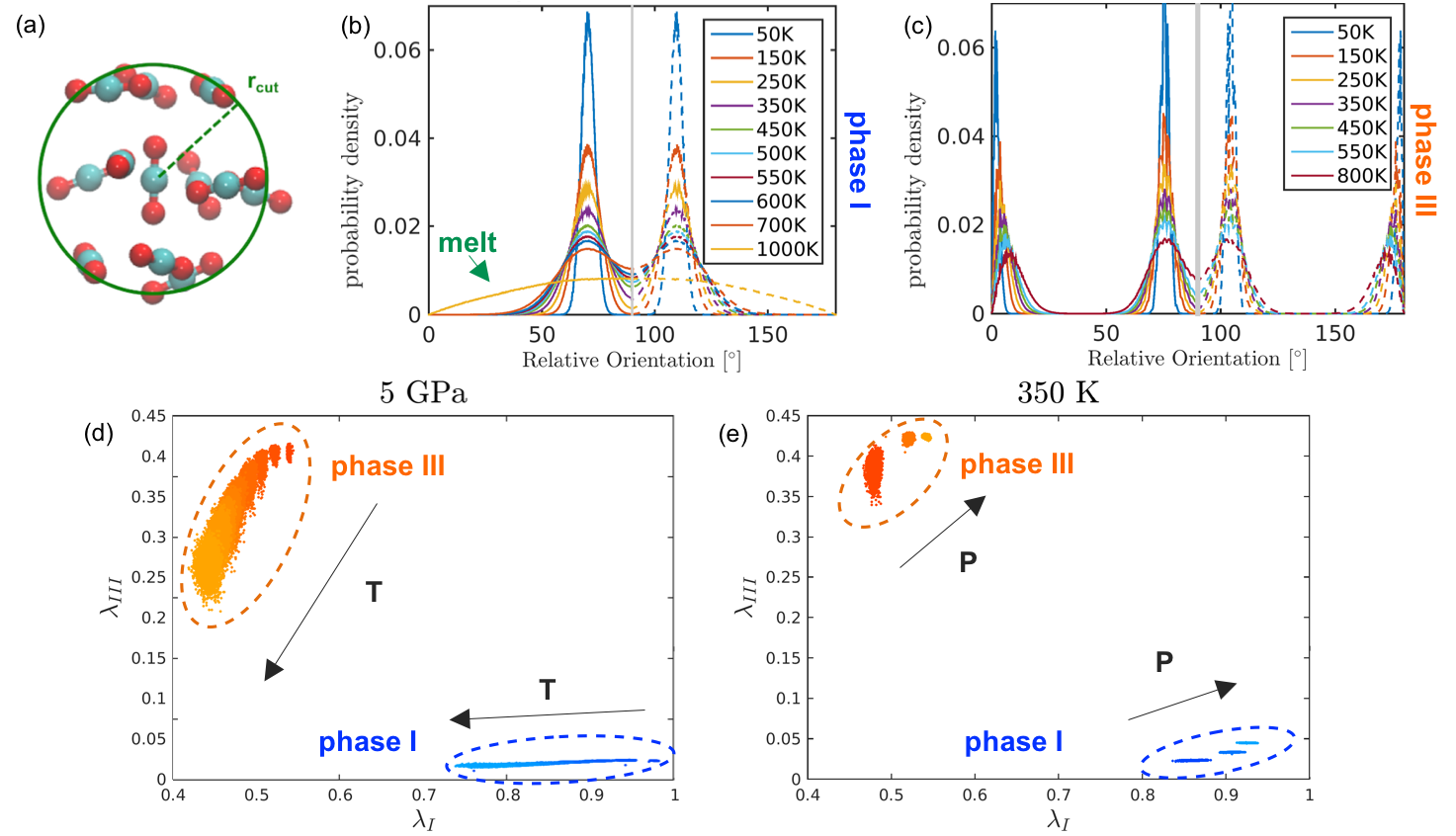
From the characterization of the local order in polymorphs I and III we can observe and compare peculiarities of the angle distribution of each phase (Figure 2 (b) and (c)), useful in the following tuning of CVs. First of all, the arrangements of phases I and III present similarities, as there is overlap between the distributions of two characteristic angles, which are however centred in different values (in around 70.2∘ and 109.8∘ for form I, while in around 75.6∘ and 104.4∘ for III). Moreover, phase III populates two additional characteristic angles, with values smaller than 10∘ and bigger than 170∘, which might relate to the presence of layers. We remark also that the melt has a sinusoidal distribution of angles, consistent with a random orientation of molecules. As a final note, increasing temperature enhances the fluctuations of the molecules in the crystal without modifying the mean value of the characteristic angles; an exception to this are the layer angles of form III that, instead, change from 1∘ to 8∘ and from 179∘ to 172∘ with growing temperature.
The number of neighbours in the first coordination shell shows, instead, a narrow distribution and the same value for the two structures, i.e. 12. Such observations lead to the tuning two CVs, namely and (see ).
Order parameter
This CV expresses the degree of phase I-likeness. The purpose of the tuning is to maximize when the crystal structure is phase I. To reach this aim, two characteristic angles, , are included, which are the ones of phase I (Table 2).
Order parameter
Similarly, the tuning of aims at maximise the parameter in presence of phase III. However, in this case we do not talk about phase III-likeness, because as we select only the specific angles that characterize layers (Table 2).
For both CVs, the cut-off is set to 4.0 Å, as it delimits the first coordination shell; the width of the Gaussians associated to the angles, , instead, is in both cases set to maximise the difference between the value of in phase I and melt and it is the same for both the characteristic angles due to the symmetry (Table 2).
| [∘] | [∘] | [∘] | ncut [-] | rcut [Å] | |
|---|---|---|---|---|---|
| 70.47 | 108.86 | 14.32 | 5 | 4 | |
| 8.02 | 171.89 | 11.46 | 5 | 4 |
The phase-space evaluated on unbiased MD simulations suggests that the CVs are effective in the distinction of separate and well-defined areas for each phase (Figure 2 (d) and (e)). Furthermore, the average order parameters can be extracted as an ensemble average.
Temperature and pressure act on the location where phases are projected in CV-space: on the one hand, increasing temperature decreases the values of both s while widening their fluctuations, consistently with the fact that the volume increases and the molecules vibrate more; on the other hand, increasing pressure leads to an increase in the absolute value of the parameters, while narrowing their distribution.
III Results
III.1 Free Energy of the I-III polymorphic transition as a function of pressure
In the following, we present the results of our study of the I - III polymorphic transition in CO2.
Firstly, we just mention that from preliminary MD unbiased simulations phase III has a smaller volume than phase I under all conditions investigated (2%, in agreement with experimental results); the volume predictions, however, slightly overestimate the experimental values (Figure LABEL:imageComp in the Supporting Information). In addition, for the same T-P settings, form I presents a lower potential energy than III: the potential energy of the system is thus not a good indicator of the relative thermodynamic stability at finite temperature. We report the outcome of MD in the Supporting Information.
Then, we discuss the results obtained from WTMetaD simulations run with the set-up discussed before.
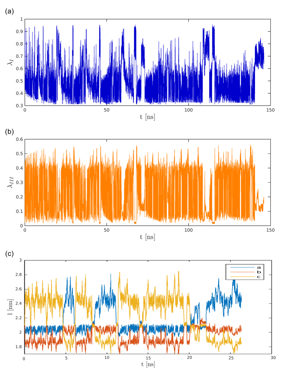
To begin with, we observe the temporal evolution of the CVs, for the explicative case at 350 K - 5 GPa (Figure 3 (a) and (b)); the other conditions investigated (Figure LABEL:imageCons in the Supporting Information) behave in a reasonably similar way. In the plots in Figure 3 it is possible to identify the system arranged in phase I as (a) is high (fluctuations between 0.7 and 0.9), (b) is below 0.05 and does not present relevant fluctuations, while the box edges (c) have the same length. The exploration of phase III’s basin, instead, shows wider fluctuations in the range of 0.36 - 0.6 for (a), and 0.1 - 0.4 for (b); the box edges, moreover, fluctuates around the unbiased average. Thanks to this clarification, it is possible to spot in Figure 3 that the system undergoes a significant number of recrossings between polymorphs I and III, in particular, four in slightly more than 5 ns at the beginning of the run. In addition, the system explores areas of the CV-space which do not represent any of these polymorphs, feature that will result more evident from the plots of the free energy surface (Figure 4). On the same surfaces it will be possible to notice the important role that the mentioned fluctuations of the CVs have on the shape of the basins for the two phases.
Furthermore, by observing the output trajectories and data of WTMetaD, we remark two interesting behaviours: on the one hand, CO2 molecules in the simulation box rearrange with a concerted motion during a phase transition; on the other hand, we find that such transition is anisotropic, meaning that each side of the box is equally likely to either elongate or shorten from I to III (Figure 3 (c)). In particular, this latter observation is important since by biasing as CVs order parameters that account for the spacial orientation of molecules, we obtain the consequential deformation of the supercell, without considering the box volume or edges as CVs, as instead done in previous MetaD works on polymorphismZykova-Timan2008 ; Martonak2003 ; Martonak2005 ; Martonak2007 ; Ceriani2004 ; Karamertzanis2008 ; Raiteri2005 ; Lukinov2015 .
Next, we present the free energy surfaces (FES) reconstructed by WTMetaD. In such FESs, the free energy is expressed as a function of the CVs: G(, ) (Figure 4 (a) and (f) to (i)). Before proceeding with the discussion, we underline that the FES at 25 GPa is not reported, as no recrossing is sampled from phase III; in addition, the results at 1 GPa are taken into account only qualitatively, since under such conditions phase III is so unstable that spontaneously evolves to I in standard MD and it is thus not possible to locate its basin.
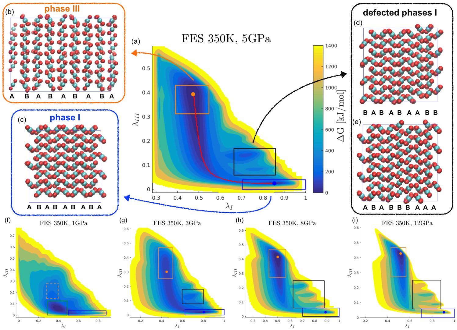
Some considerations can be drawn from the study of the FESs. First of all, the location of the minima on the FES for phases I and III is accurately close to the prediction in Figure 2(d)-(e). Moreover, phase III has a much wider basin than phase I and it develops mainly along , while phase I’s mainly along , as underlined for the temporal evolution of the CVs (Figure 3). As mentioned before, the system explores a wide area of CV-space and, in particular, the presence of black boxes in Figure 4 highlights the presence of defected phase I structures, which we shall analyse in detail later on. Relevant structural arrangements are reported in Figure 4 (b) to (e).
In order to compare the results of WTMetaD with the experimental phase diagram, we study quantitatively the relative stability between polymorphs.
Keeping in mind that the free energy is a function of the probability distribution of the CVs, it is possible to evaluate as (1):
| (1) |
Where is the probability of phase I, of phase III, and is 1/kT. The probability of each phase is computed as the integral of the distribution within the basin it occupies on the CV-space:
| (2) |
| (3) |
The integration domains are identified by coloured boxes on the FES in Figure 4 (a) and (f) to (i). In Figure 5 (a) we report relevant values over the range of pressure considered.
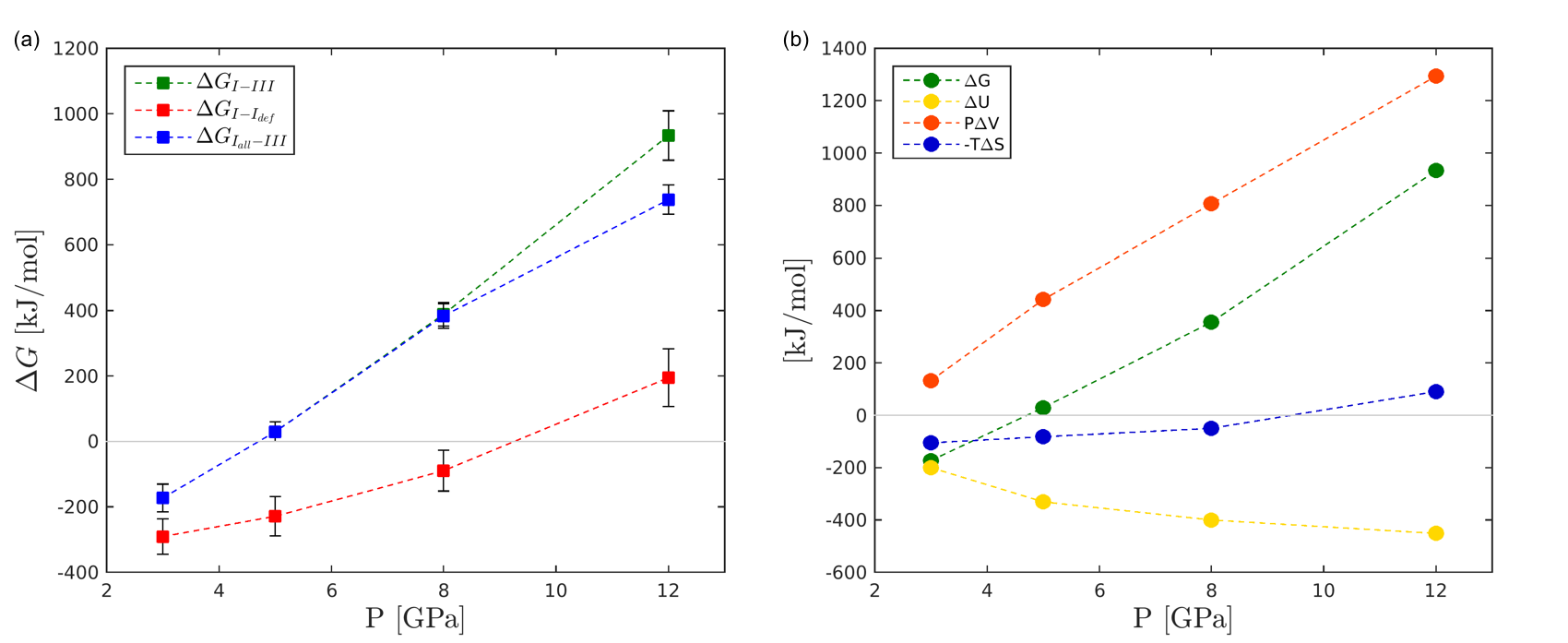
The relative stabilities in Figure 5 (a) together with the FESs in Figure 4 allow to draw some considerations on the phase diagram.
We observe that while the boundary of the solid - melt transition is in good agreement with experiments, the I - III transition pressure appears underestimated.
From the pattern shown in green in Figure 5 (a), the transition pressure at 350 K can be estimated as around 4.5 GPa. Despite underestimating the experimental value, the transition pressure agrees with literature results obtained treating CO2 as a rigid moleculeKuchta1988 ; Etters1989 ; Kuchta1993 . We also recall that commonly experimental works rather than a single value report a transition pressure interval (see Introduction), to which our estimation is closer.
Nevertheless, it is possible to notice that WTMetaD simulations are able to represent the overall trend observed in the phase diagram: increasing pressures increase the stability of phase III, while at decreasing values of pressure phase I is more stable, ultimately reaching the boundary with melt.
Since the behaviour of solid carbon dioxide is so well described, it is possible to consider a translation of the phase diagram.
A further step in the analysis of the I - III relative stability is the breakdown of the free energy in its internal energy, mechanical work and entropy contributions. With this aim, we firstly evaluate the difference in internal energy, and mechanical work, P, between phase I and phase III from the ensemble averages computed from the unbiased MD simulations; the entropy is thus obtained from the macroscopic definition of Gibbs free energy:
| (4) |
From the results in Figure 5 (b) it possible to notice some major features. First of all, the internal energy, , stabilizes form I, while P is significant in the stabilisation of phase III; in both cases their contribution becomes more relevant with growing pressures. The entropic term, instead, tends to favour form I, a part from pressure of 12 GPa.
Defected phases
As mentioned in the analysis of the CVs (Figure 3) and of the FESs (Figure 4), at pressure equal and above 3 GPa, the system evolves to new, not a priori known phases, which we recognise being defected structures I (Figure 4 (d) and (e)). Indeed, such phases are similar to phase I, being almost cubic and having comparable arrangement; however, they display packing faults, planar defects that break the orientation motif recognisable in perfect phase I. The perfect arrangement, in fact, presents the repetition of rows of CO2 that alternate the orientation respect to the Cartesian axis in a sort of ABABABAB sequence (Figure 4 (c)), while the defected phases replicate two or more lines with the same “character”, AA or BB (Figure 4 (d) and (e)). It is particularly remarkable the capability of WTMetaD to predict the production of defected structures, as, despite this phenomenon can takes place in experiments, it is “underrepresented in the current literature”Sosso2016 , due to its difficult characterization both experimentally and through modelling.
In addition, we observe that the stability of the defected phases increases at higher pressures, becoming ultimately even more stable than phase I ( in red in Figure 5 (a)).
This behaviour may be due to a more difficult expansion of volume from phase III to I under higher pressure, and thus defected phases with a smaller volume form.
To complete the analysis of these phases, we run unbiased simulations under the same T-P conditions as the related WTMetaD, and with the defected arrangement of interest as initial configuration. The results show that these forms do not spontaneously undergo any transition: the creation and correction of defects is thus an activated event.
III.2 Committor analysis
In the first part of this work, we have shown that our -order parameters are effective CVs in sampling the transition between polymorphs I and III, evaluating their relative stability, and exploring the phase space. In the following, we focus instead on the mechanism of the title transition. In particular, such analysis allows to evaluate the goodness of the CVs in the representation of the process, and to estimate quantitatively the transition pathway and the energy barrier to overcome.
First of all, we characterize the minimum free energy path (MFEP) that connects the free energy minima corresponding to phase I and III. The MFEP provides a representation in CV-space of the most probable set of intermediate states involved in the transition. Furthermore, the free energy profile along this path yields an estimate of the free energy barrier associated to the polymorphic transition. As initial estimate of the MFEP we propose an approximation obtained as the combination of the projection of FES along the CVs, more precisely, of basin I along and of basin III along , due to the observe typical L-shaped FES; further details about these approximations are provided in the Supporting Information. We then employ such path as educated guess for an optimisation routine that enables to obtain the actual MFEP from a series of trial moves, whose acceptance is based on the free energy value. The algorithm is robust and the path converges to the same route from different and less educated initial guesses.
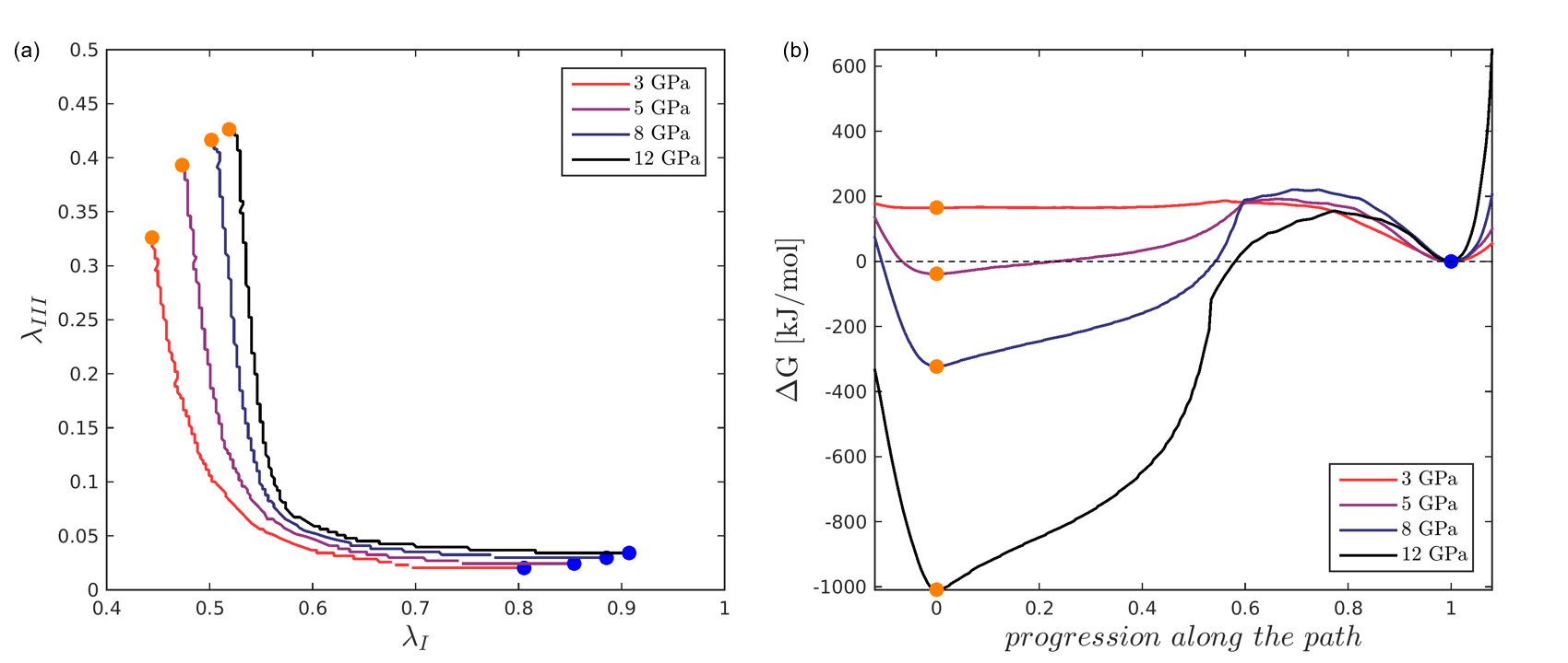
In Figure 4 (a) we report the MFEP on the FES at 350 K - 5 GPa, while in Figure 6(a) we compare transition pathways evaluated at different pressures. Interestingly, pressure only slightly affects the typical L-shape of the transition pathway, with the major difference being the location of the minima. Moreover, at this level of detail, the energy barrier to overcome from phase I to III appears similar at all pressures investigated (Figure 6(b)). We also highlight that the MFEP converges much earlier than the simulation, and no alternative routes connecting polymorphs I - III arise (Figure S7 in the Supporting Information).
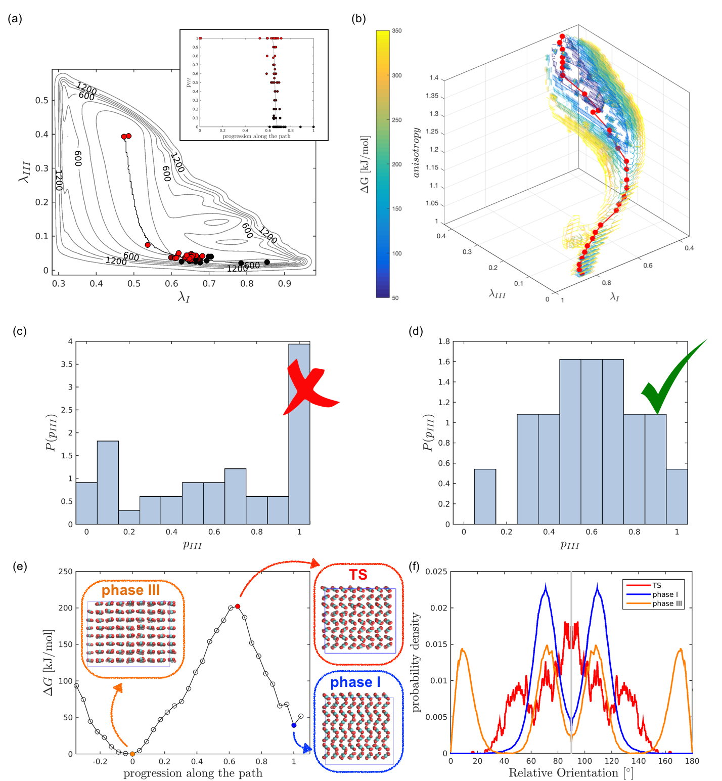
The next step towards a quantitative characterization of the transition mechanism is the validation of MFEP through a committor analysisPeters2016 ; Tuckerman2010 , with histogram test on the apparent transition state. We discuss hereafter the explicative case of 350 K - 5 GPa.
To begin with, we locate the transition state by evaluating the committor of 135 configurations extracted along the MFEP. Interestingly, configurations with committor different from zero and one are not evenly distributed along the transition pathway, but grouped in a narrow area around the saddle point, estimated in = 0.65, = 0.034; as a result, the cumulative distribution along the path resembles a very steep shape (Figure 7(a)). The behaviour shown in Figure 7(a) suggests that the order parameters alone might not be enough to account for the transition mechanism. The validation proceeds with a histogram test on the saddle point. We evaluate the committor of 41 configurations with (r) around and represent the results on a histogram (Figure 7 (c)). Such histogram shows three peaks, sign that our CVs alone are not effective reaction coordinates and other parameters need to be included in the mechanism description.
In order to identify the additional parameters to take into account, we deepen our analysis and further investigate the dependence of on properties such as potential energy, volume and box dimensions (Figure S6 in the Supporting Information). The results suggest that the deformation of the lattice plays a role in the representation of I - III transition mechanism. We define this deformation through the simulation box anisotropy, i.e. the ratio between the longest and the shortest sides of the cell; its value spans from 1 in cubic phase I to 1.35 in orthorhombic phase III. As a result, we note that only configurations r along the pathway with anisotropy of the box between 1.14 and 1.145 have committor non identical to 0 or 1, and, in particular the TS is uniquely located in = 0.65, = 0.034, = 1.1421, whise characteristic orientations are presented in Figure 7 (f). We thus repeat the histogram test on 19 configurations with CVs and anisotropy close to the TS and the outcome shows, as expected, a Gaussian shape (Figure 7 (d)): as a result, to effectively describe the mechanism of the I - III transition of solid CO2 all three parameters, namely , and , have to be taken into account.
We thus evaluateBonomi2009 the free energy as a function of the three parameters of interest: . On such FES we identify the 3D MFEP that connects phase I to phase III (Figure 7 (b)): its projection on the plane reasonably overlaps with the MFEP previously evaluated; moreover the anisotropy of the box monotonically increases from I to III, and vice versa.
Summing up the analysis carried on in this second part of the work, the transition from cubic phase I to orthorhombic phase III can be thus described as the sequence of the following actions:
-
•
The CO2 molecules firstly tend to distort the typical phase I lattice and, as a consequence, the value of decreases, with no relevant increase of (horizontal branch of the L-shaped pathway); at the same time the box starts deforming, elongating one side and reducing the others, thus increasing its anisotropy.
-
•
Then, when the deformation of the cell reaches the anysotropy threshold value of the transition state, the system completes the rearrangement to phase III; indeed, the molecules start organizing into parallel layers and the volume decreases. From this point the transformation proceeds on the vertical branch of the L-shaped pathway, with increasing for relative small variations of .
The motion of the molecules in the crystal during the transition is concerted.
From the investigation of the 3D MFEP we obtain also quantitative information about the height of the barrier for the polymorphic transformation: for a system composed by 256 CO2 molecules at 350 K - 5 GPa the transition state is located at about 202 kJ/mol (Figure 7(e)), with reference zero in phase III, i.e. the absolute minimum of the FES.
IV Conclusions
In this work we presented an investigation of the I - III polymorphic transition in carbon dioxide under pressure. Our approach combines molecular dynamics, well-tempered metadynamics and committor analysis to provide a broad insight into this phenomenon.
Firstly, we performed WTMetaD simulations at 350 K over a range of pressure (1 - 25 GPa) with two order parameters as CVs. These parameters, and , are built on the local order around each CO2 molecule and account for the reorientation of the molecules in the crystal. This feature allows to clearly distinguish in the - CV space configurations that belong to phase I or phase III and to clearly resolve amorphous configurations. Moreover, metadynamics exploration with these CVs allows to sample the formation of packing faults in phase I. Interestingly, we observe the deformation of the cell, a global rearrangement of the configuration, taking place as a consequence of enhanced sampling along and , which account for local order. This also permitted to notice that, in a I to III transition, all sides of the box have the same probability to elongate or shorten.
From the FESs resulting from WTMetaD, we evaluated the free energy difference between polymorphs; we observed that the predicted trend of the I - III relative stability over pressure is in agreement with the carbon dioxide phase diagram: increasing pressures move from the melt-phase I boundary, to the region of stability of phase I, to the one phase III. We estimate the transition pressure at 4.5 GPa, in agreement with previous literature works that considered carbon dioxide as a rigid moleculeKuchta1988 ; Etters1989 ; Kuchta1993 . Furthermore, our model suggest that the stability of the defected configurations increases with pressure. While at low pressure undefected form I is more stable than the ensemble of its defected counterparts, at high pressure the latter appears to dominate.
Alongside a description the I-III transition thermodynamics, we assess the I-III polymorphic transition mechanism for the representative case at 350 K - 5 GPa. To this aim we identify the MFEP connecting phase I to phase III in CV space and we validated the pathway carrying out committor analysis and histogram test on an ensemble of configurations corresponding to the saddle point in CV space. From this analysis it emerges that to quantitatively identify the transition mechanism we need to consider the anisotropic deformation of the supercell alongside order parameters accounting for the local arrangement of molecules. This analysis allowed to identify a reliable approximation of the transition pathway, and hence to quantify the free energy barrier associated with the transition.
Our work shows that, by combining opportunely designed order parameters with state-of-the-art enhanced sampling methods and committor analysis, we can provide an in-depth characterisation of both thermodynamics and transition mechanisms of polymorphic transformations at finite temperature.
Supplementary Material
See supplementary material for further details on the collective variables, and additional results on MFEP, committor analysis, and unbiased simulations.
Acknowledgements
The authors acknowledge EPSRC (Engineering and Physical Sciences Research Council) for PhD scholarship, and UCL Legion High Performance Computing Facility for access to Legion@UCL and associated support services, in the completion of this work.
References
References
- [1] Aurora J. Cruz-Cabeza, Susan M. Reutzel-Edens, and Joel Bernstein. Facts and fictions about polymorphism. Chem. Soc. Rev., 44(23):8619–8635, 2015.
- [2] Aurora J. Cruz-Cabeza and Joel Bernstein. Conformational Polymorphism. Chemical Reviews, 114(4):2170–2191, feb 2014.
- [3] Sarah L Price. Computational prediction of organic crystal structures and polymorphism. International Reviews in Physical Chemistry, 27:541–568, July 2008.
- [4] Sarah L Price. Crystal Energy Landscapes for Aiding Crystal Form Selection. In Computational Pharmaceutics, chapter 2, pages 7–29. John Wiley & Sons, Ltd, Chichester, UK, may 2015.
- [5] T. Zykova-Timan, P. Raiteri, and M. Parrinello. Investigating the Polymorphism in PR179: A Combined Crystal Structure Prediction and Metadynamics Study. The Journal of Physical Chemistry B, 112(42):13231–13237, oct 2008.
- [6] Oscar Valdes-Aguilera and D. C. Neckers. Aggregation phenomena in xanthene dyes. Accounts of Chemical Research, 22(5):171–177, may 1989.
- [7] Peddy Vishweshwar, Jennifer A. McMahon, Mark Oliveira, Matthew L. Peterson, and Michael J. Zaworotko. The Predictably Elusive Form II of Aspirin. Journal of the American Chemical Society, 127(48):16802–16803, dec 2005.
- [8] John Bauer, Stephen Spanton, Rodger Henry, John Quick, Walter Dziki, William Porter, and John Morris. Ritonavir: An Extraordinary Example of Conformational Polymorphism. Pharmaceutical Research, 18(6):859–866, 2001.
- [9] Andrew D. Bond. Introduction to the special issue on crystal engineering. Acta Crystallographica Section B: Structural Science, Crystal Engineering and Materials, 70(1):1–2, feb 2014.
- [10] Anthony M. Reilly, Richard I. Cooper, Claire S. Adjiman, Saswata Bhattacharya, A. Daniel Boese, Jan Gerit Brandenburg, Peter J. Bygrave, Rita Bylsma, Josh E. Campbell, Roberto Car, David H. Case, Renu Chadha, Jason C. Cole, Katherine Cosburn, Herma M. Cuppen, Farren Curtis, Graeme M. Day, Robert A. DiStasio Jr, Alexander Dzyabchenko, Bouke P. van Eijck, Dennis M. Elking, Joost A. van den Ende, Julio C. Facelli, Marta B. Ferraro, Laszlo Fusti-Molnar, Christina-Anna Gatsiou, Thomas S. Gee, René de Gelder, Luca M. Ghiringhelli, Hitoshi Goto, Stefan Grimme, Rui Guo, Detlef W M Hofmann, Johannes Hoja, Rebecca K. Hylton, Luca Iuzzolino, Wojciech Jankiewicz, Daniël T. de Jong, John Kendrick, Niek J. J. de Klerk, Hsin-Yu Ko, Liudmila N. Kuleshova, Xiayue Li, Sanjaya Lohani, Frank J J Leusen, Albert M. Lund, Jian Lv, Yanming Ma, Noa Marom, Artëm E. Masunov, Patrick McCabe, David P. McMahon, Hugo Meekes, Michael P. Metz, Alston J. Misquitta, Sharmarke Mohamed, Bartomeu Monserrat, Richard J. Needs, Marcus A. Neumann, Jonas Nyman, Shigeaki Obata, Harald Oberhofer, Artem R. Oganov, Anita M. Orendt, Gabriel I. Pagola, Constantinos C. Pantelides, Chris J. Pickard, Rafal Podeszwa, Louise S. Price, Sarah L. Price, Angeles Pulido, Murray G. Read, Karsten Reuter, Elia Schneider, Christoph Schober, Gregory P. Shields, Pawanpreet Singh, Isaac J. Sugden, Krzysztof Szalewicz, Christopher R. Taylor, Alexandre Tkatchenko, Mark E. Tuckerman, Francesca Vacarro, Manolis Vasileiadis, Alvaro Vazquez-Mayagoitia, Leslie Vogt, Yanchao Wang, Rona E. Watson, Gilles A. de Wijs, Jack Yang, Qiang Zhu, and Colin R. Groom. Report on the sixth blind test of organic crystal structure prediction methods. Acta Crystallographica Section B Structural Science, Crystal Engineering and Materials, 72(4):439–459, aug 2016.
- [11] Jack D. Dunitz and Harold A. Scheraga. Exercises in prognostication: Crystal structures and protein folding. Proceedings of the National Academy of Sciences, 101(40):14309–14311, oct 2004.
- [12] Alessandro Laio and Michele Parrinello. Escaping free-energy minima. Proceedings of the National Academy of Sciences, 99(20):12562–12566, oct 2002.
- [13] R. Martoňák, A Laio, and M Parrinello. Predicting Crystal Structures: The Parrinello-Rahman Method Revisited. Physical Review Letters, 90(7):075503, feb 2003.
- [14] Roman Martoňák, Alessandro Laio, Marco Bernasconi, Chiara Ceriani, Paolo Raiteri, Federico Zipoli, and Michele Parrinello. Simulation of structural phase transitions by metadynamics. Zeitschrift für Kristallographie - Crystalline Materials, 220(5/6):1–11, jan 2005.
- [15] Roman Martoňák, Davide Donadio, Artem R. Oganov, and Michele Parrinello. From four- to six-coordinated silica: Transformation pathways from metadynamics. Physical Review B, 76(1):014120, jul 2007.
- [16] C. Ceriani, A. Laio, E. Fois, A. Gamba, R. Martoňák, and M. Parrinello. Molecular dynamics simulation of reconstructive phase transitions on an anhydrous zeolite. Physical Review B, 70(11):113403, sep 2004.
- [17] F. Zipoli, M. Bernasconi, and R. Martoňák. Constant pressure reactive molecular dynamics simulations of phase transitions under pressure: The graphite to diamond conversion revisited. The European Physical Journal B, 39(1):41–47, may 2004.
- [18] Panagiotis G. Karamertzanis, Paolo Raiteri, Michele Parrinello, Maurice Leslie, and Sarah L. Price. The Thermal Stability of Lattice-Energy Minima of 5-Fluorouracil: Metadynamics as an Aid to Polymorph Prediction. The Journal of Physical Chemistry B, 112(14):4298–4308, apr 2008.
- [19] Paolo Raiteri, Roman Martoňák, and Michele Parrinello. Exploring Polymorphism: The Case of Benzene. Angewandte Chemie International Edition, 44(24):3769–3773, jun 2005.
- [20] Tymofiy Lukinov, Anders Rosengren, Roman Martoňák, and Anatoly B. Belonoshko. A metadynamics study of the fcc–bcc phase transition in Xenon at high pressure and temperature. Computational Materials Science, 107(April 2016):66–71, sep 2015.
- [21] Tang-Qing Yu and Mark E. Tuckerman. Temperature-Accelerated Method for Exploring Polymorphism in Molecular Crystals Based on Free Energy. Physical Review Letters, 107(1):015701, jun 2011.
- [22] Tang-Qing Yu, Pei-Yang Chen, Ming Chen, Amit Samanta, Eric Vanden-Eijnden, and Mark Tuckerman. Order-parameter-aided temperature-accelerated sampling for the exploration of crystal polymorphism and solid-liquid phase transitions. The Journal of Chemical Physics, 140(21):214109, jun 2014.
- [23] Elia Schneider, Leslie Vogt, and Mark E. Tuckerman. Exploring polymorphism of benzene and naphthalene with free energy based enhanced molecular dynamics. Acta Crystallographica Section B Structural Science, Crystal Engineering and Materials, 72(4):542–550, aug 2016.
- [24] Frédéric Datchi, Valentina M. Giordano, Pascal Munsch, and A. Marco Saitta. Structure of Carbon Dioxide Phase IV: Breakdown of the Intermediate Bonding State Scenario. Physical Review Letters, 103(18):185701, oct 2009.
- [25] Frédéric Datchi, Bidyut Mallick, Ashkan Salamat, and Sandra Ninet. Structure of Polymeric Carbon Dioxide CO2 - V. Physical Review Letters, 108(12):125701, mar 2012.
- [26] M Santoro and F a Gorelli. High pressure solid state chemistry of carbon dioxide. Chemical Society Reviews, 35(10):918, 2006.
- [27] C S Yoo, H Cynn, F Gygi, G Galli, V Iota, M Nicol, S Carlson, D. Häusermann, and C Mailhiot. Crystal Structure of Carbon Dioxide at High Pressure: “Superhard” Polymeric Carbon Dioxide. Physical Review Letters, 83(26):5527–5530, dec 1999.
- [28] C S Yoo, H Kohlmann, H Cynn, M F Nicol, V Iota, and T LeBihan. Crystal structure of pseudo-six-fold carbon dioxide phase II at high pressures and temperatures. Physical Review B, 65(10):104103, feb 2002.
- [29] J.-H. Park, C. S. Yoo, V. Iota, H. Cynn, M. F. Nicol, and T. Le Bihan. Crystal structure of bent carbon dioxide phase IV. Physical Review B, 68(1):014107, jul 2003.
- [30] Choong-Shik Yoo. Physical and chemical transformations of highly compressed carbon dioxide at bond energies. Physical Chemistry Chemical Physics, 15(21):7949, 2013.
- [31] V. M Giordano and F Datchi. Molecular carbon dioxide at high pressure and high temperature. Europhysics Letters (EPL), 77(4):46002, feb 2007.
- [32] Valentin Iota and Choong-Shik Yoo. Phase Diagram of Carbon Dioxide: Evidence for a New Associated Phase. Physical Review Letters, 86(26):5922–5925, jun 2001.
- [33] F. Datchi, G. Weck, A. M. Saitta, Z. Raza, G. Garbarino, S. Ninet, D. K. Spaulding, J. A. Queyroux, and M. Mezouar. Structure of liquid carbon dioxide at pressures up to 10 GPa. Physical Review B, 94(1):014201, jul 2016.
- [34] Valentin Iota, Choong-Shik Yoo, Jae-Hyun Klepeis, Zsolt Jenei, William Evans, and Hyunchae Cynn. Six-fold coordinated carbon dioxide VI. Nature Materials, 6(1):34–38, jan 2007.
- [35] Sean R Shieh, Ignace Jarrige, Min Wu, Nozomu Hiraoka, John S Tse, Zhongying Mi, Linada Kaci, J.-Z. Jiang, and Yong Q Cai. Electronic structure of carbon dioxide under pressure and insights into the molecular-to-nonmolecular transition. Proceedings of the National Academy of Sciences, 110(46):18402–18406, nov 2013.
- [36] K. Aoki, H. Yamawaki, M. Sakashita, Y. Gotoh, and K Takemura. Crystal Structure of the High-Pressure Phase of Solid CO2. Science, 263(5145):356–358, jan 1994.
- [37] S a Bonev, F Gygi, T Ogitsu, and G Galli. High-Pressure Molecular Phases of Solid Carbon Dioxide. Physical Review Letters, 91(6):065501, aug 2003.
- [38] B. Kuchta and R. D. Etters. Prediction of a high-pressure phase transition and other properties of solid CO2 at low temperatures. Physical Review B, 38(9):6265–6269, sep 1988.
- [39] R D Etters and Bogdan Kuchta. Static and dynamic properties of solid CO2 at various temperatures and pressures. The Journal of Chemical Physics, 90(8):4537, 1989.
- [40] Bogdan Kuchta and R. D. Etters. Generalized free-energy method used to calculate the high-pressure, high-temperature phase transition in solid CO2. Physical Review B, 47(22):14691–14695, jun 1993.
- [41] Jinjin Li, Olaseni Sode, Gregory A. Voth, and So Hirata. A solid–solid phase transition in carbon dioxide at high pressures and intermediate temperatures. Nature Communications, 4(May):2647, oct 2013.
- [42] So Hirata, Kandis Gilliard, Xiao He, Jinjin Li, and Olaseni Sode. Ab Initio Molecular Crystal Structures, Spectra, and Phase Diagrams. Accounts of Chemical Research, 47(9):2721–2730, sep 2014.
- [43] Federico A. Gorelli, Valentina M. Giordano, Pier R. Salvi, and Roberto Bini. Linear carbon dioxide in the high-pressure high-temperature crystalline phase IV. Physical Review Letters, 93(20):3–6, 2004.
- [44] H. Olijnyk, H. Däufer, H.-J. Jodl, and H. D. Hochheimer. Effect of pressure and temperature on the Raman spectra of solid CO 2. The Journal of Chemical Physics, 88(7):4204–4212, apr 1988.
- [45] Alessandro Barducci, Giovanni Bussi, and Michele Parrinello. Well-tempered metadynamics: A smoothly converging and tunable free-energy method. Physical Review Letters, 100(2):1–4, 2008.
- [46] Baron Peters. Reaction Coordinates and Mechanistic Hypothesis Tests. Annual Review of Physical Chemistry, 67(1):669–690, may 2016.
- [47] Mark E. Tuckerman. Statistical Mechanics: Theory and Molecular Simulation. OXFORD UNIVERSITY PRESS, 2010.
- [48] Alessandro Barducci, Massimiliano Bonomi, and Michele Parrinello. Metadynamics. Wiley Interdisciplinary Reviews: Computational Molecular Science, 1(5):826–843, sep 2011.
- [49] Cameron Abrams and Giovanni Bussi. Enhanced Sampling in Molecular Dynamics Using Metadynamics, Replica-Exchange, and Temperature-Acceleration. Entropy, 16(1):163–199, dec 2013.
- [50] Alessandro Laio and Francesco L Gervasio. Metadynamics: a method to simulate rare events and reconstruct the free energy in biophysics, chemistry and material science. Reports on Progress in Physics, 71(12):126601, dec 2008.
- [51] Omar Valsson, Pratyush Tiwary, and Michele Parrinello. Enhancing Important Fluctuations: Rare Events and Metadynamics from a Conceptual Viewpoint. Annual Review of Physical Chemistry, 67(1):159–184, may 2016.
- [52] Federico Giberti, Matteo Salvalaglio, and Michele Parrinello. Metadynamics studies of crystal nucleation. IUCrJ, 2(2):256–266, mar 2015.
- [53] J. J. POTOFF, J. R. ERRINGTON, and A. Z. PANAGIOTOPOULOS. Molecular simulation of phase equilibria for mixtures of polar and non-polar components. Molecular Physics, 97(10):1073–1083, nov 1999.
- [54] Jeffrey J Potoff and J Ilja Siepmann. Vapor–liquid equilibria of mixtures containing alkanes, carbon dioxide, and nitrogen. AIChE Journal, 47(7):1676–1682, jul 2001.
- [55] Cassiano G. Aimoli, Edward J. Maginn, and Charlles R.a. Abreu. Force field comparison and thermodynamic property calculation of supercritical CO2 and CH4 using molecular dynamics simulations. Fluid Phase Equilibria, 368:80–90, apr 2014.
- [56] G. Pérez-Sánchez, D. González-Salgado, M. M. Piñeiro, and C. Vega. Fluid-solid equilibrium of carbon dioxide as obtained from computer simulations of several popular potential models: The role of the quadrupole. The Journal of Chemical Physics, 138(8):084506, 2013.
- [57] S.L. Price. The structure of the homonuclear diatomic solids revisited. Molecular Physics, 62(1):45–63, sep 1987.
- [58] T Sanghi and N R Aluru. Coarse-grained potential models for structural prediction of carbon dioxide (CO 2 ) in confined environments. The Journal of Chemical Physics, 136(2):024102, jan 2012.
- [59] Giovanni Bussi, Davide Donadio, and Michele Parrinello. Canonical sampling through velocity rescaling. The Journal of Chemical Physics, 126(1):014101, 2007.
- [60] H J C Berendsen, J P M Postma, W F van Gunsteren, A DiNola, and J R Haak. Molecular dynamics with coupling to an external bath. The Journal of Chemical Physics, 81(8):3684, 1984.
- [61] M.J. Abraham, D. van der Spoel, E. Lindahl, B. Hess, and the GROMACS development Team. GROMACS User Manual version 5.1.1. www.gromacs.org, 2015.
- [62] Gareth A. Tribello, Massimiliano Bonomi, Davide Branduardi, Carlo Camilloni, and Giovanni Bussi. PLUMED 2: New feathers for an old bird. Computer Physics Communications, 185(2):604–613, feb 2014.
- [63] William Humphrey, Andrew Dalke, and Klaus Schulten. VMD – Visual Molecular Dynamics. Journal of Molecular Graphics, 14:33–38, 1996.
- [64] Matteo Salvalaglio, Thomas Vetter, Federico Giberti, Marco Mazzotti, and Michele Parrinello. Uncovering Molecular Details of Urea Crystal Growth in the Presence of Additives. Journal of the American Chemical Society, 134(41):17221–17233, oct 2012.
- [65] Matteo Salvalaglio, Thomas Vetter, Marco Mazzotti, and Michele Parrinello. Controlling and Predicting Crystal Shapes: The Case of Urea. Angewandte Chemie International Edition, 52(50):13369–13372, dec 2013.
- [66] Matteo Salvalaglio, Claudio Perego, Federico Giberti, Marco Mazzotti, and Michele Parrinello. Molecular-dynamics simulations of urea nucleation from aqueous solution. Proceedings of the National Academy of Sciences, 112(1):E6–E14, jan 2015.
- [67] Federico Giberti, Matteo Salvalaglio, Marco Mazzotti, and Michele Parrinello. Insight into the nucleation of urea crystals from the melt. Chemical Engineering Science, 121:51–59, jan 2015.
- [68] Anna Berteotti, Alessandro Barducci, and Michele Parrinello. Effect of Urea on the -Hairpin Conformational Ensemble and Protein Denaturation Mechanism. Journal of the American Chemical Society, 133(43):17200–17206, nov 2011.
- [69] Gabriele C. Sosso, Ji Chen, Stephen J. Cox, Martin Fitzner, Philipp Pedevilla, Andrea Zen, and Angelos Michaelides. Crystal Nucleation in Liquids: Open Questions and Future Challenges in Molecular Dynamics Simulations. Chemical Reviews, 116(12):7078–7116, jun 2016.
- [70] M. Bonomi, A. Barducci, and M. Parrinello. Reconstructing the equilibrium Boltzmann distribution from well-tempered metadynamics. Journal of Computational Chemistry, 30(11):1615–1621, aug 2009.