Doping of Graphene Nanoribbons via Functional Group Edge Modification
Abstract
We report on the on-surface synthesis of 7 armchair graphene nanoribbons (7-AGNRs) substituted with nitrile (CN) functional groups. The CN groups are attached to the GNR backbone by modifying the 7-AGNR precursor. While many of these groups survive the on-surface synthesis, the reaction process causes the cleavage of some CN from the ribbon backbone and the on-surface cycloisomerization of few nitriles onto pyridine rings. Scanning Tunneling Spectroscopy and Density Functional Theory reveal that CN groups behave as very efficient -dopants, significantly downshifting the bands of the ribbon, and introducing deep impurity levels associated to the nitrogen electron lone pairs.
keywords:
Scanning Tunneling Microscopy, Density Functional Theory, Graphene Nanoribbon, Doping, Functional Group, Cyano.Contributed equally to this work \altaffiliationContributed equally to this work \alsoaffiliationCentro de Física de Materiales, 20018 Donostia-San Sebastian, Spain \alsoaffiliationDonostia International Physics Center, 20018 Donostia-San Sebastian, Spain \alsoaffiliationDonostia International Physics Center, 20018 Donostia-San Sebastian, Spain \alsoaffiliationIkerbasque, Basque Foundation for Science, Bilbao, Spain
Graphene nanoribbons (GNRs) have recently arisen as potential materials capable to overcome the absence of an electronic bandgap in graphene, while maintaining many other of their structural and charge mobility properties. Narrow enough armchair graphene nanoribbons (AGNRs) possess a bandgap while zigzag graphene nanoribbons are characterized by edges hosting spin polarized states 1, 2. On surface synthesis of GNRs has demonstrated to achieve their growth with the atomic precision needed to preserve GNR electronic properties 3, 4, 5. Doping of GNRs is a key aspect to fully develop the possibilities of these nanostructures as alternative material for semiconductor applications. This bottom-up strategy for growing GNRs does not only allow tuning the electronic structure of the ribbons via width control 6, 3, 7, 8, 9, 10, 11, 12, but also opens the possibility to chemically dope them. The high precision of the on-surface built structures allows to understand atomistically the effect of dopants or functional groups in the electronic structure of the ribbon. Such effects can modify the band alignment 13, 14, 15, change the band gap 16, modify the Density of States (DOS) of a ribbon by inducing new bands 17, 18 or generate highly reflective electron scatterers 19.
Up to now, the most common approach to dope GNRs has been the chemical substitution of carbon atoms by heteroatoms in the organic precursor 13, 14, 15, 16, 17, 18. However, the on-surface synthesis strategy provides further tuning flexibility, such as the addition of functional groups to the GNR structure. The large variety of functional groups compatible with the synthesis of molecular precursors potentially adds a huge versatility to GNRs. For example, alkyne functional groups could be used as reaction centers for further on-surface reactions, such as Sonogashira 20 or Glaser 21 couplings, that could result in precise two dimensional GNR networks. Additionally, selected functional groups could be used to attach optically active centers to the GNR, such as fluorophores 22, 23 . In terms of electronic band adjustments, electron donor or withdrawal groups can dope the electronic structure of GNR while being preferential sites for the coordination of transition metal atoms.
Here we report the synthesis on a Au(111) surface of 7-armchair graphene nanoribbons (7-AGNRs) with nitrile (CN) groups substituted at the edges, which are of special interest due to their strong electron acceptor behavior. The on-surface reaction of cyano substituted dibromo bianthracene precursors ( 3, Figure 1) results in the formation of 7-AGNRs with CN groups protruding from the bay regions of the AGNR. We use high resolution Scanning Tunneling Microscopy (STM) imaging to obtain reliable information on the products of the reaction, such as the detachment of some CN functional groups during the reaction process and the on-surface formation of pyridine rings. Moreover, by means of Scanning Tunneling Spectroscopy (STS) and Density Functional Theory (DFT) calculations we show that CN functionalization induces a downshift on the ribbon bands of 0.3 eV per CN added.
1 Results and discussion
In order to introduce the CN groups in our 7-AGNR we first synthesized precursor 3 from dibromobianthracene 1 24, as depicted in Figure 1. Treatment of 2,2’-dibromo-9,9’-bianthracene (1) with CuCN substituted the Br atoms in compound 1 for CN groups present in bianthracene 2. Then 10,10’-dibromo-[9,9’-bianthracene]-2,2’-dicarbonitrile (3) was subsequently produced by means of regioselective bromination of compound 2 (see methods).

Molecular precursor 3 was sublimated at from a Knudsen cell onto a Au(111) substrate kept at room temperature. Figure 2a depicts the reaction process leading to CN functionalized 7-AGNR. Following the deposition step, the sample was annealed to for 10 minutes in order to induce the polymerization of monomer 3 by Ullmann coupling. Finally, the sample was annealed for 30 seconds at in order to trigger the final cyclodehydrogenations. It is worth noting that precursor 3 can adopt two different prochiral configurations when confined to the flat surface, which are mantained during the polymerization step. As a result, the ribbons possess an intrinsic disorder in the CN groups distributions, leading to three possible inter CN distances (Figure 2a). We have not observed any conformation where two CN share the same bay region, probably because the high steric repulsion of this substitution pattern would evolve in the fragmentation of one of the CN groups on the bay region (see Figure S1 in Supporting Information for a possible mechanistic proposal).
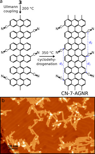
Figure 2b shows an STM overview of the resulting ribbons. The typical lenghts are around 8 to 10 nm, much shorter than average lengths for pristine 7-AGNR 3. Additionally the ribbons appear aligned in arrays in contrast with the disperse arrangement of pristine 7-AGNR 3. This points towards the presence of nitrile groups in the ribbons, which drive the clustering of the GNRs via van der Waals forces, the electric dipoles created by the nitrile groups (see below) in the edges of neighboring ribbons can also contribute to this attractive inter-ribbon interaction with the appropriate relative orientation.
However, the identification of the CN-functionalized sites in the STM images is difficult. To overcome this limitation we used a CO-functionalized tip 25, 26. As detailed in the section Methods, this allows STM imaging of GNRs with intramolecular resolution. Figure 3b shows a image of a section of a ribbon (indicated in Figure 3a) obtained using a CO-functionalized tip, resolving clearly its graphenoid backbone structure and additional features at the edges. The high resolution image allows us to characterize with high precision the edges of the ribbon and their precise functionalization. CN groups (marked with red arrows in Figure 3b) appear as linear features at the edges pointing along distinctive directions. We attribute the shadow on the CN groups to the potential landscape between CN group and the close-by H in the same bay region, to which the CO tip is sensitive 27, 28.
The high resolution images also highlight unexpected chemical processes occurring during the on-surface GNR synthesis. First, we found that a large fraction of the CN groups (about 50%) are missing from the edges, probably lost during the GNR formation steps by -bond cleavage. Second, we observed the occasional appearance of additional rings at the edges of the 7-AGNR backbone, with a similar contrast as other six-member carbon rings (Figure 3c). Moreover, these new rings present a characteristic shape at the outermost edge atoms. We propose that these new rings are pyridine rings produced through the on-surface nitrile cycloisomerization of CN groups located on GNR bay regions (Figure 3d). This on-surface cyclization, to date unreported to the best of our knowledge, is related with the copper-catalyzed synthesis of phenanthridine derivatives from biaryl-2-carbonitriles and Grignard reagents by solution chemistry 29 (see Figure S1 in Supporting Information for a possible mechanistic proposal). The resulting product can be corroborated by the high resolution dI/dV images, where the new H on the pyridine ring appears as a darker shadow, exactly as other aromatic hydrogen atoms nearby (orange arrows in Figure 3c). In contrast, a bright line in the images points to the position of the pyridinyl nitrogen atom of the heterocycle (green arrow in Figure 3c).
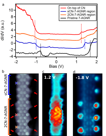
We performed STS measurements to investigate the impact of the CN edge functionalization on the electronic structure of the 7-AGNR (Figure 4a). Figure 4b shows a high resolution image of a GNR section, showing two regions with different density of remaining CN groups (marked with red arrows). One of the regions maintains the CNs only in one of the sides (labeled as sCN-7AGNR), while the other have intact all CNs at the expected sites (2CN-7AGNR). The dI/dV spectra in these regions show two steps at 1.4 (1.2) V and -1.0 (-1.1) V for the sCN (2CN) segments. In analogy to the case of pristine GNR, these steps are attributed to the onset of the second conduction band (CB+1) and of the valence band (VB), respectively 30, 12 (see Figure S2 in Supporting Information). Comparing these spectra with that of a pristine GNR segment (included in Figure 4a) we prove that the CN functionalization produces a rigid downshift of the frontier bands, which amounts to 0.3 eV in sCN sections and 0.4 eV in 2CN regions. This behavior indicates that CN groups behave as -dopants, as found for other nitrogen doped GNRs 14, 13. The spectra also indicate that the density of CN groups affects the doping strength and, consequently, the bands’ downshift. This is pictured in (constant heigth) dI/dV maps measured at 1.2 V (Figure 4c), which show a significantly larger dI/dV signal in the cyano-richer regions (the 2CN segments) due to the larger downshift of the CB+1 band.
In addition to the doping of the GNR, the CN moieties lead to a sizable accumulation of density of states in their proximity. dI/dV maps at -1.8 V (Figure 4d), a bias value well below the VB onset, find an increased conductance signal appearing mostly over the CN groups. This might suggest the existence of an impurity state similar to those observed in previous works 31 for amine and single nitrogen edge substitution. However, the calculations presented below seem to suggest that this signal should come from a rather flat band of the ribbon that strongly hybridizes with the CN group. Furthermore, dI/dV point spectra over the CN groups (red spectrum in Figure 4a), reproduce the larger occupied DOS at CN sites, but appear as a broad background, rather than a well-defined resonance.
The spectra also indicate a slight reduction of the bandgap upon addition of CN groups. Comparing to pristine 7-AGNR, the bandgap in sCN-7-AGNR and 2CN-7AGNR sections is 50 meV and 100 meV smaller, respectively. The bandgap closing is consistent with the increase of effective width of the -network, since the CN groups extend the conjugation of the 7-AGNR backbone.
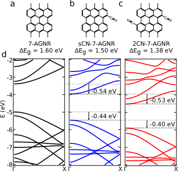
To complement the experimental picture on the impact of CN functionalization we performed first-principles calculations via DFT of freestanding 7-AGNRs with periodically arranged CN functional groups in one (Figure 5b, sCN-7AGNR) or both sides (Figure 5c, 2CN-7AGNR). Figure 5d compares the calculated band structure of pristine, sCN, and 2CN nanoribbons. The calculations reproduce both the additive downshift of the bands and the bandgap reduction upon addition of CN groups. The results show that for each CN added to the pristine ribbon, the VB downshifts 0.4 eV, while the CB and CB+1 move 0.5 eV. In our experiments, the observed band shifts are however smaller, between 0.2 eV and 0.3 eV. We attribute this to the interaction effects of the ribbon with the metal surface, such as the screening provided by the substrate. Given the calculated band downshifts, the band gap of the functionalized ribbons closes 100 meV for a sCN-7-AGNR and 220 meV for a 2CN-7-AGNR which agrees qualitatively with the experimental results. The origin of the bandgap closing is the extension of the conjugated -network due to the addition of the CN moieties. To prove this, we calculated the band structure of a similar 7-AGNR substituting the CN groups by acetylene groups, which extend the conjugate system in a similar way (2CCH-7-AGNR, Figure S3 in Supporting Information). Our results show a similar bandgap (=1.36 eV) to that of 2CN-7-AGNR, although without a downshift of the VB in this case, confirming our previous hypothesis.
The rigid downshift of the bands is an observed trend after the incorporation of electronegative species onto GNRs 14, 13. However, the band’s downshifts induced by CN groups are larger than those induced by nitrogen heterocycles. Even though the ratio between carbon and nitrogen per ribbon cell is smaller in 2CN-7-AGNR, our DFT results find a 0.5 eV downshift per CN group compared to the 0.13 eV per edge nitrogen in chevron GNRs 13. Thus, our results indicate that cyano moieties behave as more efficient n-dopants than nitrogen heterocycles.
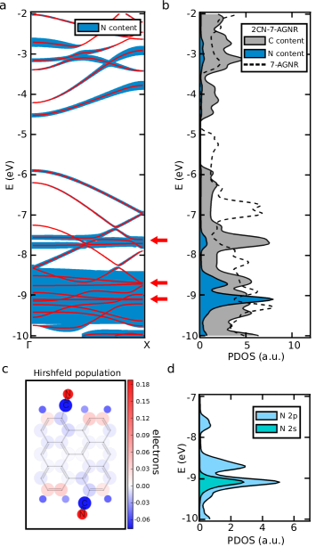
To unravel the mechanism behind the band downshift induced by the CN groups we next focus on the details of interaction between these functional groups and the ribbon. Figure 6a shows the 2CN-7-AGNR bands, with the amount of N-contribution represented by the thickness of a blue shadow. The plot shows that the the N character is widespread in the whole band structure. The origin of such strong mixing is the resonant character of the conjugation between CN and 7-AGNR mesh. The DOS is enhanced particularly at -7.7 eV due to nearly flat bands (top red arrow, Figure 6b) with strong character (Figure 6d), presumably being responsible to the dI/dV enhancement found in the spectra of Figure 4a and in the dI/dV map of Figure 4d. Moreover, states with strong N character are found at lower energy, deep inside the filled states of the ribbon (-9 eV, Figure 6b). These states, not reached in our STS spectra, have both s and p character (Figure 6d), in agreement with the nitrogen orbital hosting the lone pair. Since the localized lone pairs do not participate in the conjugation of the GNR -system, these states are regarded as the impurity levels induced by the CN groups.
Most importantly, the strong electron withdrawing character of CN induces a charge redistribution over the whole ribbon (Figure 6c) and results in sizeable dipoles at the CN sites (estimated as -2.9D, see Figure S4 in Supporting Information) pointing towards the ribbon backbone. The localized dipoles generate an electrostatic potential background approximately 1 eV lower than in pristine 7-AGNRs, which turns the ribbon more electronegative, and is consistent with the observed band downshifts (see Figure S5 in Supporting Information). Thus, our results suggest that the bands downshift is a consequence of the charge redistribution induced by the CN groups.
2 Conclusions
In summary, we have shown the growth of cyano substituted 7-AGNR and studied the impact of nitrile functional groups on the electronic structure of the ribbons. The CN groups increase the reactivity of the 7-AGNRs as inferred from the bunching of the ribbons into aligned clusters and the overall shorter ribbon lengths. STM imaging with CO-functionalized tips resolves with high precision the intramolecular structure of the ribbons and finds a significant loss of CN groups, close to 50%. Moreover, the high resolution images unveil the on-surface nitrile cycloisomerization into pyridine rings, which has not been previously reported.
By combining STS and DFT calculations we have shown that the addition of CN groups conjugated to the GNR structure reduces its bandgap, since the -network of the GNR is extended. Furthermore, we have demonstrated that CN groups behave as -dopants. The CN groups downshift the bands 0.5 eV per CN added (0.3 eV in the experiments), which is significantly more than the doping observed in substitutional nitrogen edge atoms. The downshift of the bands stems from the strong electron withdrawing character of nitrile groups, which induce dipoles at the CN sites. The charge redistribution causes a downshift of the electrostatic potential in the GNR backbone, resulting in an increased ribbon electronegativity. In conclusion, our work shows the potential of using functional groups as tools to modify the physicochemical properties of GNRs, albeit further work concerning the stability of said groups is needed.
3 Methods
Our experiments were performed in custom designed low temperature STM, under ultra-high vacuum, and at 5 K. Prior to the monomer deposition, the Au(111) single crystal surface was sputtered with Ne+ ions, typically for 10 min and then annealed in UHV at temperatures between 490 and 500, around 10 min. Gold coated tungsten tips were used for imaging and spectroscopy. CO functionalized tips were obtained by picking up a CO molecule adsorbed on top of NaCl islands deposited after the on-surface reaction was done. The NaCl was deposited to simplify the CO recognition and pickup. We measured the differential conductance spectra and maps by applying a small modulation to the sample bias and using the lock-in technique to obtain a signal proportional to from the first harmonic of the tunneling current.
High resolution images with CO-functionalized tips were obtained in constant height mode, at biases close to Fermi energy and recording the differential conductance signal. Open feedback conditions are given on top of the ribbon. Imaging close to the Pauli repulsive regime allows to obtain intramolecular resolved images 32, 33, 26, 34, 27, 35 STM images were processed and analyzed using the WSxM software 36.
Details on the synthetic procedure to obtain monomer 3 and its spectroscopic characterization are given in the Supporting Information.
Our first-principles simulations where performed via DFT as implemented in SIESTA code.37 We used van der Waals density functional of Dion et al.38 with the modified exchange by Klimeš, Bowler and Michaelides39. A double-zeta basis set was used to expand the valence-electron wave functions, while the core electrons were described by non-conserving Troullier-Martins pseudopotentials40. An energy cutoff of 350 Ry was used for real space integrations and the orbital radii were defined using a 30 meV energy shift.37 We used a sampling of 50 k-points along the periodic direction. All structures were fully optimized with the conjugate gradient method until all forces were lower than 10 meV/Å. A unit cell length of 8.64 Å , 8.65 Å and 8.67 Å were obtained for pristine 7-AGNR, sCN-7-AGNR and 2CN-7-AGNR, respectively, in the periodic direction (Figure 5a), while in the other directions 40 Å (perpendicular to the ribbon plane) and 50 Å (in the ribbon plane) cell sizes were considered. To check the convergence of the calculated positions of the electronic levels with respect to vacuum, we performed calculations using even larger inter-ribbon distances (90 Å and 100 Å, respectively, along the perpendicular or parallel to the ribbon plane) and we found the highest differences to be lower than 50 meV. The atomic population analysis were performed using the Hirshfeld scheme for partitioning the electron density 41.
4 Acknowledgements
The authors thank D.G. de Oteyza for his insights on GNR doping. This work was supported by FP7 FET-ICT “Planar Atomic and Molecular Scale devices” (PAMS) project (funded by the European Commission under contract No. 610446), by the Spanish Ministerio de Economía y Competitividad (MINECO) (cooperative grant No. MAT2016-78293 and grant FIS 2015-62538-ERC) and the Basque Government (Dep. de Educación and UPV/EHU, Grant No. IT-756-13, and Dep. Industry, Grant PI_2015_1_42).
5 Supporting Information
Possible mechanism for CN cleavage and pyridine formation. Wavefunctions of 2CN-7-AGNR bands. Band structure of acetylene functionalized 7-AGNR and their derived bandgaps. Charge redistribution plots and edge dipole estimation. Calculation and plot of the averaged electrostatic potential. Molecular precursor synthesis and characterization.
References
- Fujita et al. 1996 Fujita, M.; Wakabayashi, K.; Nakada, K.; Kusakabe, K. Peculiar Localized State at Zigzag Graphite Edge. Journal of the Physical Society of Japan 1996, 65, 1920–1923
- Wakabayashi et al. 2010 Wakabayashi, K.; Sasaki, K.-i.; Nakanishi, T.; Enoki, T. Electronic states of graphene nanoribbons and analytical solutions. Science and Technology of Advanced Materials 2010, 11, 054504
- Cai et al. 2010 Cai, J.; Ruffieux, P.; Jaafar, R.; Bieri, M.; Braun, T.; Blankenburg, S.; Muoth, M.; Seitsonen, A. P.; Saleh, M.; Feng, X. et al. Atomically precise bottom-up fabrication of graphene nanoribbons. Nature 2010, 466, 470–473
- Ruffieux et al. 2012 Ruffieux, P.; Cai, J.; Plumb, N. C.; Patthey, L.; Prezzi, D.; Ferretti, A.; Molinari, E.; Feng, X.; Müllen, K.; Pignedoli, C. A. et al. Electronic Structure of Atomically Precise Graphene Nanoribbons. ACS Nano 2012, 6, 6930–6935
- Ruffieux et al. 2016 Ruffieux, P.; Wang, S.; Yang, B.; Sánchez-Sánchez, C.; Liu, J.; Dienel, T.; Talirz, L.; Shinde, P.; Pignedoli, C. A.; Passerone, D. et al. On-surface synthesis of graphene nanoribbons with zigzag edge topology. Nature 2016, 531, 489–492
- Lipton-Duffin et al. 2009 Lipton-Duffin, J. A.; Ivasenko, O.; Perepichka, D. F.; Rosei, F. Synthesis of Polyphenylene Molecular Wires by Surface-Confined Polymerization. Small 2009, 5, 592–597
- Chen et al. 2013 Chen, Y.-C.; de Oteyza, D. G.; Pedramrazi, Z.; Chen, C.; Fischer, F. R.; Crommie, M. F. Tuning the Band Gap of Graphene Nanoribbons Synthesized from Molecular Precursors. ACS Nano 2013, 7, 6123–6128
- Basagni et al. 2015 Basagni, A.; Sedona, F.; Pignedoli, C. A.; Cattelan, M.; Nicolas, L.; Casarin, M.; Sambi, M. Molecules-Oligomers-Nanowires-Graphene Nanoribbons: A Bottom-Up Stepwise On-Surface Covalent Synthesis Preserving Long-Range Order. Journal of the American Chemical Society 2015, 137, 1802–1808
- Zhang et al. 2015 Zhang, H.; Lin, H.; Sun, K.; Chen, L.; Zagranyarski, Y.; Aghdassi, N.; Duhm, S.; Li, Q.; Zhong, D.; Li, Y. et al. On-Surface Synthesis of Rylene-Type Graphene Nanoribbons. Journal of the American Chemical Society 2015, 137, 4022–4025
- Kimouche et al. 2015 Kimouche, A.; Ervasti, M. M.; Drost, R.; Halonen, S.; Harju, A.; Joensuu, P. M.; Sainio, J.; Liljeroth, P. Ultra-narrow metallic armchair graphene nanoribbons. Nature Communications 2015, 6, 10177
- Liu et al. 2015 Liu, J.; Li, B.-W.; Tan, Y.-Z.; Giannakopoulos, A.; Sanchez-Sanchez, C.; Beljonne, D.; Ruffieux, P.; Fasel, R.; Feng, X.; Müllen, K. Toward Cove-Edged Low Band Gap Graphene Nanoribbons. Journal of the American Chemical Society 2015, 137, 6097–6103
- Talirz et al. 2017 Talirz, L.; Söde, H.; Dumslaff, T.; Wang, S.; Sanchez-Valencia, J. R.; Liu, J.; Shinde, P.; Pignedoli, C. A.; Liang, L.; Meunier, V. et al. On-Surface Synthesis and Characterization of 9-Atom Wide Armchair Graphene Nanoribbons. ACS Nano 2017, 11, 1380–1388
- Cai et al. 2014 Cai, J.; Pignedoli, C. A.; Talirz, L.; Ruffieux, P.; Söde, H.; Liang, L.; Meunier, V.; Berger, R.; Li, R.; Feng, X. et al. Graphene nanoribbon heterojunctions. Nature Nanotechnology 2014, 9, 896–900
- Bronner et al. 2013 Bronner, C.; Stremlau, S.; Gille, M.; Brauße, F.; Haase, A.; Hecht, S.; Tegeder, P. Aligning the Band Gap of Graphene Nanoribbons by Monomer Doping. Angewandte Chemie International Edition 2013, 52, 4422–4425
- Zhang et al. 2014 Zhang, Y.; Zhang, Y.; Li, G.; Lu, J.; Lin, X.; Du, S.; Berger, R.; Feng, X.; Müllen, K.; Gao, H.-J. Direct visualization of atomically precise nitrogen-doped graphene nanoribbons. Applied Physics Letters 2014, 105, 023101
- Nguyen et al. 2016 Nguyen, G. D.; Toma, F. M.; Cao, T.; Pedramrazi, Z.; Chen, C.; Rizzo, D. J.; Joshi, T.; Bronner, C.; Chen, Y.-C.; Favaro, M. et al. Bottom-Up Synthesis of N = 13 Sulfur-Doped Graphene Nanoribbons. The Journal of Physical Chemistry C 2016, 120, 2684–2687
- Kawai et al. 2015 Kawai, S.; Saito, S.; Osumi, S.; Yamaguchi, S.; Foster, A. S.; Spijker, P.; Meyer, E. Atomically controlled substitutional boron-doping of graphene nanoribbons. Nature Communications 2015, 6, 8098
- Cloke et al. 2015 Cloke, R. R.; Marangoni, T.; Nguyen, G. D.; Joshi, T.; Rizzo, D. J.; Bronner, C.; Cao, T.; Louie, S. G.; Crommie, M. F.; Fischer, F. R. Site-Specific Substitutional Boron Doping of Semiconducting Armchair Graphene Nanoribbons. Journal of the American Chemical Society 2015, 137, 8872–8875
- Carbonell-Sanromà et al. 2017 Carbonell-Sanromà, E.; Brandimarte, P.; Balog, R.; Corso, M.; Kawai, S.; Garcia-Lekue, A.; Saito, S.; Yamaguchi, S.; Meyer, E.; Sánchez-Portal, D. et al. Quantum Dots Embedded in Graphene Nanoribbons by Chemical Substitution. Nano Letters 2017, 17, 50–56
- Sanchez-Sanchez et al. 2015 Sanchez-Sanchez, C.; Orozco, N.; Holgado, J. P.; Beaumont, S. K.; Kyriakou, G.; Watson, D. J.; Gonzalez-Elipe, A. R.; Feria, L.; Fernández Sanz, J.; Lambert, R. M. Sonogashira Cross-Coupling and Homocoupling on a Silver Surface: Chlorobenzene and Phenylacetylene on Ag(100). Journal of the American Chemical Society 2015, 137, 940–947
- Gao et al. 2013 Gao, H.-Y.; Franke, J.-H.; Wagner, H.; Zhong, D.; Held, P.-A.; Studer, A.; Fuchs, H. Effect of Metal Surfaces in On-Surface Glaser Coupling. The Journal of Physical Chemistry C 2013, 117, 18595–18602
- Chong et al. 2016 Chong, M. C.; Reecht, G.; Bulou, H.; Boeglin, A.; Scheurer, F.; Mathevet, F.; Schull, G. Narrow-Line Single-Molecule Transducer between Electronic Circuits and Surface Plasmons. Physical Review Letters 2016, 116, 036802
- Chong et al. 2016 Chong, M. C.; Sosa-Vargas, L.; Bulou, H.; Boeglin, A.; Scheurer, F.; Mathevet, F.; Schull, G. Ordinary and Hot Electroluminescence from Single-Molecule Devices: Controlling the Emission Color by Chemical Engineering. Nano Letters 2016, 16, 6480–6484
- de Oteyza et al. 2016 de Oteyza, D. G.; García-Lekue, A.; Vilas-Varela, M.; Merino-Díez, N.; Carbonell-Sanromà, E.; Corso, M.; Vasseur, G.; Rogero, C.; Guitián, E.; Pascual, J. I. et al. Substrate-Independent Growth of Atomically Precise Chiral Graphene Nanoribbons. ACS Nano 2016, 10, 9000–9008
- Gross et al. 2009 Gross, L.; Mohn, F.; Moll, N.; Liljeroth, P.; Meyer, G. The Chemical Structure of a Molecule Resolved by Atomic Force Microscopy. Science 2009, 325, 1110–1114
- Kichin et al. 2011 Kichin, G.; Weiss, C.; Wagner, C.; Tautz, F. S.; Temirov, R. Single Molecule and Single Atom Sensors for Atomic Resolution Imaging of Chemically Complex Surfaces. Journal of the American Chemical Society 2011, 133, 16847–16851
- Hapala et al. 2014 Hapala, P.; Kichin, G.; Wagner, C.; Tautz, F. S.; Temirov, R.; Jelínek, P. Mechanism of high-resolution STM/AFM imaging with functionalized tips. Physical Review B 2014, 90, 085421
- Hämäläinen et al. 2014 Hämäläinen, S. K.; van der Heijden, N.; van der Lit, J.; den Hartog, S.; Liljeroth, P.; Swart, I. Intermolecular Contrast in Atomic Force Microscopy Images without Intermolecular Bonds. Physical Review Letters 2014, 113, 186102
- Zhang et al. 2010 Zhang, L.; Ang, G. Y.; Chiba, S. Copper-Catalyzed Synthesis of Phenanthridine Derivatives under an Oxygen Atmosphere Starting from Biaryl-2-carbonitriles and Grignard Reagents. Organic Letters 2010, 12, 3682–3685
- Söde et al. 2015 Söde, H.; Talirz, L.; Gröning, O.; Pignedoli, C. A.; Berger, R.; Feng, X.; Müllen, K.; Fasel, R.; Ruffieux, P. Electronic band dispersion of graphene nanoribbons via Fourier-transformed scanning tunneling spectroscopy. Physical Review B 2015, 91, 045429
- Cervantes-Sodi et al. 2008 Cervantes-Sodi, F.; Csányi, G.; Piscanec, S.; Ferrari, A. C. Edge-functionalized and substitutionally doped graphene nanoribbons: Electronic and spin properties. Physical Review B 2008, 77, 165427
- Temirov et al. 2008 Temirov, R.; Soubatch, S.; Neucheva, O.; Lassise, A. C.; Tautz, F. S. A novel method achieving ultra-high geometrical resolution in scanning tunnelling microscopy. New Journal of Physics 2008, 10, 053012
- Weiss et al. 2010 Weiss, C.; Wagner, C.; Kleimann, C.; Rohlfing, M.; Tautz, F. S.; Temirov, R. Imaging Pauli Repulsion in Scanning Tunneling Microscopy. Physical Review Letters 2010, 105, 086103
- Kichin et al. 2013 Kichin, G.; Wagner, C.; Tautz, F. S.; Temirov, R. Calibrating atomic-scale force sensors installed at the tip apex of a scanning tunneling microscope. Physical Review B 2013, 87, 081408
- Krejčí et al. 2017 Krejčí, O.; Hapala, P.; Ondráček, M.; Jelínek, P. Principles and simulations of high-resolution STM imaging with a flexible tip apex. Physical Review B 2017, 95, 045407
- Horcas et al. 2007 Horcas, I.; Fernández, R.; Gómez-Rodríguez, J. M.; Colchero, J.; Gómez-Herrero, J.; Baro, A. M. WSXM: A software for scanning probe microscopy and a tool for nanotechnology. Review of Scientific Instruments 2007, 78, 013705
- Soler et al. 2002 Soler, J. M.; Artacho, E.; Gale, J. D.; García, A.; Junquera, J.; Ordejón, P.; Sánchez-Portal, D. The SIESTA method for ab initio order-N materials simulation. J. Phys.: Condens. Matter 2002, 14, 2745–2779
- Dion et al. 2004 Dion, M.; Rydberg, H.; Schröder, E.; Langreth, D. C.; Lundqvist, B. I. Van der Waals Density Functional for General Geometries. Phys. Rev. Lett. 2004, 92, 246401
- Klimeš et al. 2010 Klimeš, J.; Bowler, D. R.; Michaelides, A. Chemical accuracy for the van der Waals density functional. J. Phys.: Condens. Matter 2010, 22, 022201
- Troullier and Martins 1991 Troullier, N.; Martins, J. L. Efficient pseudopotentials for plane-wave calculations. Phys. Rev. B 1991, 43, 1993–2006
- Hirshfeld 1977 Hirshfeld, F. L. Bonded-atom fragments for describing molecular charge densities. Theoretica chimica acta 1977, 44, 129–138