Multislip Friction with a Single Ion
Abstract
A trapped ion transported along a periodic potential is studied as a paradigmatic nanocontact frictional interface. The combination of the periodic corrugation potential and a harmonic trapping potential creates a one-dimensional energy landscape with multiple local minima, corresponding to multistable stick-slip friction. We measure the probabilities of slipping to the various minima for various corrugations and transport velocities. The observed probabilities show that the multislip regime can be reached dynamically at smaller corrugations than would be possible statically, and can be described by an equilibrium Boltzmann model. While a clear microscopic signature of multislip behavior is observed for the ion motion, the frictional force and dissipation are only weakly affected by the transition to multistable potentials.
Stick-slip friction is a ubiquitous non-equilibrium dynamical process that occurs at the interface between surfaces across a wide range of length scales scholz ; urbakh ; bormuth ; mo ; urbakh2 ; vanossi . The term stick-slip describes the system’s response to an applied shear force: the surfaces slip out of a local minimum in the interface energy landscape, and stick into a new lower-energy minimum, releasing heat in the process.
Recent advances in atomic force microscopy (AFM) have extended the study of stick-slip friction to the atomic scale, where atom-by-atom slips occur at the interface between a probe tip and a periodic substrate binnig ; carpick ; carpick2 ; krylov ; meyer ; gnecco ; riedo ; liu ; li ; zhao ; jansen ; barel . For a single-atom probe, the number of local minima in the probe-substrate interaction potential is determined by the ratio of the periodic substrate potential to the spring constant with which the probe is bound to its support object. As the load on the probe is increased, or equivalently, the periodic substrate potential is deepened, the system transitions from a bistable regime (where the probe deterministically single-slips from the first minimum to the second) to a multistable regime (where a probe can stochastically multislip to one of several local minima). This has been demonstrated in AFM simulations nakamura ; dong ; tshiprut ; evstigneev and experiments mate ; medyanik ; roth where single-slip and multislip events have been clearly differentiated. However, in the absence of control over dissipation rates and the microscopic energy landscape, it is difficult to tie the observations to ab initio friction models.
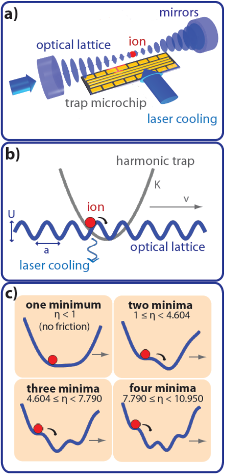
Following theoretical proposals tosatti ; tosatti2 ; shepelyansky ; haffner , we have recently demonstrated a trapped-ion friction emulator with extensive control over all microscopic interface parameters science ; velocity ; aubry . In analogy to AFM, the emulator features a small probe (one or several trapped ions) transported over a periodic substrate potential created by an optical standing wave (Figs. 1a,b) enderlein ; linnet ; karpa . To date, we have used the emulator to study the velocity-dependence of nanofriction velocity , as well as the interplay between superlubricity science ; shinjo ; hirano ; dienwiebel ; socoliuc and the Aubry transition aubry . These studies and a recent study of zig-zag ion chains mehlstaubler have focused on the single-slip regime.
In this Letter, we study multislip friction in deep substrate potentials. We observe the ion fluorescence associated with slip events, from which we directly extract the temperature- and velocity-dependent probabilities for the ion to localize in one of the available local minima. We find that at finite rethermalization times following a slip, the multislip regime can be reached dynamically at smaller corrugations than would be possible statically. We also find that the probabilities agree well with a simple Boltzmann model, despite the dynamical nature of the process. Remarkably, the average frictional energy dissipation and the maximal static friction force are mostly unaffected by the transition from the single-slip to the multislip regime, increasing approximately linearly with the depth of the substrate potential.
The potential energy landscape experienced by the ion is produced by the combination of an electrostatic harmonic potential provided by a linear Paul trap cetina and a sinusoidal optical lattice karpa ; science ; velocity ; aubry . The potential energy of the ion at position is given by the Prandtl-Tomlinson model P ; T :
| (1) |
The first term is attributed to the harmonic trap at position , corresponding to a spring with constant ( is the mass of the 174Yb+ ion and kHz is the axial vibrational frequency of the harmonic trap). The second term is due to the AC Stark shift of the lattice with period nm science . The number of local minima in is determined by the corrugation parameter , with the vibrational frequency at the lattice minima. By adjusting the optical-lattice amplitude to a maximum of MHz, we change up to MHz, and thus tune in the range . Values of interest include and , which mark the transition to potentials with , , and local minima, respectively (Fig. 1c).
The ion is transported by adjusting the potentials of the trap electrodes so as to translate the harmonic-trap position . Thus, we drive the ion over the optical lattice at constant average velocity , forcing the ion to slip over lattice maxima (Fig. 1c). During the transport, the ion is continuously laser cooled via Raman sideband cooling to a typical temperature of K ( in the range to for in the range to ) karpa , and observed via the fluorescence emitted during the cooling process. For a stationary ion, the fluorescence peaks when its stable minimum becomes an inflection point, the moment when the ion is closest to the maximum of the optical-lattice potential karpa . After the ion slips over a lattice maximum, its fluorescence falls exponentially while it cools and localizes into a new local minimum.
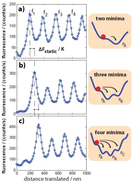
Initializing the ion in the global potential minimum, and then transporting through consecutive slip events, we observe a series of fluoresence peaks; the relative heights of these peaks differentiate single-slip from multislip behavior. For an ion undergoing single-slips, a series of equally-spaced fluorescence peaks of equal height is observed, as the ion always localizes in the adjacent minimum after every slip event (Fig. 2a). The transition to the multislip regime manifests itself as fluorescence peaks of different heights, associated with random localizations in more distant minima (Fig. 2b,c).
To see why the two slip modes result in different peak height distributions, we note that the fluorescence traces are averaged over multiple repetitions of the initialization-transport experiment. The more likely an ion is to slip at a particular time, the higher the associated averaged fluorescence peak. The ion is initialized in the global potential minimum; when this minimum vanishes due to trap translation, the ion will always slip and fluoresce. Thus, the first peak is the largest. After this initial slip, if the ion localizes in the adjacent minimum (single-slip), then further trap translation by one lattice period will cause the ion to slip and fluoresce again, and we will observe . If it localizes instead in the next-adjacent minimum (multislip), then a fluorescence peak will not appear until translation by . A finite probability of next-adjacent localization will result in a reduced peak and a higher peak (see Fig. 2b,c).
The relationship between the localization probabilities (where denotes localization in the adjacent minimum, the next-adjacent minimum, etc.) and the peak height distribution is given by supplemental :
| (2) | ||||
Note that the probability distribution is extracted directly from the observed peak heights without making assumptions about the localization process.
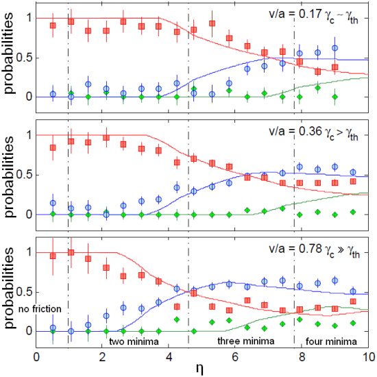
By measuring fluorescence patterns like the ones shown in Fig. 2 for different corrugation parameters and transport velocities , we extract localization probabilities over a range of experimental conditions (Fig. 3). For all three velocities shown, multislip behavior (second-next neighbor slip probability ) is observed in the multistable regime (). This is a consequence of the underdamping of the system (), which guarantees that an ion, following a slip, can sample the full potential landscape before it recools at rate s-1 and localizes in a minimum. More surprising is the appearance of multislip events before the corrugation is deep enough to create multiple static minima (in the region of bistable potential , where a third minimum should not yet exist). This is the case for the two fastest transport speeds but not for the slowest transport. This can be readily explained by the change in the energy landscape during the recooling time : by the time the ion is sufficiently cooled to localize, another potential minimum may have opened up if . Thus, we find that the multislip regime can be reached dynamically at smaller corrugations than would be possible statically.
The experimental data shown in Fig. 3 is overlayed with a theoretical model that takes into account the system’s competing rates (transport rate and recooling rate ). Our model’s central assumption is that an ion is more likely to localize in a lower-energy minimum and that this effect can be described by a quasi-equilibrium Boltzmann probability . Here, is the potential energy of the th minimum at time when the ion localizes, and is the temperature of the ion at that time. To model its temperature, we note that an ion has some potential energy at the slipping point. This is converted into kinetic energy and dissipated exponentially by laser cooling: . Our model’s free parameter is the localization time, found to be ) s by fitting the model to the data.
We note that the dataset with the slowest transport speed is fitted with lower confidence by the model above . This discrepancy is the signature of another dynamical rate of the system, the thermal hopping rate s-1, observed previously in Refs. velocity ; jinesh ; sang . Thermal hopping across a barrier due to the ion’s finite temperature dominates at the slowest transport speed, where (the thermolubric regime velocity ). Its effect on the fluorescence signal is to smooth the peak height distribution, which causes us to overestimate the value of and underestimate supplemental . Evidently, the relationship between fluorescence and probability (Eq. 2) is strictly valid only for faster transport speeds, where thermal hopping is negligible ().
Thermal hopping also affects the observed slip probability to the third minimum , which for large and large speed is distinctly smaller than predicted. The third minimum, most distant from the slipping point, has the smallest potential barrier and the longest dwell time before the minimum disappears, making the ion most susceptible to thermal hopping out of that minimum, even at fast transport speeds. Because a thermal hop at a random time does not result in an (averaged) fluorescence peak, we undervalue the localization probability .
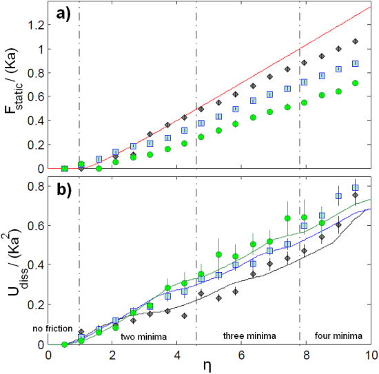
Figs. 2 and 3 show clear delineations of single-slip and multislip behavior as a function of . Interestingly, purely frictional quantities, like the maximal static friction force and energy dissipated per slip , do not reveal clear signatures of the transition. Fig. 4a shows that exerted by the lattice on the ion increases monotonically with the corrugation parameter , without any discernable changes near the critical values for multistability ( is determined from the observed hysteretic shift of the fluorescence peaks as a function of trap translation, see Fig. 2 and Ref. science ). Fig. 4b shows the calculated average energy dissipated per slip by laser cooling, determined from the measured slip probabilites and the calculated energy landscape supplemental . Like , the dissipated energy increases monotonically with the corrugation parameter , and shows little dependence on the number of potential minima in the energy landscape for . This can be attributed to the cancelation of two competing effects: if an ion slips to the second-next minimum rather than to the next minimum, it will release more heat, as the second-next minimum is lower in energy by the time the ion localizes. On the other hand, an ion slipping to a more distant minimum will wait longer before it slips again. Thus, to leading order, the dissipation is independent of the slipping mode. To higher order, the slope is slightly reduced for a potential with more minima, as the reduction in slip frequency overpowers the smaller increase in dissipated energy per slip. This trend is visible in Fig. 4b, where the slope is reduced at the transition between single-slip and multislip behavior.
In this work, we have measured the slip probabilities for stick-slip friction in a multistable energy landscape, and have shown that the dynamic stick-slip process can be described by a quasi-equilibrium model. We suggest that the model’s predictive power could be used in nanopositioning applications: by tuning system parameters, a probe could be engineered to multislip to a specific potential minimum. In the future, these studies could be extended to the quantum regime in order to study quantum annealing in a multistable energy landscape.
This work was supported in part by the NSF, the NSF CUA, and the MURI program through ONR.
References
- (1) C.H. Scholz. Nature 391, 37-42 (1998).
- (2) M. Urbakh, J. Klafter, D. Gourdon, J. Israelachvili. Nature 430, 525-528 (2004).
- (3) V. Bormuth, V. Varga, J. Howard, E. Schäffer. Science 325, 870-3 (2009).
- (4) Y. Mo, K.T. Turner, and I. Szlufarska. Nature 457, 1116-1119 (2009).
- (5) M. Urbakh and E. Meyer. Nature Materials 9, 8-10 (2010).
- (6) A. Vanossi, N. Manini, M. Urbakh, S. Zapperi, E. Tosatti. Reviews of Modern Physics 85, 529 (2013).
- (7) G. Binnig, C.F. Quate, Ch. Gerber. Physical Review Letters 56, 930-933 (1986).
- (8) R.W. Carpick and M. Salmeron. Chemical Reviews 97, 1163-1194 (1997).
- (9) E. Gnecco, R. Bennewitz, T. Gyalog, Ch. Loppacher, M. Bammerlin, E. Meyer, H.-J. Güntherodt. Physical Review Letters 84, 1172-1175 (2000).
- (10) I. Szlufarska, M. Chandross, R.W. Carpick. Journal of Physics D 41, 123001 (2008).
- (11) E. Riedo, E. Gnecco, R. Bennewitz, E. Meyer, H. Brune. Physical Review Letters 91, 084502 (2003).
- (12) X. Zhao, S.R. Phillpot, W.G. Sawyer, S.B. Sinnott, S.S. Perry. Physical Review Letters 102, 186102 (2009).
- (13) L. Jansen, H. Hölscher, H. Fuchs, A. Schirmeisen. Physical Review Letters 104, 256101 (2010).
- (14) I. Barel, M. Urbakh, L. Jansen, A. Schirmeisen. Physical Review B 84, 115417 (2011).
- (15) S.Y. Krylov and J.W.M. Frenken. Physica Status Solidi (b) 251, 711-736 (2014).
- (16) E. Meyer and E. Gnecco. Friction 2, 106-113 (2014).
- (17) Q. Li, Y. Dong, D. Perez, A. Martini, R.W. Carpick. Physical Review Letters 106, 126101 (2011).
- (18) Xin-Z. Liu, Z. Ye, Y. Dong, P. Egberts, R.W. Carpick, A. Martini. Physical Review Letters 114, 146102 (2015).
- (19) J. Nakamura, S. Wakunami, A. Natori. Physical Review B 72, 235415 (2005).
- (20) Z. Tshiprut, S. Zelner, M. Urbakh. Physical Review Letters 102, 136102 (2009).
- (21) Y. Dong, D. Perez, A.F. Voter, A. Martini. Tribology Letters 42, 99-107 (2011).
- (22) M. Evstigneev and P. Reimann. Physical Review B 87, 205441 (2013).
- (23) C.M. Mate, G.M. McClelland, R. Erlandsson, S. Chiang. Physical Review Letters 59, 1942-1946 (1987).
- (24) S.N. Medyanik, W.K. Liu, I.-H. Sung, R.W. Carpick. Physical Review Letters 97, 136106 (2006).
- (25) R. Roth, Th. Glatzel, P. Steiner, E. Gnecco, A. Baratoff, E. Meyer. Tribology Letters 39, 63-69 (2010).
- (26) A. Benassi, A. Vanossi, E. Tosatti. Nature Communications 2, 236 (2011).
- (27) D. Mandelli, A. Vanossi, E. Tosatti. Physical Review B 87, 195418 (2013).
- (28) I. García-Mata, O.V. Zhirov, D.L. Shepelyansky. European Physical Journal D 41, 325-330 (2007).
- (29) T. Pruttivarasin, M. Ramm, I. Talukdar, A. Kreuter, H. Häffner. New Journal of Physics 13, 075012 (2011).
- (30) D. Gangloff, A. Bylinskii, I. Counts, W. Jhe, V. Vuletić. Nature Physics 11, 915-919 (2015).
- (31) A. Bylinskii, D. Gangloff, V. Vuletić. Science 348, 1115-1118 (2015).
- (32) A. Bylinskii, D. Gangloff, I. Counts, V. Vuletić. Nature Materials 15, 717-721 (2016).
- (33) M. Enderlein, T. Huber, C. Schneider, T. Schaetz. Physical Review Letters 109, 233004 (2012).
- (34) R.B. Linnet, I.D. Leroux, M. Marciante, A. Dantan, M. Drewsen. Physical Review Letters 109, 233005 (2012).
- (35) L. Karpa, A. Bylinskii, D. Gangloff, M. Cetina, V. Vuletić. Physical Review Letters 111, 163002 (2013).
- (36) K. Shinjo and M. Hirano. Surface Science 283, 473 (1993).
- (37) M. Hirano, K. Shinjo, R. Kaneko, Y. Murata. Physical Review Letters 78, 1448 (1997).
- (38) M. Dienwiebel, G.S. Verhoeven, N. Pradeep, J.W.M. Frenken, J.A. Heimberg, H.W. Zandbergen. Physical Review Letters 92, 126101 (2004).
- (39) A. Socoliuc, R. Bennewitz, E. Gnecco, E. Meyer. Physical Review Letters 92, 134301 (2004).
- (40) J. Kiethe, R. Nigmatullin, D. Kalincev, T. Schmirander, T.E. Mehlstaubler. Nature Communications 8, 15364 (2017).
- (41) M. Cetina, A. Bylinskii, L. Karpa, D. Gangloff, K.M. Beck, Y. Ge, M. Scholz, A.T. Grier, I. Chuang, V. Vuletić. New Journal of Physics 15, 053001 (2013).
- (42) L. Prandtl. Z. Angew. Math. Mech. 8, 85 (1928).
- (43) G. A. Tomlinson. Philos. Mag. 7, 905 (1929).
- (44) K.B. Jinesh, S.Y. Krylov, H. Valk, M. Dienwiebel, J.W.M. Frenken. Physical Review B 78, 155440 (2008).
- (45) Y. Sang, M. Dubé, M. Grant. Physical Review Letters 87, 174301 (2001).
- (46) Supplemental Material.