Understanding In-line Probing Experiments by Modeling Cleavage of Non-reactive RNA Nucleotides
Abstract
Ribonucleic acid (RNA) is involved in many regulatory and catalytic processes in the cell. The function of any RNA molecule is intimately related with its structure. In-line probing experiments provide valuable structural datasets for a variety of RNAs and are used to characterize conformational changes in riboswitches. However, the structural determinants that lead to differential reactivities in unpaired nucleotides have not been investigated yet. In this work we used a combination of theoretical approaches, i.e., classical molecular dynamics simulations, multiscale quantum mechanical/molecular mechanical calculations, and enhanced sampling techniques in order to compute and interpret the differential reactivity of individual residues in several RNA motifs including members of the most important GNRA and UNCG tetraloop families. Simulations on the multi ns timescale are required to converge the related free-energy landscapes. The results for uGAAAg and cUUCGg tetraloops and double helices are compared with available data from in-line probing experiments and show that the introduced technique is able to distinguish between nucleotides of the uGAAAg tetraloop based on their structural predispositions towards phosphodiester backbone cleavage. For the cUUCGg tetraloop, more advanced ab initio calculations would be required. This study is the first attempt to computationally classify chemical probing experiments and paves the way for an identification of tertiary structures based on the measured reactivity of non-reactive nucleotides.
1 Introduction
Ribonucleic acid (RNA) participates in several kinds of cellular processes, which involve the transmission of genetic information, the synthesis of proteins, the cellular differentiation and development, the regulation of gene expression and enzyme-like catalysis (Strulson et al., 2012; Kung et al., 2013; Sarkies and Miska, 2014). Detailed information about RNA secondary structures is a preliminary step required for tertiary structure determination and, ultimately, for understanding RNA function (Walter, 2009). Identification of specific small RNA motifs, like RNA tetraloops, is particularly important as they stabilize larger RNA structures and can be involved in RNA-RNA or RNA-protein interactions (Hall, 2015). Indirect information about RNA secondary structure is usually obtained by chemical probing experiments (Xu and Culver, 2009; Weeks, 2010; Kubota et al., 2015). Among those, selective 2’-hydroxyl (2’-OH) acylation characterized by primer extension (SHAPE, Merino et al., 2005) and in-line probing (Soukup and Breaker, 1999) experiments provide sequence independent structural information on RNA at single-nucleotide resolution (Weeks, 2010).
In-line probing characterizes backbone mobility by structural dependent phosphodiester cleavage, which breaks RNA molecules into segments at distinct positions (Soukup and Breaker, 1999). Unpaired nucleotides within single stranded RNA regions are often unstable and degrade over time (Reynolds et al., 1996; Welch et al., 1997). The chemical reaction, termed as an internal trans-esterification, starts with 2’-OH attack of neighboring phosphate moiety and results with 2’,3’-cyclic phosphate and 5’-hydroxyl termini (Soukup and Breaker, 1999; Lilley, 2003). The same RNA backbone cleavage (or RNA degradation) is performed by self-catalytic systems called RNA enzymes (ribozymes, Doudna and Cech, 2002; Scott, 2007; Lilley and Eckstein, 2008) and by ribonuclease A (RNase A, Raines, 1998), although in these cases with significantly higher rate constants. By comparing nucleotides from various RNA motifs, Soukup and Breaker observed a relation between the cleavage rate constant and the in-line attack angle of the scissile phosphate, i.e., the angle between O2’ oxygen, the adjacent phosphorus, and O5’ oxygen (Soukup and Breaker, 1999). Later, they defined the ability to bring the active site towards the in-line attack conformation (the in-line attack angle close to 180∘) as one of catalytic strategies for the phosphodiester backbone cleavage used by ribozymes (Breaker et al., 2003; Emilsson et al., 2003). Since then, in-line probing is routinely used in studies of riboswitches, where the binding of a small molecule (ligand) results in a conformational change of the whole RNA molecule (Mandal and Breaker, 2004; Regulski and Breaker, 2008; Montange and Batey, 2008; Garst et al., 2011). In general, chemical probing experiments are typically employed to identify unpaired and flexible nucleotides, allowing one to choose among different predicted secondary structures. However, it must be noticed that not all the unpaired nucleotides are usually reactive. The reactivity pattern of unpaired nucleotide could in principle provide a wealth of information that is usually discarded. To the best of our knowledge, the pattern of reactivity of specific motifs have never been analyzed in detail.
Computational techniques are an established tool for the investigation of structural and dynamical properties of RNA at an atomistic level (Schlick, 2010; Cheatham and Case, 2013; Šponer et al., 2013) and could in principle allow for an investigation and interpretation of reactivity patterns in RNA. In particular, quantum mechanical/molecular mechanical (QM/MM, Warshel and Levitt, 1976) approaches, where only the reactive portion of the system is described at the QM level, have been used to characterize cleavage reactions within catalytic RNA systems (Banáš et al., 2008; Nam et al., 2008b ; Nam et al., 2008a ; Lee et al., 2008; Mlýnský et al., 2011; Rosta et al., 2011; Xu et al., 2012; Gu et al., 2013; Ganguly et al., 2014; Mlýnský et al., 2015; Dubecký et al., 2015; Zhang et al., 2015; Thaplyal et al., 2015; Casalino et al., 2016). Single point QM/MM calculations evaluate potential energy surfaces neglecting entropic contributions. The reconstruction of free-energy surfaces (FES) along the reaction pathway requires combination of QM/MM calculations with molecular dynamics (MD) simulations and enhanced sampling techniques (Palermo et al., 2015). In this context, semiempirical (SE) methods allow for a reasonable compromise between accuracy and efficiency (Christensen et al., 2016), allowing statistically converged FES to be computed. Two SE methods have been carefully parametrized for reactions involving the phosphate moiety (Nam et al., 2007; Yang et al., 2008).
In this paper we combine QM/MM calculations and enhanced sampling methods to model phosphodiester backbone cleavage of nucleotides from three model systems: one tetraloop from each of the GNRA and UNCG family (R = purine and N = any nucleotide) and a double stranded RNA (dsRNA). Regions undergoing the cleavage reaction were described by a DFTB3 SE method, which allowed us to perform simulations on tens-of-ns timescales and obtain converged free-energy landscapes. Our calculations required to design a putative reaction pathway with a number of restraints in order to overcome persisting shortcomings within parameterization of the DFTB3. However, we were able to differentiate among nucleotides within simple motifs by comparing their activation free-energy barriers. Computational results were validated against available experimental data from in-line probing measurements. To our knowledge, this represents the first attempt to design computations in order to understand and mimic in-line probing experiments and, more generally, to investigate the phosphodiester backbone cleavage within non-reactive RNA nucleotides, i.e., not considering the active centers of RNA catalytic motifs.
2 Results
We performed combined QM/MM-MetaD simulations (see Materials and Methods) and reconstructed FES relative to the phosphodiester cleavage reaction for nucleotides within three simple RNA motifs: uGAAAg tetraloop, dsRNA (GC-duplex) and cUUCGg tetraloop. For the tetraloops we computed the reactivity for the unpaired residues and for the closing base pairs. For the duplex we chose 3 consecutive non-terminal residues from each strand (Figure S1 in the Supporting Information (SI)). We thus analyzed relative differences in reactivities among 6 nucleotides for each system by comparing cleavage barriers along reaction pathway designed to be equivalent for all nucleotides from different RNA motifs. Since our intention was not to give insight into the mechanism of phoshodiester backbone cleavage, possible involvement of other RNA groups, water molecules, and ions was omitted and any mechanistic interpretation deliberately neglected.
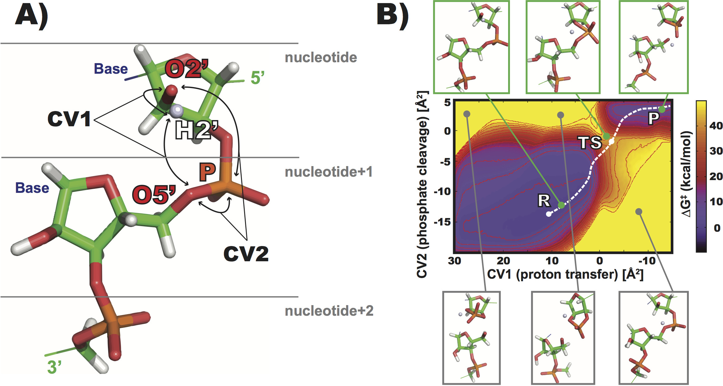
The computed FES profiles mapped changes along two tracked distance-based collective variables (CVs, Figure 1A) to describe the proton transfer and the phosphodiester cleavage. The most relevant information that we want to extract is the cleavage barrier, which was estimated as explained in Materials and Methods. The FES displays two energy minima containing reactant (R) and product (P) state geometries (Figure 1B). The R state minimum is very broad because the simulation is allowed to sample all the possible geometries including different orientations of the active 2’-OH group. This is necessary for the accurate estimation of barriers. On the contrary, we restrained the extensive separation of the RNA molecule after the cleavage reaction, leading to a narrower free-energy minimum associated to P state. The complete exploration of P state geometries would make convergence very difficult and is irrelevant for a proper estimation of the cleavage barrier.
The FES profiles for nucleotides within three different RNA motifs (uGAAAg and cUUCGg tetraloops, GC-duplex) are qualitatively very similar among each other (see Figure S2 in SI for all computed FES). The R and transition states (TS) have slightly different locations for particular nucleotides within each RNA motif, but no relevant correlations. For instance, purine/pyrimidine or tetraloop/duplex differences, were not detected. The computed barriers after 40 ns-long QM/MM-MetaD simulations were in the range between 40.0 and 44.5 kcal/mol. Note that the reaction coordinates are affected by the applied restraints and approximations, which forced the proton from the 2’-OH to be kept around the direct pathway (Figure 1). Furthermore, the reported cleavage barriers are expected to be overestimated due to the complete exclusion of scenarios involving proton transfer through nonbridging oxygens (nbO) of the adjacent phosphate and/or through other proximal RNA groups. However, this allows for a consistent estimation of values and their comparison across nucleotides within different RNA motifs.
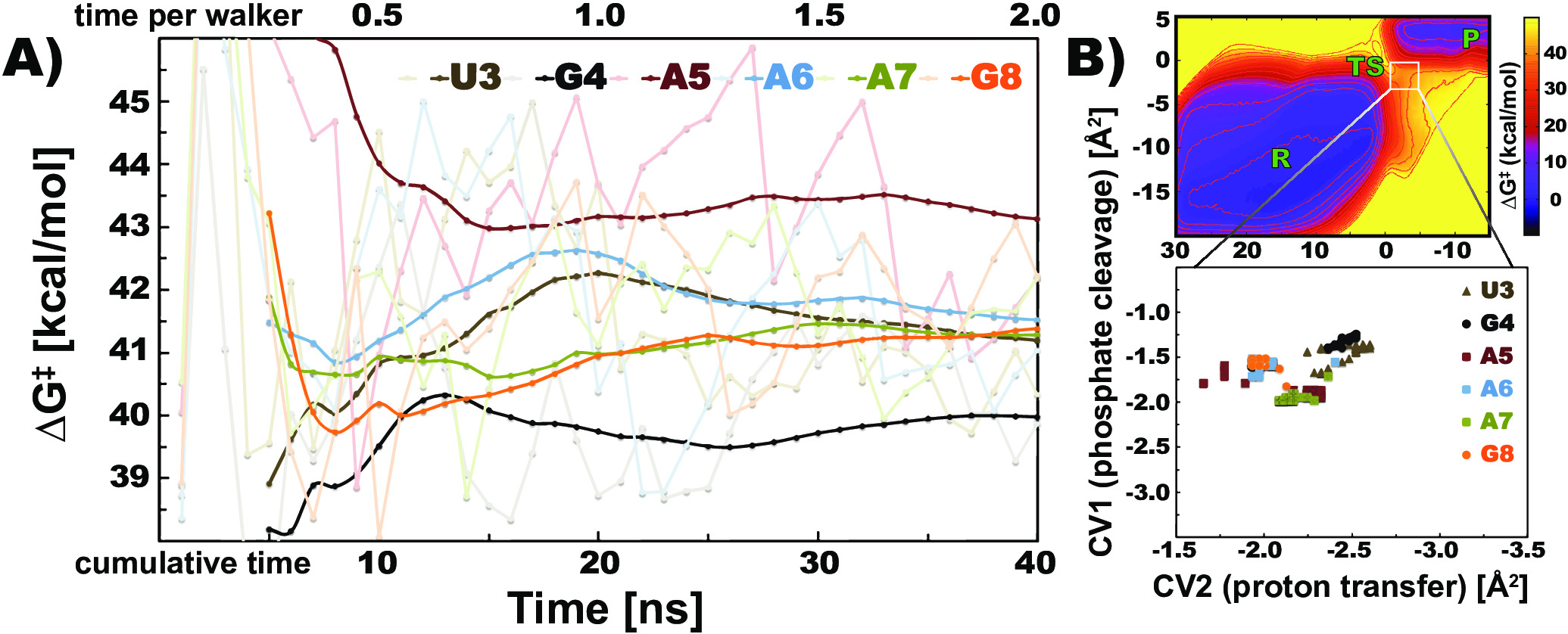
We tested carefully the statistical convergency of computed phosphodiester cleavage barriers. The analysis showed that barriers of 6 investigated nucleotides within uGAAAg tetraloop fluctuate within a few kcal/mol over the time of the simulation (shaded lines at Figure 2A), making it difficult to differentiate among nucleotides. All those barriers were estimated by using the final bias potential. In order to increase the accuracy of the method, we then calculated from time-averaged bias potentials. This latter approach gives a smoother convergence, which enables nucleotides to be clearly distinguished. The resulting barriers of uGAAAg nucleotides are clearly converged after 40 ns of cumulative simulated time (Figure 2A). Note that even initial estimated averages (7 ns of total simulated time) show clear differences within among tested nucleotides. The GC-duplex was used as a control simulation because the computed barriers are expected to be identical for three equivalent nucleotides. Our approach shows that the computed differences of equivalent G and C nucleotides are negligible after 40 ns, i.e., up to 0.4 and 0.5 kcal/mol, respectively (Figure S3A in SI). We also analyzed the location of TS during different stages of the simulations because the estimation of depends on the position of TS and R states on the FES. The TS positions of all nucleotides within uGAAAg tetraloop are located within a small region in the CV space (Figure 2B). The variance in positions of R states were even smaller (data not shown). The same trend was observed for the nucleotides within GC-duplex (Figure S3B in SI), whereas differences in TS positions on FES were slightly larger for nucleotides within cUUCGg tetraloop (Figure S4B in SI).
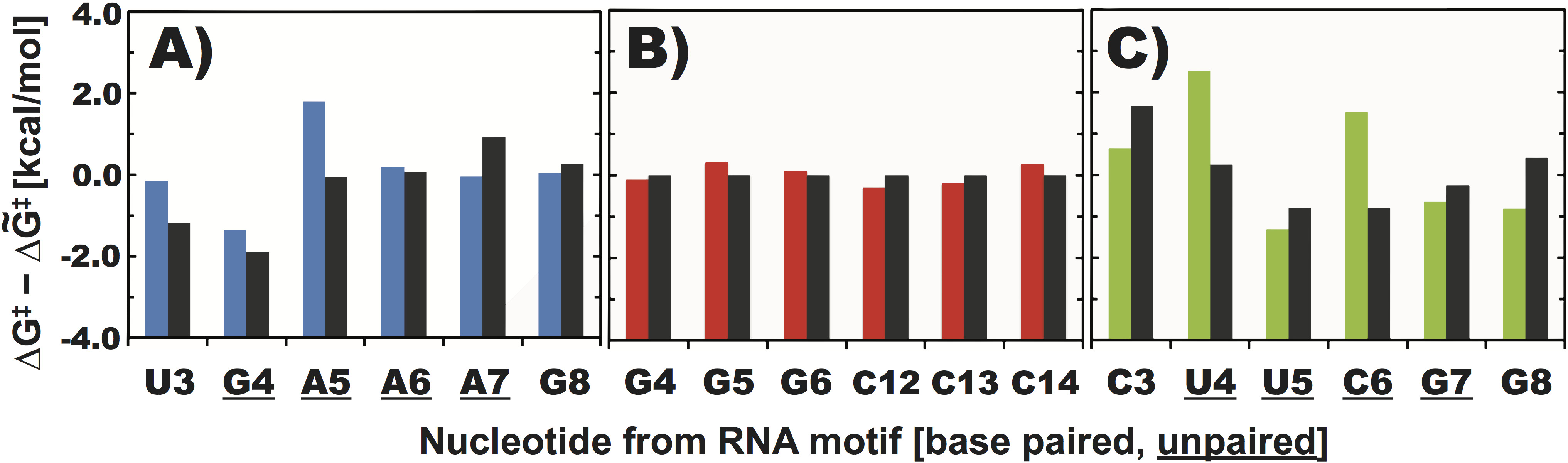
The phosphodiester backbone cleavage barriers are generally between 41 and 42 kcal/mol, but some nucleotides showed intrinsic differences. G4 within uGAAAg tetraloop revealed the lowest barrier among all the nucleotides (40 kcal/mol, Figure S2 in SI). The following A5 showed a significantly higher barrier of 43.1 kcal/mol, whereas the barriers for the remaining nucleotides were comparable (41.5 kcal/mol). All the nucleotides within the GC-duplex revealed comparable barriers between 40.9 and 41.5 kcal/mol. Among cUUCGg nucleotides, U5 showed lowest barriers (40.6 kcal/mol), whereas U4 and C6 provided significantly higher barriers of 44.5 and 43.5 kcal/mol, respectively (Figures S2 and S4A in SI).
We then compared the computed barriers against data from the in-line probing measurements. The experimental reactivities (pseudo free-energies) were derived from available polyacrylamide gel electrophoresis (PAGE) datasets, quantified and normalized separately according to the scheme described in Materials and Methods. We observed good agreement between computed and experimental reactivities for nucleotides within the uGAAAg tetraloop. Our computations overestimated the barriers for U3 and A5 nucleotides, but led the an overall correct trend (Figure 3A). However, a similar comparison for the nucleotides within cUUCGg tetraloop revealed some differences. U5, C6, and G7 were identified as reactive nucleotides according to the experiment (Strauss et al., 2012), but the computed barrier for C6 was significantly higher, suggesting that the nucleotide is non-reactive (Figure 3C). Note that the in-line probing data for a uniform GC-duplex are not available, but that reactivity of paired residues is typically lower than reactivity for unpaired residues (Soukup and Breaker, 1999; Kulshina et al., 2009; Erion and Strobel, 2011; Strauss et al., 2012; Nelson et al., 2013; Hickey and Hammond, 2014; Furukawa et al., 2015).
3 Discussion
In this paper, we used QM/MM-MetaD calculations to classify in-line probing experiments characterized by the phosphodiester backbone cleavage reaction. To this aim, we calculated the free-energy profiles modeling the RNA backbone cleavage for 18 nucleotides within two RNA tetraloops and a dsRNA. The computed obtained from the QM/MM-MetaD calculation is expected to be related to the reactivity of the particular nucleotide as observed in in-line probing experiments. The aim of our study was not to predict absolute reactivities but to explain differential reactivities observed among nucleotides within the same or from different RNA motifs. To minimize the error in differential estimates, we forced the system to explore a similar reaction pathway for each nucleotide and prolonged simulations in the tens-of-ns timescale. The approach provides converged and consistent results for all the nucleotides. Results were then assessed by comparing barriers of identical nucleotides within a GC-duplex motif and by analyzing reference in-line probing reactivities from PAGE gels.
We included a number of artificial restraints, which were required in order to automatize the computational protocol, i.e., to allow the straightforward comparison of various nucleotides within distinct RNA motifs. Restraints improved stability of the simulations namely by preventing spurious rupture of bonds and by excluding several unphysical geometries detected during phosphodiester cleavage reaction. However, all the backbone dihedrals, sugar puckers, glycosidic bonds, as well as base pairing and stacking were left free to rearrange. We actually observed significant dynamics during our QM/MM-MetaD simulations. This is important since the essence of in-line probing experiments is to quantify the effects of RNA structural fluctuations on phosphodiester cleavage rate. The introduction of restraints is nevertheless expected to affect the computed barriers, and thus the respective cleavage rates, in two specific ways. Firstly, restraints forced the cleavage reaction to proceed through the designed reaction pathway that is similar for all nucleotides and likely different from the validated in-line attack reaction pathway. The possible contributions of other reaction pathways, that could be different between one nucleotide to the other, were thus ignored. As a result, the reactivity trends estimated as differences of barriers could in principle be compressed. Secondly, all the computed barriers are expected to be overestimated by excluding the scenario where the proton from 2’-OH group is transferred through nbO atoms. Considering the typical timescale of in-line probing experiments (hours/days), the barriers derived from the estimated rate constants using the Eyring equation are expected to be in range from 26 to 32 kcal/mol (Soukup and Breaker, 1999), i.e., by 7 to 13 kcal/mol lower than the herein reported barriers. Both those issues are very difficult to tackle since they depend on intrinsic deficiencies of the DFTB3 parameterizations. Despite the fact that DFTB and other SE methods improved significantly during last decade (Huang et al., 2014; Christensen et al., 2016), their general application towards chemical reactions remains challenging. In particular, recent studies carefully assessed the performance and revealed limitations of DFTB methods in description of phosphoranes and phosphoryl transfers (Mlýnský et al., 2014; Gaus et al., 2014; Huang et al., 2015; Lu et al., 2016). In this study, we still opted for the usage of DFTB3 because we did not aim for an accurate description of states along phosphodiester cleavage reaction. We rather focused on relative differences of barriers among different nucleotides, forcing the reaction to proceed through the same pathway for all the analyzed nucleotides. We expect such an approach to be more robust and less sensitive to the applied QM method. QM/MM-MetaD simulations at least on the several-ns timescale are required to converge these FES computations to a level allowing for the discrimination of reactivity patterns, ruling out more accurate QM methods such as DFT or ab initio.
Here, we ranked different RNA nucleotides, i.e., base paired/unpaired, purine/pyrimidine, from duplex and tetraloops by their tendency to undergo phosphodiester backbone cleavage. Nucleotide reactivities reported by in-line probing experiments were used as a reference. A number of experimental datasets for specific motifs from different RNA systems are available (Figure S5 in SI, Soukup and Breaker, 1999; Kulshina et al., 2009; Erion and Strobel, 2011; Strauss et al., 2012; Furukawa et al., 2015), but their quantitative estimation is often affected by unclear signals (Erion and Strobel, 2011; Hickey and Hammond, 2014; Furukawa et al., 2015) and/or participations in a tertiary interaction within complex RNA structure (Soukup and Breaker, 1999; Kulshina et al., 2009; Erion and Strobel, 2011). Hence, we used a single specific experiment providing distinct signals for all nucleotides as a reference of each of the tetraloop motifs (Strauss et al., 2012; Nelson et al., 2013). We observed that nucleotides within cUUCGg tetraloop revealed some differences between predicted and calculated reactivities (Figure 3C). Calculated barriers of U4 and C6 nucleotides are significantly overestimated, resulting in a poor correlation between theory and experiment (R=0.17, Figure 4A). We notice that (i) the FES profiles for cUUCGg nucleotides are statistically converged (Figure S4A in SI), (ii) experimental reactivities for nucleotides within cUUCGg motif reveal similar trends among different systems (Figure S5B in SI), and (iii) the procedure of quantification of experimental reactivities, i.e., using digitalized images (see Materials and Methods) provide almost identical profile against the raw count data (a comparison for uGAAAg tetraloop is reported, Figure S6 in SI). Thus, the poor correlation between computed barriers and experimental reactivities for cUUCGg suggests possible limitations of our approach. We carefully inspected the structures along the cleavage reaction and found that the reactive 2’-OH of U5 and, especially, U4 established H-bonds with other RNA groups outside the QM region (described by the empirical force field, Figure S7 in SI). Such interactions could result in over-stabilization of R states, leading to the overestimation of the computed barriers by the current approach. Furthermore, cleavage site of C6 favored rare conformations with high in-line attack angle. Such geometry is not favorable for the mechanism enforced herein and would require to explore the in-line attack reaction pathway, where nbO atoms (and/or external RNA groups, water molecules) are involved in the proton transfer. This was not possible due to deficiencies within DFTB3 parameterization (see SI for details). One possible way to improve the results for cUUCGg would require the number of atoms within QM region (described by DFTB3) to be increased. However, we identified that RNA groups forming those interactions are typically belonging to nucleotides located further away along the sugar-phosphate backbone, making the calculation unfeasible.
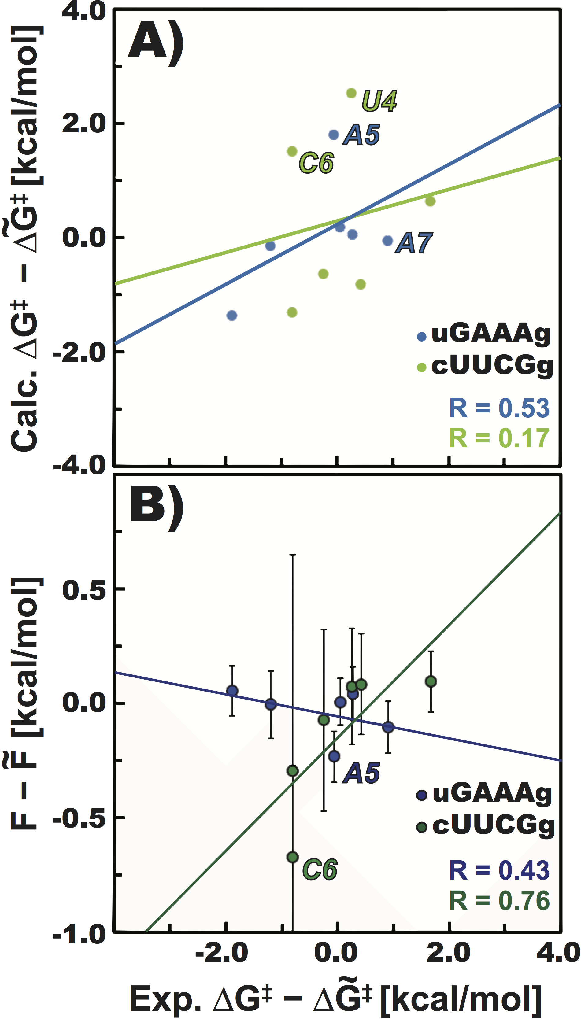
On the other hand, the agreement between theory and experiment is satisfactory for both GC-duplex and uGAAAg tetraloop. In the former case, nucleotides revealed very similar barriers, which is expected for three consecutive equivalent G and C within dsRNA. The fact that each of these barriers was obtained with a totally independent simulation further confirms the low statistical error and hence the reproducibility of our computational approach. The possible differences between purine and pyrimidine nucleotides were negligible for this motif (within the error of our approach, Figure S3A in SI). However, we observed that those barriers are comparable with cleavage barriers of several unpaired residues within tetraloop motifs. This is unexpected, since the reactivity of paired nucleotides is generally lower (Reynolds et al., 1996; Welch et al., 1997; Soukup and Breaker, 1999). We speculate that such behavior is caused by the number of restraints used in our computations, although it could also be linked to the approximations in the SE method used. In the uGAAAg tetraloop, QM/MM-MetaD simulations can clearly predict that the unpaired G4 is significantly more reactive than the other nucleotides. Simulations are long enough to consider this difference statistically significant. Other nucleotides have higher barriers, in agreement with the lower reactivity observed in experiments (Figure 3A). Plotting computational and experimental reactivities against each other revealed that the two unpaired nucleotides (A5 and A7) are worsening the correlation (R=0.53) due to slightly overestimated (A5) and underestimated (A7) barriers (Figure 4A). It is worth noting that the overall stability and flexibility of the tetraloop motifs can be affected by the nature of the closing base pair (Proctor et al., 2002; Blose et al., 2009). For this reason, we explicitly replaced the G-C base pair observed in the crystal structure with the wobble G-U pair found in the sequence used in the reference in-line probing experiments.
Our results can be also compared against predictions made using the approach introduced by Soukup and Breaker, where a correlation between geometrical parameter (in-line fitness) and cleavage rates was proposed (Soukup and Breaker, 1999). The fitness parameter combines the in-line attack angle and the distance between O2’ and the adjacent phosphorus. Interestingly, the fluctuations of the in-line attack angle were proposed as a proxy for the chemical reactivity of individual nucleotides (Kirmizialtin et al., 2015). We searched among high-resolution X-ray structures (3.5 Å) of RNA molecules from the RCSB Protein Data Bank (PDB, Berman et al., 2000) and used the baRNAba tool (Bottaro et al., 2014) to extract representative uGAAAg tetraloops. The average fitness values for those nucleotides are anti-correlated with the experimental reactivities derived from gels (Figure 4B), showing that the in-line fitness formula (Soukup and Breaker, 1999) derived from static X-ray structures cannot reproduce the experimental reactivity pattern for this motif. This is not surprising, since the in-line fitness was not designed for differentiating among random nucleotides, but rather for the specific identification of highly reactive nucleotides within catalytic centers of ribozymes (see Figure 8 in the original paper, Soukup and Breaker, 1999).
Inspection of the starting structure used for uGAAAg computations (PDB ID 4QLM, Ren and Patel, 2014) revealed accidental syn orientation of the unpaired A5 and A7, which surprisingly improved the correlation between in-line fitness and experimental reactivity (R=0.85, Figure S8A in SI). We recall that considering the structures extracted from the PDB as well as solution experiments (Heus and Pardi, 1991; Jucker et al., 1996; Bottaro et al., 2016), the syn conformation is rare and unexpected for nucleotides within uGAAAg tetraloop. Since we did not find any apparent crystallographic contact that may invoke those reorientations, it appears likely that the higher flexibility of unpaired bases affected the refinement and resulted in poor electron densities for unpaired nucleotides located away from the important (binding) centers of the ydaO riboswitch. Syn/anti flips of A5, A6 and A7 nucleotides also occurred during classical MD simulation used for the system equilibration. Remarkably, all nucleotides revealed the correct anti conformation after 100 ns of the simulation time, i.e., in the structure used for subsequent QM/MM-MetaD calculations, indicating that the MM force field used was able to recover the expected native structure. We notice that the syn/anti flips of all unpaired nucleotides from uGAAAg tetraloop were also occasionally detected during QM/MM-MetaD simulations (in timescale of tens to hundreds of ps), despite the fact that the starting structure contained all bases in correct anti conformation. This may suggest that possible anti/syn reorientation might induce the phosphodiester backbone cleavage by enabling more favorable ribose pucker state (C2’-endo) for the initial nucleophilic attack and/or different conformations of the adjacent phosphate. Interestingly, functional nucleotides within catalytic centers of RNAs are frequently found in syn conformation (Sokoloski et al., 2011).
In conclusion, we presented an approach to characterize the reactivity in RNA motifs. We employed QM/MM calculations with semi-empirical methods, in combination with multiple-walkers metadynamics, to compute the free-energy barriers associated with phosphodiester backbone cleavage in generic, non-catalytic nucleotides. The computational protocol is fast and robust, though limited by the currently available parameters for the DFTB3 method. Remarkably, our procedure was able to reproduce and explain differential reactivities in a common RNA tetraloop (uGAAAg). However, reactivities in another tetraloop (cUUCGg) were more difficult to classify. Our results suggest that better DFTB3 parameters would be required for appropriate modeling of phosphodiester cleavage reactions of this system and our protocol may serve as a benchmark for the further improvements of the semiempirical method. To the best of our knowledge, this study represents the first computational approach for the interpretation and classification of chemical probing experiments. The introduced procedure could be applied to more complex RNA motifs, providing the initial step for fast and cheap distinguishing among several experimentally suggested RNA structures.
4 Materials and Methods
Initial structures of RNA motifs were taken from crystal structures, i.e., PDB ID 4QLM (uGAAAg tetraloop, Ren and Patel, 2014), 1QCU (dsRNA, Klosterman et al., 1999) and 4JF2 (cUUCGg tetraloop, Liberman et al., 2013). Tetraloop motifs contain 10 nucleotides and dsRNA duplex consists of 8 G-C basepairs (see Figure S1 and Methods section in SI for structures and details). The QM region included two ribose rings and two phosphates (Figure 1A). We used the DFTB3 method (Gaus et al., 2011), as implemented in GROMACS 5.0 (Abraham et al., 2015; Kubař et al., 2015) in combination with PLUMED (Tribello et al., 2014). Recent corrections (Huang et al., 2014) that improve the description of ribose rings (sugar-puckers) were additionally applied using PLUMED. Bases were described at the MM level by AMBER ff14 (Cornell et al., 1995; Wang et al., 2000; Pérez et al., 2007; Zgarbová et al., 2011) in order to handle all of them at the same level of theory. We explicitly tested the performance of all available DFTB3 parameter sets, i.e., MIO (Gaus et al., 2011), 3OB (Gaus et al., 2014), and 3OB-OPhyd (Gaus et al., 2014). After accurate validations we opted for the MIO set and all results reported herein were calculated by that setup. To avoid spurious reactions and unphysical geometries we had to enforce specific reaction pathways with a number of artificial restraints to disallow the rupture of bonds not involved in the cleavage reaction. These restraints might penalize the reactive in-line attack geometry and the enforced pathway is likely to be different from the reaction monitored by in-line probing experiments (see SI for details).
Well-tempered metadynamics (MetaD, Laio and Parrinello, 2002; Barducci et al., 2008) under the multiple-walker algorithm (Raiteri et al., 2006) was used to accelerate the phosphodiester cleavage and to estimate the associated FES. Two CVs were employed (Figure 1A): one to describe the proton transfer and the other to describe the phosphodiester cleavage. FES were computed either considering the final bias potential or considering time-averages of the bias potential (Micheletti et al., 2004) and convergence was monitored during different stages of the simulation. Further discussion and justification for the time-averaging procedure can be found in the Methods section of SI. The activation free-energy () barrier of the phosphodiester backbone cleavage for the particular nucleotide was extracted from computed FES by localizing the saddle point (TS) on the reaction coordinate, i.e., the most convenient path (requiring the lowest energies) connecting two areas with minimal energies on the FES, corresponding to R and P state geometries (Figure 1).
Experimental reactivities for specific nucleotides within uGAAAg and cUUCGg tetraloops were quantified by analyzing PAGE data. We took digitalized images extracted from the original papers (Nelson et al., 2013; Strauss et al., 2012) and we integrated the color density present in the area of the image corresponding to each nucleotide. Subsequently, pseudo free-energy reactivities were derived using an approach similar to the one developed for SHAPE experiments (Low and Weeks, 2010): , where is the signal intensity from gels and kcal/mol. Intensities were normalized by shifting the medians of experimental reactivities to match the one of the calculated barriers for each tetraloop motif. We notice that all the considered systems were studied using identical settings and analysis procedures so as to allow for an unbiased comparison.
5 Supplemental Material
Supplemental material is available for this article and contains detailed Methods section, preliminary QM/MM-MetaD runs and additional figures showing structures of investigated RNA motifs, FES’s for all compared nucleotides, convergence of barriers for GC-duplex and cUUCGg tetraloop, quantitation of experimental cleavage patterns, sample structures of R state for U4 and U5 nucleotides, additional correlation between in-line fitness and nucleotide reactivity, and sample PLUMED input file.
6 Acknowledgement
Tomáš Kubař is acknowledged for providing early access and support in using the DFTB3 implementation for GROMACS. Philip C. Bevilacqua is acknowledged for carefully reading the manuscript and providing useful suggestions. Ronald R. Breaker and James W. Nelson are acknowledged for providing raw experimental datasets. Sandro Bottaro and Alejandro Gil-Ley are also acknowledged for help with setting up trajectory analysis and preparation of DFTB3 input in GROMACS, respectively. The research leading to these results has received funding from the European Research Council under the European Union’s Seventh Framework Programme (FP/ 2007-2013) / ERC Grant Agreement n. 306662, S-RNA-S.
References
- Abraham et al., 2015 Abraham, M. J., Murtola, T., Schulz, R., Páll, S., Smith, J. C., Hess, B., and Lindahl, E. (2015). GROMACS: High performance molecular simulations through multi-level parallelism from laptops to supercomputers. SoftwareX, 1:19–25.
- Banáš et al., 2008 Banáš, P., Rulíšek, L., Hánošová, V., Svozil, D., Walter, N. G., Šponer, J., and Otyepka, M. (2008). General base catalysis for cleavage by the active-site cytosine of the hepatitis delta virus ribozyme: QM/MM calculations establish chemical feasibility. J. Phys. Chem. B, 112(35):11177–11187.
- Barducci et al., 2008 Barducci, A., Bussi, G., and Parrinello, M. (2008). Well-tempered metadynamics: A smoothly converging and tunable free-energy method. Phys. Rev. Lett., 100(2):020603.
- Berman et al., 2000 Berman, H. M., Westbrook, J., Feng, Z., Gilliland, G., Bhat, T. N., Weissig, H., Shindyalov, I. N., and Bourne, P. E. (2000). The protein data bank. Nucleic Acids Res., 28(1):235–242.
- Blose et al., 2009 Blose, J. M., Proctor, D. J., Veeraraghavan, N., Misra, V. K., and Bevilacqua, P. C. (2009). Contribution of the closing base pair to exceptional stability in RNA tetraloops: Roles for molecular mimicry and electrostatic factors. J. Am. Chem. Soc., 131(24):8474–8484.
- Bottaro et al., 2014 Bottaro, S., Di Palma, F., and Bussi, G. (2014). The role of nucleobase interactions in RNA structure and dynamics. Nucleic Acids Res., 42(21):13306–13314.
- Bottaro et al., 2016 Bottaro, S., Gil-Ley, A., and Bussi, G. (2016). RNA folding pathways in stop motion. Nucleic Acids Res., 44(12):5883–5891.
- Breaker et al., 2003 Breaker, R. R., Emilsson, G. M., Lazarev, D., Nakamura, S., Puskarz, I. J., Roth, A., and Sudarsan, N. (2003). A common speed limit for RNA-cleaving ribozymes and deoxyribozymes. RNA, 9(8):949–957.
- Casalino et al., 2016 Casalino, L., Palermo, G., Rothlisberger, U., and Magistrato, A. (2016). Who activates the nucleophile in ribozyme catalysis? An answer from the splicing mechanism of group II introns. J. Am. Chem. Soc., 138(33):10374–10377.
- Cheatham and Case, 2013 Cheatham, T. E. and Case, D. A. (2013). Twenty-five years of nucleic acid simulations. Biopolymers, 99(12):969–977.
- Christensen et al., 2016 Christensen, A. S., Kubař, T., Cui, Q., and Elstner, M. (2016). Semiempirical quantum mechanical methods for noncovalent interactions for chemical and biochemical applications. Chem. Rev., 116(9):5301–5337.
- Cornell et al., 1995 Cornell, W. D., Cieplak, P., Bayly, C. I., Gould, I. R., Merz, K. M., Ferguson, D. M., Spellmeyer, D. C., Fox, T., Caldwell, J. W., and Kollman, P. A. (1995). A second generation force field for the simulation of proteins, nucleic acids, and organic molecules. J. Am. Chem. Soc., 117(19):5179–5197.
- Doudna and Cech, 2002 Doudna, J. A. and Cech, T. R. (2002). The chemical repertoire of natural ribozymes. Nature, 418(6894):222–228.
- Dubecký et al., 2015 Dubecký, M., Walter, N. G., Šponer, J., Otyepka, M., and Banáš, P. (2015). Chemical feasibility of the general acid/base mechanism of glms ribozyme self-cleavage. Biopolymers, 103(10):550–562.
- Emilsson et al., 2003 Emilsson, G. M., Nakamura, S., Roth, A., and Breaker, R. R. (2003). Ribozyme speed limits. RNA, 9(8):907–918.
- Erion and Strobel, 2011 Erion, T. V. and Strobel, S. A. (2011). Identification of a tertiary interaction important for cooperative ligand binding by the glycine riboswitch. RNA, 17(1):74–84.
- Furukawa et al., 2015 Furukawa, K., Ramesh, A., Zhou, Z., Weinberg, Z., Vallery, T., Winkler, W. C., and Breaker, R. R. (2015). Bacterial riboswitches cooperatively bind Ni2+ or Co2+ ions and control expression of heavy metal transporters. Mol. Cell, 57(6):1088–1098.
- Ganguly et al., 2014 Ganguly, A., Thaplyal, P., Rosta, E., Bevilacqua, P. C., and Hammes-Schiffer, S. (2014). Quantum mechanical/molecular mechanical free energy simulations of the self-cleavage reaction in the hepatitis delta virus ribozyme. J. Am. Chem. Soc., 136(4):1483–1496.
- Garst et al., 2011 Garst, A. D., Edwards, A. L., and Batey, R. T. (2011). Riboswitches: Structures and mechanisms. Cold Spring Harbor Perspect. Biol., 3(6):a003533.
- Gaus et al., 2011 Gaus, M., Cui, Q., and Elstner, M. (2011). DFTB3: Extension of the self-consistent-charge density-functional tight-binding method (SCC-DFTB). J. Chem. Theory Comput., 7(4):931–948.
- Gaus et al., 2014 Gaus, M., Lu, X., Elstner, M., and Cui, Q. (2014). Parameterization of DFTB3/3OB for sulfur and phosphorus for chemical and biological applications. J. Chem. Theory Comput., 10(4):1518–1537.
- Gu et al., 2013 Gu, H., Zhang, S., Wong, K.-Y., Radak, B. K., Dissanayake, T., Kellerman, D. L., Dai, Q., Miyagi, M., Anderson, V. E., York, D. M., Piccirilli, J. A., and Harris, M. E. (2013). Experimental and computational analysis of the transition state for ribonuclease A-catalyzed RNA 2’-O-transphosphorylation. Proc. Natl. Acad. Sci. U.S.A., 110(32):13002–13007.
- Hall, 2015 Hall, K. B. (2015). Mighty tiny. RNA, 21(4):630–631.
- Heus and Pardi, 1991 Heus, H. A. and Pardi, A. (1991). Structural features that give rise to the unusual stability of RNA hairpins containing GNRA loops. Science, 253(5016):191–194.
- Hickey and Hammond, 2014 Hickey, S. F. and Hammond, M. C. (2014). Structure-guided design of fluorescent s-adenosylmethionine analogs for a high-throughput screen to target SAM-I riboswitch RNAs. Chem. Biol., 21(3):345–356.
- Huang et al., 2014 Huang, M., Giese, T. J., Lee, T.-S., and York, D. M. (2014). Improvement of DNA and RNA sugar pucker profiles from semiempirical quantum methods. J. Chem. Theory Comput., 10(4):1538–1545.
- Huang et al., 2015 Huang, M., Giese, T. J., and York, D. M. (2015). Nucleic acid reactivity: Challenges for next-generation semiempirical quantum models. J. Comput. Chem., 36(18):1370–1389.
- Jucker et al., 1996 Jucker, F. M., Heus, H. A., Yip, P. F., Moors, E. H., and Pardi, A. (1996). A network of heterogeneous hydrogen bonds in GNRA tetraloops. Journal of Mol. Biol., 264(5):968–980.
- Kirmizialtin et al., 2015 Kirmizialtin, S., Hennelly, S. P., Schug, A., Onuchic, J. N., and Sanbonmatsu, K. Y. (2015). Integrating molecular dynamics simulations with chemical probing experiments using SHAPE-FIT. Methods Enzymol., 553:215–234.
- Klosterman et al., 1999 Klosterman, P. S., Shah, S. A., and Steitz, T. A. (1999). Crystal structures of two plasmid copy control related RNA duplexes: An 18 base pair duplex at 1.20 å resolution and a 19 base pair duplex at 1.55 å resolution. Biochemistry, 38(45):14784–14792.
- Kubař et al., 2015 Kubař, T., Welke, K., and Groenhof, G. (2015). New QM/MM implementation of the DFTB3 method in the gromacs package. J. Comput. Chem., 36(26):1978–1989.
- Kubota et al., 2015 Kubota, M., Tran, C., and Spitale, R. C. (2015). Progress and challenges for chemical probing of RNA structure inside living cells. Nat. Chem. Biol., 11(12):933–941.
- Kulshina et al., 2009 Kulshina, N., Baird, N. J., and Ferré-D’Amaré, A. R. (2009). Recognition of the bacterial second messenger cyclic diguanylate by its cognate riboswitch. Nat. Struct. Mol. Biol., 16(12):1212–1217.
- Kung et al., 2013 Kung, J. T., Colognori, D., and Lee, J. T. (2013). Long noncoding RNAs: Past, present, and future. Genetics, 193(3):651–669.
- Laio and Parrinello, 2002 Laio, A. and Parrinello, M. (2002). Escaping free-energy minima. Proc. Natl. Acad. Sci. U.S.A., 99(20):12562–12566.
- Lee et al., 2008 Lee, T.-S., López, C. S., Giambasu, G. M., Martick, M., Scott, W. G., and York, D. M. (2008). Role of Mg2+ in hammerhead ribozyme catalysis from molecular simulation. J. Am. Chem. Soc., 130(10):3053–3064.
- Liberman et al., 2013 Liberman, J. A., Salim, M., Krucinska, J., and Wedekind, J. E. (2013). Structure of a class II preQ1 riboswitch reveals ligand recognition by a new fold. Nat. Chem. Biol., 9(6):353–355.
- Lilley, 2003 Lilley, D. M. (2003). The origins of RNA catalysis in ribozymes. Trends Biochem. Sci., 28(9):495–501.
- Lilley and Eckstein, 2008 Lilley, D. M. J. and Eckstein, F. (2008). Ribozymes and RNA Catalysis, volume 10. Royal Society of Chemistry.
- Low and Weeks, 2010 Low, J. T. and Weeks, K. M. (2010). SHAPE-directed RNA secondary structure prediction. Methods, 52(2):150–158.
- Lu et al., 2016 Lu, X., Fang, D., Ito, S., Okamoto, Y., Ovchinnikov, V., and Cui, Q. (2016). QM/MM free energy simulations: recent progress and challenges. Mol. Simul., 42(13):1056–1078.
- Mandal and Breaker, 2004 Mandal, M. and Breaker, R. R. (2004). Gene regulation by riboswitches. Nat. Rev. Mol. Cell Biol., 5(6):451–463.
- Merino et al., 2005 Merino, E. J., Wilkinson, K. A., Coughlan, J. L., and Weeks, K. M. (2005). RNA structure analysis at single nucleotide resolution by selective 2’-hydroxyl acylation and primer extension (SHAPE). J. Am. Chem. Soc., 127(12):4223–4231.
- Micheletti et al., 2004 Micheletti, C., Laio, A., and Parrinello, M. (2004). Reconstructing the density of states by history-dependent metadynamics. Phys. Rev. Lett., 92(17):170601.
- Mlýnský et al., 2014 Mlýnský, V., Banáš, P., Šponer, J., van der Kamp, M. W., Mulholland, A. J., and Otyepka, M. (2014). Comparison of it ab initio, DFT, and semiempirical QM/MM approaches for description of catalytic mechanism of hairpin ribozyme. J. Chem. Theory Comput., 10(4):1608–1622.
- Mlýnský et al., 2011 Mlýnský, V., Banáš, P., Walter, N. G., Šponer, J., and Otyepka, M. (2011). QM/MM studies of hairpin ribozyme self-cleavage suggest the feasibility of multiple competing reaction mechanisms. J. Phys. Chem. B, 115(47):13911–13924.
- Mlýnský et al., 2015 Mlýnský, V., Walter, N. G., Šponer, J., Otyepka, M., and Banáš, P. (2015). The role of an active site Mg2+ in HDV ribozyme self-cleavage: Insights from QM/MM calculations. Phys. Chem. Chem. Phys., 17(1):670–679.
- Montange and Batey, 2008 Montange, R. K. and Batey, R. T. (2008). Riboswitches: Emerging themes in RNA structure and function. Annu. Rev. Biophys., 37:117–133.
- Nam et al., 2007 Nam, K., Cui, Q., Gao, J., and York, D. M. (2007). Specific reaction parametrization of the AM1/d Hamiltonian for phosphoryl transfer reactions: H, O, and P atoms. J. Chem. Theory Comput., 3(2):486–504.
- 50 Nam, K., Gao, J., and York, D. M. (2008a). Electrostatic interactions in the hairpin ribozyme account for the majority of the rate acceleration without chemical participation by nucleobases. RNA, 14(8):1501–1507.
- 51 Nam, K., Gao, J., and York, D. M. (2008b). Quantum mechanical/molecular mechanical simulation study of the mechanism of hairpin ribozyme catalysis. J. Am. Chem. Soc., 130(14):4680–4691.
- Nelson et al., 2013 Nelson, J. W., Sudarsan, N., Furukawa, K., Weinberg, Z., Wang, J. X., and Breaker, R. R. (2013). Riboswitches in eubacteria sense the second messenger c-di-AMP. Nat. Chem. Biol., 9(12):834–839.
- Palermo et al., 2015 Palermo, G., Cavalli, A., Klein, M. L., Alfonso-Prieto, M., Dal Peraro, M., and De Vivo, M. (2015). Catalytic metal ions and enzymatic processing of DNA and RNA. Acc. Chem. Res., 48(2):220–228.
- Pérez et al., 2007 Pérez, A., Marchán, I., Svozil, D., Šponer, J., Cheatham, T. E., Laughton, C. A., and Orozco, M. (2007). Refinement of the AMBER force field for nucleic acids: Improving the description of / conformers. Biophys. J., 92(11):3817–3829.
- Proctor et al., 2002 Proctor, D. J., Schaak, J. E., Bevilacqua, J. M., Falzone, C. J., and Bevilacqua, P. C. (2002). Isolation and characterization of a family of stable RNA tetraloops with the motif YNMG that participate in tertiary interactions. Biochemistry, 41(40):12062–12075.
- Raines, 1998 Raines, R. T. (1998). Ribonuclease A. Chem. Rev., 98(3):1045–1066.
- Raiteri et al., 2006 Raiteri, P., Laio, A., Gervasio, F. L., Micheletti, C., and Parrinello, M. (2006). Efficient reconstruction of complex free energy landscapes by multiple walkers metadynamics. J. Phys. Chem. B, 110(8):3533–3539.
- Regulski and Breaker, 2008 Regulski, E. E. and Breaker, R. R. (2008). In-line probing analysis of riboswitches. Post-Transcriptional Gene Regulation, pages 53–67.
- Ren and Patel, 2014 Ren, A. and Patel, D. J. (2014). c-di-AMP binds the ydao riboswitch in two pseudo-symmetry-related pockets. Nat. Chem. Biol., 10(9):780–786.
- Reynolds et al., 1996 Reynolds, M. A., Beck, T. A., Say, P. B., Schwartz, D. A., Dwyer, B. P., Daily, W. J., Vaghefi, M. M., Metzler, M. D., Klem, R. E., and Arnold, L. J. (1996). Antisense oligonucleotides containing an internal, non-nucleotide-based linker promote site-specific cleavage of RNA. Nucleic Acids Res., 24(4):760–765.
- Rosta et al., 2011 Rosta, E., Nowotny, M., Yang, W., and Hummer, G. (2011). Catalytic mechanism of RNA backbone cleavage by ribonuclease H from quantum mechanics/molecular mechanics simulations. J. Am. Chem. Soc., 133(23):8934–8941.
- Sarkies and Miska, 2014 Sarkies, P. and Miska, E. A. (2014). Small RNAs break out: The molecular cell biology of mobile small RNAs. Nat. Rev. Mol. Cell Biol., 15(8):525–535.
- Schlick, 2010 Schlick, T. (2010). Molecular Modeling and Simulation: An Interdisciplinary Guide, volume 21. Springer Science & Business Media.
- Scott, 2007 Scott, W. G. (2007). Ribozymes. Curr. Opin. Struct. Biol., 17(3):280–286.
- Sokoloski et al., 2011 Sokoloski, J. E., Godfrey, S. A., Dombrowski, S. E., and Bevilacqua, P. C. (2011). Prevalence of syn nucleobases in the active sites of functional RNAs. RNA, 17(10):1775–1787.
- Soukup and Breaker, 1999 Soukup, G. A. and Breaker, R. R. (1999). Relationship between internucleotide linkage geometry and the stability of RNA. RNA, 5(10):1308–1325.
- Strauss et al., 2012 Strauss, B., Nierth, A., Singer, M., and Jäschke, A. (2012). Direct structural analysis of modified RNA by fluorescent in-line probing. Nucleic Acids Res., 40(2):861–870.
- Strulson et al., 2012 Strulson, C. A., Molden, R. C., Keating, C. D., and Bevilacqua, P. C. (2012). RNA catalysis through compartmentalization. Nat. Chem., 4(11):941–946.
- Thaplyal et al., 2015 Thaplyal, P., Ganguly, A., Hammes-Schiffer, S., and Bevilacqua, P. C. (2015). Inverse thio effects in the hepatitis delta virus ribozyme reveal that the reaction pathway is controlled by metal ion charge density. Biochemistry, 54(12):2160–2175.
- Tribello et al., 2014 Tribello, G. A., Bonomi, M., Branduardi, D., Camilloni, C., and Bussi, G. (2014). PLUMED 2: New feathers for an old bird. Comput. Phys. Commun., 185(2):604–613.
- Šponer et al., 2013 Šponer, J., Šponer, J. E., Mládek, A., Banáš, P., Jurečka, P., and Otyepka, M. (2013). How to understand quantum chemical computations on DNA and RNA systems? A practical guide for non-specialists. Methods, 64(1):3–11.
- Walter, 2009 Walter, N. G. (2009). The blessing and curse of RNA dynamics: Past, present, and future. Methods, 49(2):85–86.
- Wang et al., 2000 Wang, J., Cieplak, P., and Kollman, P. A. (2000). How well does a restrained electrostatic potential (RESP) model perform in calculating conformational energies of organic and biological molecules? J. Comput. Chem., 21(12):1049–1074.
- Warshel and Levitt, 1976 Warshel, A. and Levitt, M. (1976). Theoretical studies of enzymic reactions: Dielectric, electrostatic and steric stabilization of the carbonium ion in the reaction of lysozyme. Journal of Mol. Biol., 103(2):227–249.
- Weeks, 2010 Weeks, K. M. (2010). Advances in RNA structure analysis by chemical probing. Curr. Opin. Struct. Biol., 20(3):295–304.
- Welch et al., 1997 Welch, M., Majerfeld, I., and Yarus, M. (1997). 23S rRNA similarity from selection for peptidyl transferase mimicry. Biochemistry, 36(22):6614–6623.
- Xu et al., 2012 Xu, J., Zhang, J. Z., and Xiang, Y. (2012). Ab initio QM/MM free energy simulations of peptide bond formation in the ribosome support an eight-membered ring reaction mechanism. J. Am. Chem. Soc., 134(39):16424–16429.
- Xu and Culver, 2009 Xu, Z. and Culver, G. M. (2009). Chemical probing of RNA and RNA/protein complexes. Methods Enzymol., 468:147–165.
- Yang et al., 2008 Yang, Y., Yu, H., York, D., Elstner, M., and Cui, Q. (2008). Description of phosphate hydrolysis reactions with the self-consistent-charge density-functional-tight-binding (SCC-DFTB) theory. 1. Parameterization. J. Chem. Theory Comput., 4(12):2067–2084.
- Zgarbová et al., 2011 Zgarbová, M., Otyepka, M., Šponer, J., Mládek, A., Banáš, P., Cheatham III, T. E., and Jurečka, P. (2011). Refinement of the Cornell et al. nucleic acids force field based on reference quantum chemical calculations of glycosidic torsion profiles. J. Chem. Theory Comput., 7(9):2886–2902.
- Zhang et al., 2015 Zhang, S., Ganguly, A., Goyal, P., Bingaman, J. L., Bevilacqua, P. C., and Hammes-Schiffer, S. (2015). Role of the active site guanine in the glms ribozyme self-cleavage mechanism: Quantum mechanical/molecular mechanical free energy simulations. J. Am. Chem. Soc., 137(2):784–798.