Skin Cancer Detection and Tracking
using Data Synthesis and Deep Learning
1 Introduction
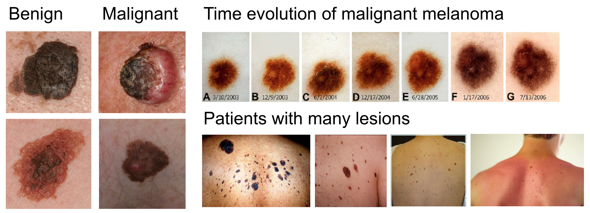
Dermatology is a medical field which stands to be heavily augmented by the use of artificial intelligence techniques. Diseases are visually screened for, and many disease diagnoses are performed strictly with an in-clinic visual examination. Discerning between skin lesions is difficult - the difference between skin cancer (melanoma, carcinoma) and benign lesions (nevi, seborrheic keratosis) is minute (Figure 1). With 5.4 million cases of skin cancer diagnosed each year in the United States alone, the need for quick and effective clinical screenings is rising [1, 2]. Patients with skin cancer tend to be afflicted with many moles, and so one of the challenges in skin cancer screenings is identifying them amongst a myriad of benign lesions. Another key element of these diagnoses is based on inspecting temporal changes in lesions - a fast changing lesion is more likely to be malignant. As such, patients and providers need tools to support this at scale.
Recent advances in detection and tracking using CNNs [3, 4, 5] has the potential to augment healthcare providers by (1) detecting points of malignancy, and (2) finding corresponding lesions across images, allowing them to be tracked temporally. However, the primary challenge in using traditional detection techniques is working in a low-data regime without the availability of high volumes of annotated and labeled data - the largest existing open-source skin cancer dataset of photographic images is the Edinburgh Dermofit dataset, containing 1,300 biopsied images. To overcome this we develop a domain-specific data synthesis technique which stitches small single-lesion images onto large body images. Both the body images and the skin lesions images are heavily augmented with various techniques, and the lesions are blended onto the bodies using Poisson image editing [6].
For large-scale lesion detection, we use this synthetic data to train a fully-convolutional CNN on the task of pixel-wise classification between three classes: background, benign lesion, malignant lesion, then post-processing to generate region proposals. For image-to-image tracking, a network is trained (using image pairs containing altered lesion and pose positions) to output pixel-wise positional shift.
Our method demonstrates a working end-to-end CNN system capable of tackling two critical diagnostic tasks with superior performance to baseline techniques. It is trained with very little original data and thus the techniques demonstrated here can be easily transferred to other data-limited domains.
2 Data Synthesis
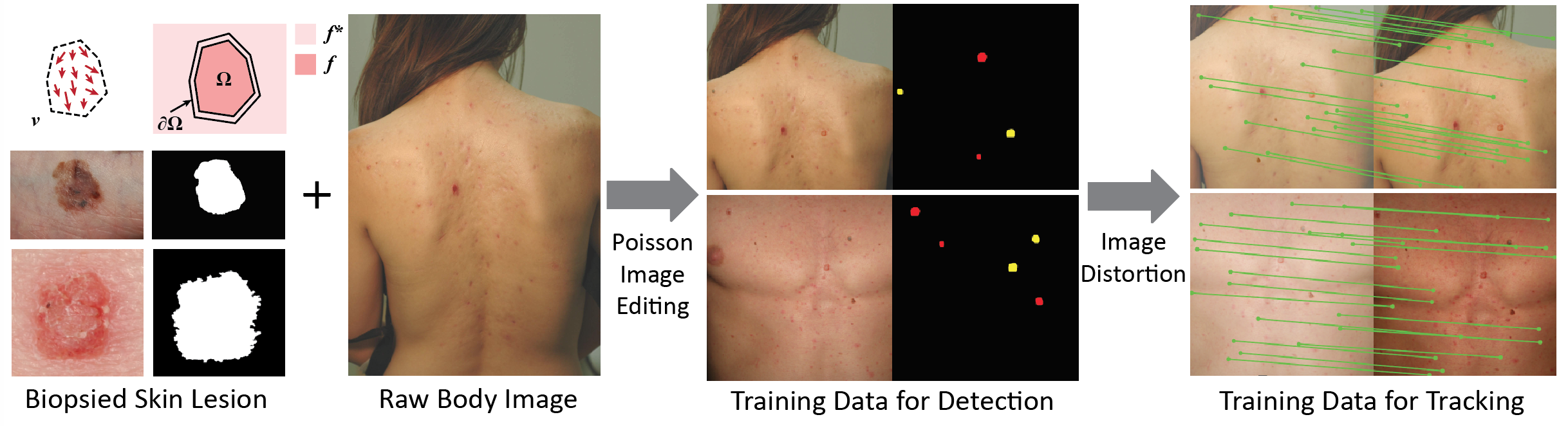
We create a domain-specific data augmentation technique for generating synthetic images from two low-data sources: high-quality lesion images (1,300 biopsy-proven cancers and moles), and body images (400 back, leg, and chest images) whose skin regions have been manually segmented. We first generate images for detection and then further augment them for tracking. These images are intended to mimic the real-world clinical case of patients exhibiting many lesions, some possibly malignant, with the need to track them over time.
Detection data is generated in two steps: (1) A blending position on the body image is chosen using local feature matching between a lesion image and the body image, (2) the lesion image is blended into the body image using Poisson image editing. For tracking, data is generated by further augmenting detection images - that is, given a detection image, we create a pair of images from it. The purpose here is to recreate temporal images (which will exhibit changes in lesion shape/size, as well as body changes) and to force the network to learn pixel-wise correspondence by focusing on the distortion in local texture information.
3 System Pipeline
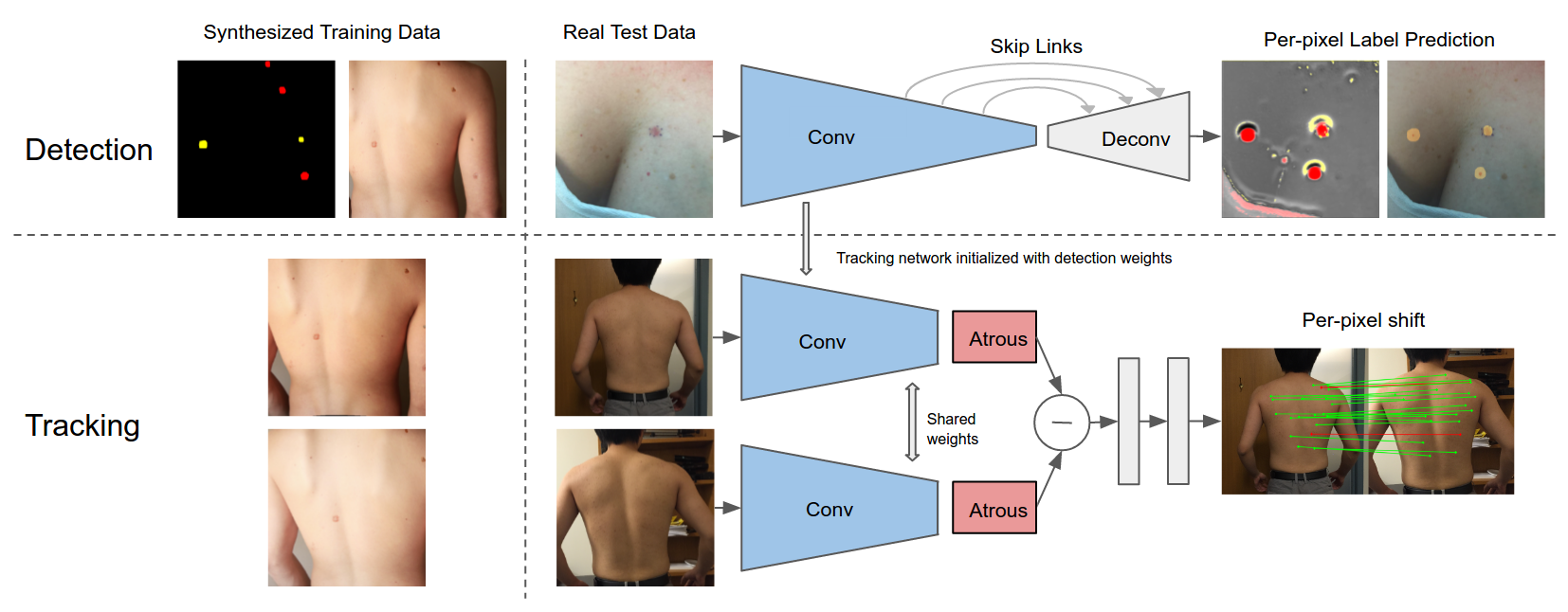
Our system is composed of two parts: the first detects malignant and benign skin lesions, the second tracks them across images. The detection network is trained with the synthetic images (skin lesion + body images) described in the previous section, using pixel-wise labels. Once the detection network is trained to convergence, its weights are used to initialize the tracking network. This network is then trained on image-pairs formed from the detection data.
The detection component is intended to highlight to a clinician the potentially malignant lesions on a given input image. Providers are often faced with body regions containing a multitide of lesions and discerning malignancy is a challenging task. Our system feedforwards an input image through the CNN, outputs a pixel-wise heatmap over the five classes of interest, and then uses post-processing techniques to make the heatmap more human-interpretable. Examples of raw prediction results and post-processed images are shown in Figure 4.
The tracking component of our system is intended to find pixel-wise correspondence between two images of the same body part, in order to track lesions over time. Shown in Figure 3, the tracking network is an adaptation of the detection network. During the feedforward pass the two images are fed through the net, which then outputs a 2D vector field of correspondences.
4 Experiments and Results
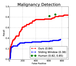
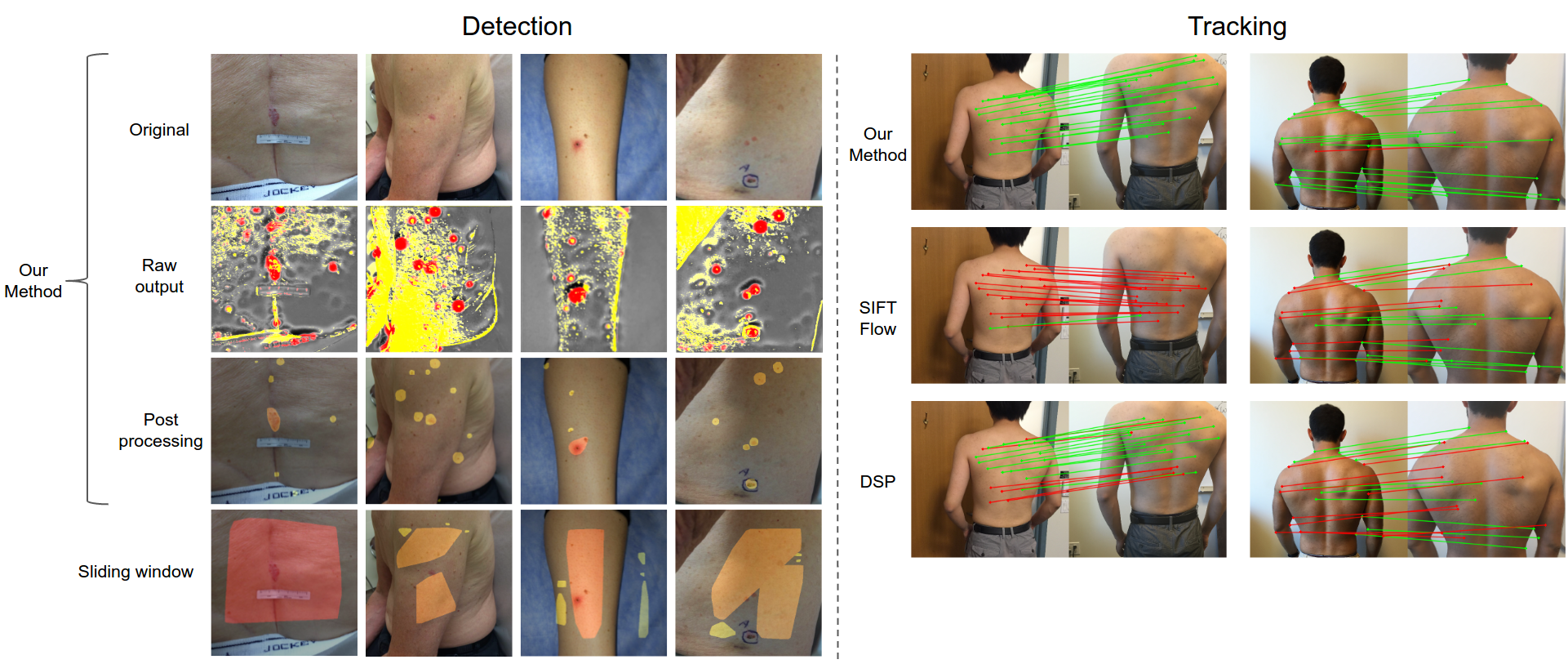
Example results for detection are shown in the first three rows of Figure 4, whose columns show examples as they are processed from input images to raw network output to post-processed results. Our method is compared to a baseline sliding window method (bottom row of Figure 4). For quantitative comparison, we test detection using 108 clinical images, and include results of two non-expert humans trained on Edinburgh Dermofit dataset to detect malignant lesions. The generated receiver-operator curve shown in the left part of Figure 5.
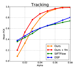
For tracking, we compare our results to two baseline techniques: SIFTFlow, and Deformable Spatial Pyramids (DSP) [7, 8]. Example image results are shown in Figure 4, where green lines show correctly predicted correspondences, and red lines show incorrect predictions. For quantitative comparison, we evaluate tracking accuracy using the percentage-of-correct-keypoints (PCK) metric. Our test set is composed of 260 pairs of correspondence labels manually annotated on temporal image pairs. These image pairs vary in pose, background, distance, viewpoint, and illumination condition. Results are plotted in Figure 5, in comparison to both baselines.
5 Conclusion
Here we show large-scale detection and tracking of skin lesions across images using FCN in a low-data regime using domain-specific data augmentation. In the absence of large amounts of labeled and annotated data, we generate high volumes of synthetic data using 1,300 biopsy-proven clinical images of skin lesions and 400 body images. Skin lesion images are blended onto body images, heavily augmented with a variety of techniques, and used to train a detection network. We demonstrate human-interpretable detection with this method, and demonstrate superior performance over baseline. We then further augment the data and generate image pairs with pixel-wise correspondence between them, and use this to train a tracking network whose architecture is partially composed of the detection network and initialized with its trained weights, outperforming both SIFTFlow and DSP. The networks are trained on synthetic data and tested on real-world data.
AI systems of this sort have the potential to improve the way healthcare is practiced, which may extend outside of the clinic. Algorithms such as these could aid providers at spotting suspicious lesions amongst benign ones, and at observing temporal changes in lesions that may signify malignancies.
References
- Rogers et al. [2015] Howard W Rogers, Martin A Weinstock, Steven R Feldman, and Brett M Coldiron. Incidence estimate of nonmelanoma skin cancer (keratinocyte carcinomas) in the us population, 2012. JAMA dermatology, 151(10):1081–1086, 2015.
- Stern [2010] Robert S Stern. Prevalence of a history of skin cancer in 2007: results of an incidence-based model. Archives of dermatology, 146(3):279–282, 2010.
- Girshick et al. [2014] Ross Girshick, Jeff Donahue, Trevor Darrell, and Jitendra Malik. Rich feature hierarchies for accurate object detection and semantic segmentation. In Proceedings of the IEEE Conference on Computer Vision and Pattern Recognition (CVPR), 2014.
- Jifeng Dai [2016] Kaiming He Jian Sun Jifeng Dai, Yi Li. R-FCN: Object detection via region-based fully convolutional networks. arXiv preprint arXiv:1605.06409, 2016.
- Kanazawa et al. [2016] Angjoo Kanazawa, David W. Jacobs, and Manmohan Chandraker. Warpnet: Weakly supervised matching for single-view reconstruction. In The IEEE Conference on Computer Vision and Pattern Recognition (CVPR), June 2016.
- Pérez et al. [2003] Patrick Pérez, Michel Gangnet, and Andrew Blake. Poisson image editing. ACM Trans. Graph., 22(3):313–318, July 2003. ISSN 0730-0301. doi: 10.1145/882262.882269. URL http://doi.acm.org/10.1145/882262.882269.
- Liu et al. [2008] Ce Liu, Jenny Yuen, Antonio Torralba, Josef Sivic, and William T Freeman. Sift flow: Dense correspondence across different scenes. In European conference on computer vision, pages 28–42. Springer, 2008.
- Kim et al. [2013] Jaechul Kim, Ce Liu, Fei Sha, and Kristen Grauman. Deformable spatial pyramid matching for fast dense correspondences. In The IEEE Conference on Computer Vision and Pattern Recognition (CVPR), June 2013.