Enhanced responsiveness in asynchronous irregular neuronal networks
Abstract
Networks of excitatory and inhibitory neurons display asynchronous irregular (AI) states, where the activities of the two populations are balanced. At the single cell level, it was shown that neurons subject to balanced and noisy synaptic inputs can display enhanced responsiveness. We show here that this enhanced responsiveness is also present at the network level, but only when single neurons are in a conductance state and fluctuation regime consistent with experimental measurements. In such states, the entire population of neurons is globally influenced by the external input. We suggest that this network-level enhanced responsiveness constitute a low-level form of sensory awareness.
pacs:
87.10.+e, 05.40.+j, 02.50.FzNetworks of neurons are capable of displaying asynchronous and irregular (AI) states of activity, where neurons fire in an apparent stochastic fashion, and with very low levels of synchrony. This property was found in networks of excitatory and inhibitory neurons, in a balanced state, first by binary neurons VV96 ; VV98 , and subsequently by more complex models such as spiking neurons Amit97 ; Brunel2000 ; Vogels2005 ; Destexhe2009 ; Yger2011 .
In parallel, a characterization of the noisy background activity seen in cortical neurons in vivo Pare98 showed that these neurons are in a high-conductance (HC) state DP99 ; Destexhe2003 . HC states were first shown in vivo under anesthesia, then confirmed in the awake brain Steriade2001 ; Rudolph2007 ; Petersen . HC states were also largely investigated in vitro by a number of studies using the dynamic-clamp technique. These studies have collectively shown that the HC state confers to neurons enhanced responsiveness, which is due to a tight interplay between conductances and fluctuations Destexhe2001 ; Chance2002 ; Rauch ; Wolfart2005 ; Prescott ; Zerlaut2016 .
In the present paper, we attempt to unify these two points of view by showing that AI states in networks can display enhanced responsiveness properties, but only if neurons display the correct conductance state and fluctuation regime.
Figure 1 compares different network models displaying AI states, with experimental measurements. The measurements of excitatory and inhibitory conductances in neurons is of primary importance, as we will show here. Conductance measurements in cortical neurons in awake cats are depicted in Fig. 1A. There was a very wide range of conductance values measured from cell to cell, but when normalized to the estimated resting conductance, approximately symmetric distributions were obtained Rudolph2007 ; NeuroComp2007 . The total excitatory conductance is slightly lower than the resting conductance, while inhibitory conductances are in general about 1.5 times larger than the leak (Fig. 1A).
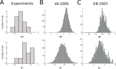
These measured conductances were compared to a well-known model of AI state consisting of 5000 randomly-connected leaky integrate and fire neurons (80% excitatory, 20% inhibitory) Vogels2005 . We computed the total excitatory and inhibitory conductances seen in different neurons of this model. It appeared that, in this 5000-neuron network, when normalized to the resting conductance, the relative conductances in this model were considerably larger than physiological measurements, up to about 20 times larger (Fig. 1B). Another model of AI state NeuroComp2007 , consisting of 16,000 randomly-conected neurons, but with smaller synaptic weights, did not display such an aberrant conductance state, and could generate a conductance state much closer to physiological measurements NeuroComp2007 (Fig. 1C).
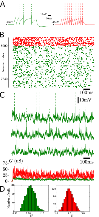
Such a physiologically plausible conductance state was also obtained in a more realistic network of excitatory and inhibitory cells, where the adaptation of excitatory cells was taken into account (Fig. 2). This model used two types of cells, the “regular spiking” (RS) and “fast spiking” (FS) neurons, which correspons to the typical firing patterns seen in vitro McCormick85 . These cell types were modeled using the Adaptive Exponential integrate and fire model Brette2005 (Fig. 2A; see details in Appendix). A network of 8000 RS and 2000 FS cells generated AI states with the typical higher frequency firing of inhibitory neurons (Fig. 2B). In this network, the conductance state of individual cells was consistent with experimental measurements in awake animals (Fig. 2C). The conductance distributions obtained in this network model (Fig. 2D) are close to experimental values (compare with (Fig. 1A).

Interestingly, comparing these networks in term of their responsiveness, revealed that the conductance state of the network is very important. When an external input was given to a randomly chosen sample of cells, the network in a physiologically plausible AI state could display a remarkable sensitivity to this external input (Fig. 3). However, in a network displaying an aberrant conductance state, this response was not present. The response was also absent in a quiescent state, or in an oscillatory state in the same network (Fig. 3B). This shows that the AI state, if with correct conductance and fluctuations, displays an enhanced responsiveness to external inputs, and is able to collectively detect inputs of very small amplitude, which normally would have been subthreshold.
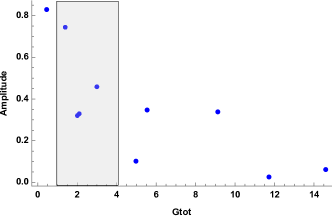
We have done additional simulations to explore how responsiveness depends on conductance state, by considering random sets of parameters around the AI state in Fig. 3A. This exploration showed that the responsiveness highly depends on the conductance state of the network (Fig. 4). The response was quantified as the area of the evoked population response (as in Fig. 3B), represented against the total conductance, as calculated from distributions (as in Fig. 2D). One can see that the response is indeed high for physiological conductance states (gray area in Fig. 4) and approaches to zero for aberrant conductance states. This result explains the difference of responsiveness between the two networks shown in Fig. 3B (top). However, it is important to keep in mind that the response also depends on the level of fluctuations and the average Vm level, which can be quantified by considering distributions of the standard deviation of the membrane potential () and of the mean Vm of the cells (not shown). The responsiveness can also be understood using a phenomenological model, as shown recently Reig2015 .
This phenomenon of network-level responsiveness is very similar to the enhanced responsiveness that was found earlier at the single neuron level Ho2000 . In single-cell studies, the presence of synaptic “noise” conferred an anhanced sensitivity to the neuron. It was also shown that having the correct conductance state and fluctuation regime of the cell is important, it sets the response to a realistic level, and enhanced responsiveness was present using the conductances measured experimentally, as well as their level of fluctuations. Enhanced responsiveness was also found in noisy networks, either in feedforward networks where all cells display a high-conductance state Ho2000 , or in chaotic recurrent networks where the enhanced responsiveness was quantified in terms of information transport Destexhe94 ; Destexhe-Contreras2006 . One of these studies was the first to demonstrate that networks in a chaotic state display enhanced responsiveness Destexhe94 . We complement here these previous studies by showing that enhanced responsiveness is present in recurrent networks displayin AI states, which are presumably chaotic VV98 ; VV98 . In addition, we show that this property highly depends on conductance state, and that physiologically plausible conductance state are particularly responsive. This underlies the importance to work with conductance-based models, as current-based models are unlikely to display the correct responsiveness, and cannot be checked against conductance measurements, so are also unconstrained.
Thus, the present results suggest that the conductance state of a network is a fundamental property to understand its responsiveness, which emphasize the importance of conductance measurements in vivo. We suggest that HC states have a universal aspect, in the sense that the conductance and fluctuation level measured in vivo are the fundamental parameters that neural networks should reproduce, to yield responsiveness properties relevant to neural function.
Finally, let us emphasize that in the AI state, the network responds instantaneously to an input, and this input can occur at any time, which presents evident useful computational properties. A particularly interesting property is that, when submitted to successive presentations of the same stimulus, different neurons respond on each trial, showing that it is the whole network that is globally “aware” of this stimulus. We therefore propose that such network-level responsiveness during AI states implements a low-level form of sensory awareness in networks of neurons. The fact that this occurs in AI states is consistent with the observation that cerebral cortex is in a “desynchronized” state in awake and attentive states (reviewed in Niedermeyer ; Steriade-book ). A recent review concluded that desynchonized cortical activity is so far the best correlate of conscious states Koch-Nat-Rev , but no mechanism was given. We propose here such a possible mechanism to link these high-level aspects to elementary biophysical properties of neurons.
Acknowledgments: Research funded by the CNRS and the European Community (Human Brain Project H2020-720270). We thank Yann Zerlaut for many discussions and help with the simulations.
References
- (1) van Vreeswijk C, Sompolinsky H (1996) Chaos in neuronal networks with balanced excitatory and inhibitory activity. Science 274: 1724-1726.
- (2) van Vreeswijk, C., & Sompolinsky, H. (1998). Chaotic balanced state in a model of cortical circuits. Neural Comput. 10: 1321-1371.
- (3) Amit, D.J. and Brunel, N. (1997) Model of global spontaneous activity and local structured activity during delay periods in the cerebral cortex. Cerebral cortex 7: 237-252.
- (4) Brunel, N. (2000) Dynamics of sparsely connected networks of excitatory and inhibitory spiking neurons. J. Computational Neurosci. 8: 183-208.
- (5) Vogels, T.P., and Abbott, L.F. (2005) Signal propagation and logic gating in networks of integrate-and-fire neurons. J. Neurosci. 25: 10786-10795.
- (6) Destexhe, A. (2009) Self-sustained asynchronous irregular states and up–down states in thalamic, cortical and thalamocortical networks of nonlinear integrate-and-fire neurons. J. Computational Neurosci. 27: 493-506.
- (7) Yger, P., El Boustani, S., Destexhe, A. and Fregnac, Y. (2011) Topologically invariant macroscopic statistics in balanced networks of conductance-based integrate-and-fire neurons. J. Computational Neurosci. 31: 229-245.
- (8) Paré, D., Shink, E., Gaudreau, H., Destexhe, A. and Lang, E.J. (1998) Impact of spontaneous synaptic activity on the resting properties of cat neocortical neurons in vivo. J. Neurophysiol. 79: 1450-1460.
- (9) Destexhe A and Paré D. (1999) Impact of network activity on the integrative properties of neocortical pyramidal neurons in vivo. J. Neurophysiol. 81: 1531-1547.
- (10) Destexhe, A., Rudolph, M. and Paré, D. (2003) The high-conductance state of neocortical neurons in vivo. Nature Reviews Neurosci. 4: 739-751.
- (11) Steriade, M., Timofeev, I., and Grenier, F. (2001) Natural waking and sleep states: a view from inside neocortical neurons. J. Neurophysiol. 85: 1969-1985.
- (12) Crochet, S., and Petersen, C.C. (2006) Correlating whisker behavior with membrane potential in barrel cortex of awake mice. Nature neurosci. 9: 608-610.
- (13) Rudolph, M., Pospischil, M., Timofeev, I. and Destexhe, A. (2007) Inhibition determines membrane potential dynamics and controls action potential generation in awake and sleeping cat cortex. J. Neurosci. 27: 5280-5290.
- (14) Destexhe, A., Rudolph, M., Fellous, J. M. and Sejnowski, T. J. (2001) Fluctuating synaptic conductances recreate in vivo-like activity in neocortical neurons. Neuroscience 107: 13-24.
- (15) Chance, F. S., Abbott, L. F and Reyes, A. D. (2002) Gain modulation from background synaptic input. Neuron 35: 773-782.
- (16) Rauch, A., La Camera, G., Luscher, H. R., Senn, W. and Fusi, S. (2003) Neocortical pyramidal cells respond as integrate-and-fire neurons to in vivo-like input currents. J. Neurophysiol. 90: 1598-1612.
- (17) Wolfart, J., Debay, D., Le Masson, G., Destexhe, A. and Bal, T. (2005) Synaptic background activity controls spike transfer from thalamus to cortex. Nature Neurosci. 8: 1760-1767.
- (18) Prescott, S. A., Ratté, S., De Koninck, Y., and Sejnowski, T. J. (2008) Pyramidal neurons switch from integrators in vitro to resonators under in vivo-like conditions. J. Neurophysiol. 100: 3030-3042.
- (19) Zerlaut, Y., Telenczuk, B., Deleuze, C., Bal, T., Ouanounou, G. and Destexhe, A. (2016) Heterogeneous firing rate response of mice layer V pyramidal neurons in the fluctuation-driven regime. J. Physiol. 594: 3791-3808.
- (20) El Boustani, S., Pospischil, M., Rudolph-Lilith, M. and Destexhe, A. (2007) Activated cortical states: experiments, analyses and models. J. Physiol. Paris 101: 99-109.
- (21) McCormick, D.A., Connors, B.W., Lighthall, J.W. and Prince, D.A. (1985) Comparative electrophysiology of pyramidal and sparsely spiny stellate neurons of the neocortex. J. Neurophysiol. 54: 782-806.
- (22) Brette, R. and Gerstner, W. (2005) Adaptive exponential integrate-and-fire model as an effective description of neuronal activity J. Neurophysiol. 94: 3637-3642.
- (23) Hô, N. and Destexhe, A. (2000) Synaptic background activity enhances the responsiveness of neocortical pyramidal neurons. J. Neurophysiol. 84: 1488-1496.
- (24) Reig, R., Zerlaut, Y., Vergara, R., Destexhe, A. and Sanchez-Vives, M. (2015) Gain modulation of synaptic inputs by network state in auditory cortex in vivo. J. Neurosci. 35: 2689-2702.
- (25) Destexhe, A. (1994) Oscillations, complex spatiotemporal behavior and information transport in networks of excitatory and inhibitory neurons. Physical Review E 50: 1594-1606.
- (26) Destexhe, A. and Contreras, D. (2006) Neuronal computations with stochastic network states. Science 314: 85-90.
- (27) Niedermeyer, E., and da Silva, F. L. (Eds.). (2005) Electroencephalography: basic principles, clinical applications, and related fields. Lippincott Williams and Wilkins, New York.
- (28) Steriade, M. M., and McCarley, R. W. (2013) Brainstem control of wakefulness and sleep, Springer, New York.
- (29) Koch, C., Massimini, M., Boly, M., and Tononi, G. (2016) Neural correlates of consciousness: progress and problems. Nature Reviews Neurosci. 17: 307-321.
Appendix
Details of the network model
The network model used here consisted of excitatory (RS) and inhibitory (FS) neurons described by the Adaptive Exponential integrate and fire model Brette2005 , for which the equations for the membrane potential and the adaptation current read:
where is the membrane potential, is the membrane capacitance, is the resting conductance and its reversal potential, is the synaptic current, and are threshold parameters. is the adaptation current, which evolved according to a time constant and increases by a value at each spike . FS cells correspond to the same model with =0.
The cellular parameters were, for RS cells: =10nS, =200pF, =5ms, =-70mV, =-50mV, =-70mV, =2mV, =4nS, =20pA, =500ms. FS cells were descrivbed by a leaky integrate and fire model with =10nS, =200pF, =5ms, =-70mV, =-50mV, =-70mV. Network parameters: 4000 RS and 1000 FS cells, connectd randomly with probability of 5%, and with synaptic weights of =1nS and =5nS (with respective reversal potentials of =0mV, =-80mV). The synaptic conductances were decaying exponentials with a time constant of 5 ms.
An external drive was present in all neurons and consisted of 4000 independent Poisson processes (rate of 4 Hz), projected over the 5000 neurons with 5% connection probability (weight of 1nS).
The external stimulus of Fig 3 consisted of 40 synchronous EPSPs of 1nS, spread over 40 randomly-choosen excitatory neurons within the population.