Steady State Visually Evoked Potentials detection using a single electrode consumer-grade EEG device for BCI applications
Abstract
Brain-Computer Interfaces (BCIs) implement a direct communication pathway between the brain of an user and an external device, as a computer or a machine in general. One of the most used brain responses to implement non-invasive BCIs is the so called steady-state visually evoked potential (SSVEP). This periodic response is generated when an user gazes to a light flickering at a constant frequency. The SSVEP response can be detected in the user’s electroencephalogram (EEG) at the corresponding frequency of the attended flickering stimulus. In SSVEP based BCIs, multiple stimuli, flickering at different frequencies, are commonly presented to the user, where to each stimulus is associated a command for an actuator. One of the limitations to a wider adoption of BCIs is given by the need of EEG acquisition devices and software tools which are commonly not meant for end-user usage. In this work, exploiting state-of-the-art software tools, the use of a low cost easy to wear single electrode EEG device is demonstrated to be exploitable to implement simple SSVEP based BCIs. The obtained results, although less impressive than the ones obtainable with professional EEG equipment, are interesting in view of practical low cost BCI applications meant for end-users.
I Introduction
Brain-Computer Interfaces (BCIs) implement a direct communication pathway between the brain of an user and an external device, as a computer or a machine in general [1]. The purpose of a BCI is to translate a detectable brain state of the user in a command for an actuator, providing the user with a real-time feedback. BCIs may record the brain activity of their users with different methods, but the most used is electroencephalography (EEG). EEG recording of the neural activity allow for non-invasive recordings with a relatively high time resolution and the use of relatively inexpensive devices, with respect to other methods.
Initial research regarding BCIs aimed to provide mobility-impaired users with a tool capable of translating a thought or a will into a command for an external device or a prosthetic limb. Nevertheless, more recently, BCIs are gaining attention also as new means to interact with computers and other devices for healthy subjects too [2].
In literature, different BCI modalities have been successfully adopted and differentiate between them accordingly to the kind of underlying brain process that is investigated to detect features associated to brain states. The most popular are the P300 or Event Related Potentials (ERP), Motor Imagery (MI) or Event Related Synchronization/De-synchronization (ERS/ERDS) and Steady-State Visually Evoked Potentials.
BCIs could be further divided into three main categories, with smooth boundaries [3]; every modality may fit inside one of these category, accordingly to how it is used, although not every modality is suitable for every category:
-
•
Active BCIs derive their outputs from brain activity which is directly consciously controlled by the user, independently from external events, for controlling an application.
-
•
Reactive BCIs derive their outputs from brain activity arising in reaction to external stimulation, which is indirectly modulated by the user for controlling an application.
-
•
Passive BCIs derive their outputs from arbitrary brain activity without the purpose of voluntary control, for enriching a human-computer interaction with implicit information.
The performance of a consciously controlled BCI (Active or Reactive) is commonly given in terms of its Information Transfer Rate (ITR). The ITR value has been introduced in order to take into account both the speed of a BCI in detecting a user command and its accuracy in detecting the correct command [4]. BCI research is consequently strongly focused on the improvement of the detection accuracy and the time needed to issue a command. Both of these factors are strongly dependent on the acquired signals quality, as well as the efficiency of the feature extraction algorithms, machine learning methods and the potential training of the users.
Other performance metrics for a BCI could be its cost in terms of hardware, or its ease of use, concerning the time to wear the electrodes system and the training time needed for calibrations or for the user to learn to modify her/his brain activity to issue commands.
One of the limitations to a wider adoption of BCIs is given by the need of data acquisition devices which are commonly not meant for end-user usage, requiring trained technicians to use them and moreover being orders of magnitude more expensive than ordinary interaction devices. Furthermore, only recently general purpose BCI software frameworks are being developed in order to provide user friendly tools for non-programmers.
The aim of this work is indeed to exploit state-of-the-art software tools and feature extraction algorithms in order to investigate the feasibility of an easy to use BCI, minimizing the hardware cost and the user time needed for the system set up.
In particular a low cost easy to wear single electrode EEG device is investigated in order to implement a Reactive BCI based on the SSVEP response detection.
II SSVEP based BCIs
In this research work the SSVEP modality, commonly used to implement Reactive BCIs, has been chosen because of its high reachable ITR, the short calibration time needed, the low number of EEG electrodes required and also for the low BCI illiteracy showed [5], granting high usability for most of the users, also in out-of-laboratory environments. In fact, in the last years the SSVEP response has been adopted widely for the implementation of Reactive BCIs [6].
Steady-state VEP (SSVEP) are a particular case of Visually Evoked Potentials (VEP), where the visual stimulus is presented several times at a frequency at least higher than 3.5Hz, but more commonly higher than 6Hz. In this case, a quasi-sinusoidal periodic response called SSVEP can be observed in the scalp recorded EEG signals, in particular over the occipital brain region, where the visual cortex resides.
The stimulus presented to elicit a SSVEP response is commonly a flickering light and accordingly to the flickering frequency, in the EEG signals acquired from an user looking at the stimulus an increase in the power at the corresponding frequency (and harmonics) can be detected. Consequently in the case of SSVEP, the presence of the response can be detected analyzing the frequency spectrum of the recorded signal.
In presence of multiple stimuli, flickering at different frequencies in the visual field of the user, the increase of the signal power in the same frequencies of the stimuli is more pronounced in the single frequency corresponding to the single stimulus the user is gazing at.
Thanks to this observation, Reactive BCIs can be implemented using this modality, since the user, gazing at a particular stimulus chosen from the presented ones, can issue to the system a command previously associated to the stimulus.
For BCI applications, stimuli are commonly presented by LED lights, or by shapes on a regular computer monitor, flickering at frequencies ranging between 6Hz and 40Hz. The provided visual stimuli characteristics are known to be highly relevant concerning the SSVEP response amplitude, indeed their frequency stability as well as their size (in terms of user visual angle), color, duty cycle, modulation depth, etc. have to be carefully controlled to obtain a stronger response and thus an easier detection [7].
Data acquisition is commonly performed placing electrodes over the visual cortex in the occipital region of the user’s brain using multiple electrodes EEG devices.
The SSVEP signal processing could be very simple and lot of SSVEP based BCIs implemented so far simply filter the raw EEG signals with narrow band-pass filters, centered on the stimuli frequencies, to later estimate the signal power in the frequency regions corresponding to the different stimuli. Features corresponding to the power in the different frequency bands can then be used to detect the gazed target using a previously trained classifier.
Anyhow, it has been demonstrated that better performances can be obtained using more sophisticated methods as: evaluating the signal power also on higher harmonics for each stimulation frequency [8]; merging the information coming from different electrodes using spatial filters [9, 10]; or using as features SNR indexes representing the ratio between the SSVEP response power and the stimulus-uncorrelated brain activity occurring in the same frequency band [10].
The adoption of sophisticated signal processing methods do not commonly introduce complexity from the user point of view, on the other side it commonly improve the BCI performance, thus, as will be highlighted in this work, it permits to obtain a fair performance also when using lower quality EEG signals.
III Towards practical BCIs
Conductive gel based EEG electrodes represent the state-of-the-art in terms of signals acquisition quality [11], despite of this, the montage of EEG cups adopting them commonly requires a trained technician and moreover users need to wash their hair after usage. Just the fact that the user would need another person to mount the EEG headset on her/him is a strong limitation for end-user applications, as is also the high cost of electroencephalographers with respect to other interaction devices.
Custom hardware is being investigated [12] in order to provide easier to use devices by means of dry electrodes or salted-water based electrodes and also industrial companies are working towards this direction (as Emotiv, Neurosky, Biosemi, g.Tec, etc.). To exploit easy to wear EEG devices is indeed one of the most challenging research directions towards practical BCIs in the last years [13, 14].
Professional general purpose EEG headsets, implementing dry electrodes, are already available (e.g. the g.Tec g.SAHARA system), but from their cost and their complexity (e.g. the need to choose electrodes positions, connect the electrodes to the amplifiers, etc.) is clear that they are still meant to be used mainly by BCI and clinical researchers, but not end-users.
New kind of EEG devices, meant to improve the usability by non-trained end-users, recently appeared on the market; they are commonly characterized by a rigid structure where electrodes are fixed on a pre-determined position and could be worn by the user himself without the need of external help. These devices commonly adopt dry electrodes or water-based electrode technologies in order to not slime the user’s hair, renouncing to the higher signal quality given by the use of conductive gel, but improving users’ comfort.
A famous commercial device of this kind is the Emotiv EPOC 111http://www.emotiv.com/epoc/ which is a salted-water based 14-electrodes system, that for example has been recently successfully used to implement SSVEP based BCIs [14, 15].
One of the less expensive devices of this kind is the Mindset single dry electrode system provided by Neurosky Inc. which looks like regular headphones, a part from an additional arm holding a dry EEG electrode to be positioned on the forehead. It was designed to provide an additional interaction mean for generic computer application, e.g. computer games, implementing Active BCIs where the user can learn, using a bio-feedback approach, how to control her/his own brain activity and thus the features extracted from it. The features extracted from the raw EEG data are computed by a proprietary algorithm, which is known to compute indexes based on relative clinical band power ratios, but no more details were disclosed about it by its manufacturer. Interestingly raw EEG data can be read-out from this device as well.
The Mindset is clearly not designed to detect SSVEP responses, but in view of more practical BCIs, it would be very interesting if the Mindset could be used also for SSVEP based BCIs, thanks to its extremely low cost and for its ease to be wear. In particular, in contrast to the Active BCI its manufacturer had in mind, it would be very interesting to be able to use it for Reactive BCIs based on the SSVEP modality, since it would avoid the need of subject training. Applications where a generic naive user could just wear it in order to be able to use the BCI, would be possible.
Anyhow, to use the Mindset for this purpose various challenges has to be faced: having a single electrode it will not allow to exploit spatial filtering algorithms [9]; having a dry electrode it will provide a noisier signal than gel or water based ones [11] and moreover, as already mentioned, wearing it as indicated by its manufacturer, the electrode would be positioned on the forehead (roughly at Fp1 with respect to the 10-20 positioning system) and thus very far from the visual cortex where SSVEP responses are more intense [6].
IV Material and Methods
IV-A Stimuli presentation
In the performed experiments, the flickering stimuli presentation was provided on a regular LCD computer monitor (HP LP2065) thanks to a custom developed C++/OpenGL software [16]. The used flickering stimulus was a squared white patch subtending of the user visual angle. To achieve an optimal stimuli presentation, the flickering frequency control was synchronized to the vertical screen refresh as suggested in [17].
The duty cycle of the stimulus was tuned for best performances according to the results discussed in [18] and was for the stimulation frequency and for the one (since in this case a duty cycle is not possible due to the integer odd number of frames given a screen refresh).
IV-B Data acquisition
The EEG signal acquisition was performed using the MindSet device already mentioned. Its single electrode is designed to be positioned on the forehead, roughly at the Fp1 position. It acquires the EEG signal band-pass filtered between and at a sampling rate of , digitizing it at . It can be connected to a computer for data acquisition using a Bluetooth connection. It incorporates a notch filter to remove power-line artifacts and implements proprietary algorithms for further signal cleaning and feature extraction. In addition to the single acquisition electrode, the MindSet has also three other contacts positioned over the left ear of the subject, which are used as ground and reference electrodes. In particular, a part from the raw filtered EEG signal, it provides also proprietary dimensionless features representing the power strength in the clinical frequency bands and also two sampled signals called e-Sense Attention and e-Sense Meditation values. The Attention and Meditation values are computed thanks to a proprietary algorithm and very few information are available about their actual meaning. In the manufacturer intention, the subject wearing this device should be able to learn to control these two values in order to be able to use Active BCI applications after several hours of training.
Data acquisition and software triggering handling, was performed using the OpenVibe software framework [19]. Interestingly the MindSet has an automatic on-line check to detect the contact quality between the skin and the electrodes, that, instead of an impedance value as is commonly used, returns a SNR between what a proprietary algorithm considers as the EEG signal and what it identifies as artifacts. This value can be acquired by OpenVibe too and displayed in real-time to the user while wearing the device.
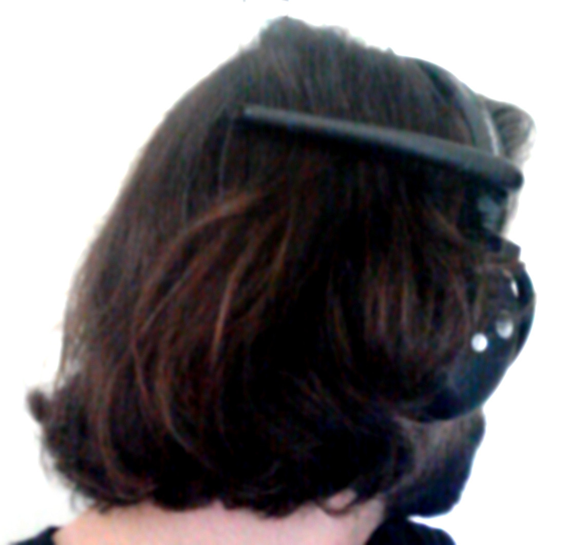
To overcome the wrong positioning of the MindSet’s electrode for SSVEP response detection, experiments was conducted using the MindSet on the subjects head, wearing it “reversed”, letting the single electrode to be positioned backward and the reference/ground electrodes over the right ear. In this manner, as shown in Fig. 1, the electrode is roughly positioned near P2 (according to the extended 10-20 system), which is a much more suitable location to detect the SSVEP response, although not the optimal one [6].
Being the MindSet electrode meant to be positioned on the forehead, its shape is not appropriate to have a connection to the scalp where hair is present. To overcome this problem a droplet of conductive electrode gel was used to improve the contact. Although consequences in terms of impedance could not be assessed due to the proprietary hardware (electrode-skin impedance could not be measured and the amplifier input impedance is unknown), experimental results, as exposed in the next section, confirm that this procedure improve the acquisition signal quality. In order to have a similar impedance also on ground and reference electrodes, a very small amount of gel was positioned also on them.
Despite of the use of a little amount of conductive gel, wearing the MindSet remains much easier than wearing ordinary gel based EEG devices and it could be easily done by the subject himself with no need of external help. Moreover, the small amount of gel to be used, do not force the subject to have a shower right after the use of the device.
The user, as already mentioned, could use the automatic impedance checker, implemented in the device, in order to assess the quality of the electrodes contact, which was reported in real-time and displayed by the acquisition software.
Montage of the device was in the range of about , according to the hair volume of the user.
The study was conducted in accordance with the recommendations of the Declaration of Helsinki and before the experiment, all participants signed the informed consent.
IV-C Signal Processing
The features describing the SSVEP response intensity were computed using the test statistic described in [10], where one of the best performing spatial filtering methods [9] known as Minimum Energy Combination was proposed. Having the MindSet a single electrode, spatial filtering is not possible, but the same approach discussed in [10] was used to separate the SSVEP response contribution from the stimulus-uncorrelated brain activity.
The acquired EEG signal can be modeled as shown in Eq. 1, adapted from the multi-channel signal model described in [10].
| (1) |
The first component of is the actual SSVEP response of interest, which is characterized by a set of sinusoids with frequency and its harmonics, each of which has a specific amplitude and phase . The second component of the model is a set of signals , scaled by the weighting factors , which are unrelated to the SSVEP response and comprise concurrent brain activity and internal as external artifacts. These signals are present. Eventually, the last component is a measurement noise component.
In vector form, the model can be expressed as and an estimate of the stimulus-uncorrelated component can be obtained as shown in Eq. 2.
| (2) |
Given this model, the actual statistic to be used as a feature, can be computed as shown in Eq. 3:
| (3) |
where is the estimated SSVEP power for the -th harmonic frequency in channel signal and is an estimate of the noise and uncorrelated brain activity in the same frequency. In other words, the statistic estimates how many time larger is the SSVEP response power compared to the case where no visual stimulus is present, averaging the SNRs ratios across harmonics.
The power in the -th harmonic is estimated as , while, in order to avoid the need of calibration data acquired with no stimuli presentation and also to take into account the nonstationarity of the noise, the noise power is estimated on the same data segment, containing the SSVEP response, used to compute .
The SSVEP contribution is therefore removed from the signal as shown in Eq. 2 to later fit an auto-regressive model of order and use the fitted models to interpolate the noise power in the SSVEP frequencies. The models are fitted using the Wiener-Khinchin theorem for computing the autocovariance of the signal and then solving the Yule-Walker equations using a Levinson-Durbin recursion [10]. This yields the parameters as well as an estimate of the variance of the white noise driving the process. Once fitted the model to the signal , the noise level estimated at the -th harmonic is given by:
| (4) |
where are the signal samples, is the harmonic number, is the stimulation frequency in , is the sampling frequency in and .
V Performed experiments
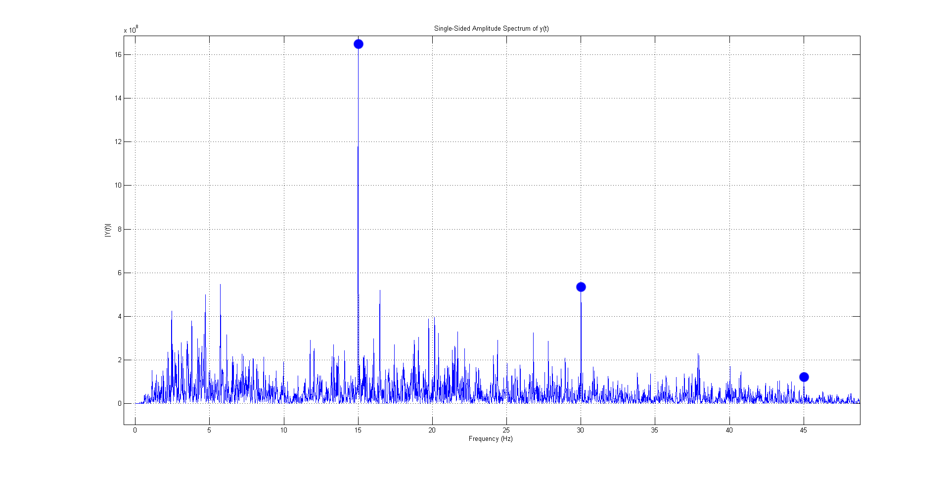
As a preliminary test to assess the possibility to record a SSVEP response using the MindSet unconventionally wore, as shown in Fig. 1, one subject was visually stimulated with a flickering white patch displayed on a screen and the PSD of its acquired EEG signal was computed off-line taking the FFT of the signal auto-correlation. As shown in Fig. 2, the proposed setup led to a clear detection of a SSVEP response to the stimulation; peaks at the fundamental frequency and first harmonics are visible.
Knowing that the SSVEP response could be recorded with the MindSet, further experiments have been conducted to identify the shortest signal length able to lead to a classification accuracy high enough for BCI applications. Having BCIs to be quasi real-time by definition, a SSVEP response detection has to be performed using at most few seconds long signal windows. The ITR is indeed dependent on the accuracy, but on the detection speed as well.
In a similar fashion as performed in [10], multiple subjects were visually stimulated for for each trial for two different stimulation frequencies, chosen as and , while recording their EEG with the MindSet. Four trials have been recorded for each subject in order to have a total of + recording for each stimulation frequency.
Off-line analysis was then performed, computing the test statistic reported in Eq. 3 setting , for both the stimulation frequencies, for each signal window, irrespective of the actual stimulation frequency. The same operation was performed for non-overlapping signal windows and then again, using non-overlapping signal windows.
Each signal window could be labeled with three values: , the actual stimulation frequency and the two features representing the detected SSVEP response respectively for the two stimulation frequencies.
Information coming from the first trial for each subject and each stimulation frequency was used to train a simple linear least squares binary classifier, while information coming from the second trial was used to test it. The datasets were not randomized between trials, since in an actual BCI application the training data would be acquired before the BCI use, so the same approach has been followed.
VI Results
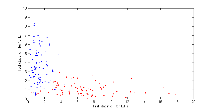
Data points computed using two second signal windows, for one subject, are reported in Fig. 3, where the points color represents the actual stimulation frequency during the epoch, while the points coordinates are the computed features .
As can be seen, a linear classification between the epochs acquired under the two different stimulation frequencies seems to be feasible, despite of the short signal window (considering the used acquisition device).
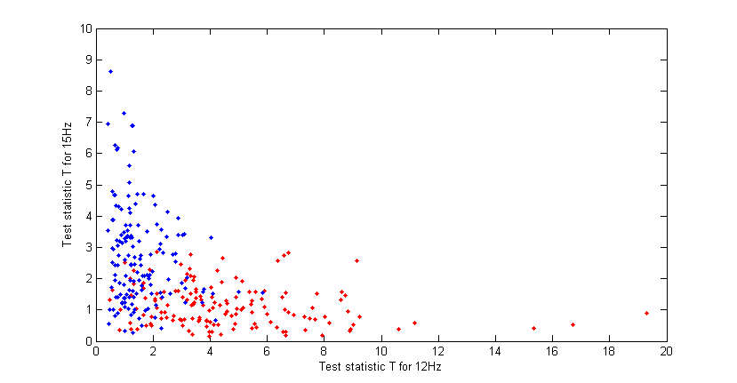
Using the same approach, the data points computed using one second signal window, on the same dataset are reported in Fig. 4.
As can be seen, as expected using a shorter signal window, in this case, a linear classification would produce a lower accuracy, but nevertheless it seems to be feasible.
The classifier training was performed using the first trial acquired for each of the two frequencies, while the remaining two trials where used as test sets; results for the different subjects are reported in Tab. I.
| windows | windows | |
|---|---|---|
| Subject 1 | ||
| Subject 2 | ||
| Subject 3 | ||
| Subject 4 | ||
| Subject 5 | ||
| Subject 6 |
According to the reported results, for 5 subjects out of 6 the SSVEP response can be detected with a reasonable accuracy. As expected, using 2 seconds epochs lead to better results for all the subject a part for the 6th one. For the 6th subject, a manual inspection of the data points revealed that the two point clouds, relative to the two stimulation frequencies, are not separable for all the acquired trials. The reason may be a SSVEP based BCI illiteracy of the subject, a very low attention payed to the flickering target or a particularly inefficient electrode location for the particular subject.
Concerning the classification accuracy, it is worth to mention that it has been computed using non-overlapping windows, but, when implementing SSVEP based BCIs, is a common practice to compute the SSVEP response index (e.g. the frequencies power or the test statistic, as in this case) for sliding windows and then to evaluate the computed value for several subsequent windows. This leads to a smoother output, lowering the effect of false-positive detections which may be computed in a single signal window. In this work this approach was not used to compute the values in Tab. I, since it would have been not a fair way to evaluate the classification accuracy, in the sense that multiple points would have been computed from the same parts of acquired signal.
VII Conclusion and future works
With the previously described experiment has been demonstrated that using a popular single electrode consumer-grade EEG acquisition device is possible to detect a SSVEP response. Moreover, despite of the not optimal electrode position and its physical shape, has been demonstrated that, using a state-of-the-art signal processing technique, the signal window length needed to accurately detect the SSVEP response could be short enough for BCI applications.
Moreover, in an actual SSVEP based BCI application adopting the proposed method, the real-time feedback, which was not presented in the performed experiments, as already demonstrated in other contexts [20], should increase the users’ SSVEP response intensity and thus enhance the detection accuracy and/or shorten the needed signal windows.
The reported results highlight the feasibility to implement a simple SSVEP based BCI using the MindSet device and the presented signal processing method. This is interesting due to the wide diffusion and affordable cost of this device.
Even more interestingly, this work highlights the possibility to design new simple single electrode devices with a more suitable electrode position for the SSVEP detection and also adopting a specific electrode shape to let it be positioned where hair is present without the need of conductive gel.
References
- [1] J. R. Wolpaw, N. Birbaumer, D. J. McFarland, G. Pfurtscheller, T. M. Vaughan et al., “Brain-computer interfaces for communication and control,” Clinical neurophysiology, vol. 113, no. 6, pp. 767–791, 2002.
- [2] J. van Erp, F. Lotte, and M. Tangermann, “Brain-Computer Interfaces: Beyond Medical Applications,” Computer, vol. 45, no. 4, pp. 26–34, 2012.
- [3] T. O. Zander, C. Kothe, S. Jatzev, and M. Gaertner, Enhancing human-computer interaction with input from active and passive brain-computer interfaces, ser. Human-Computer Interaction Series. Springer, 2010, pp. 181–199.
- [4] D. J. McFarland, W. A. Sarnacki, J. R. Wolpaw et al., “Brain-computer interface (BCI) operation: optimizing information transfer rates,” Biological psychology, vol. 63, no. 3, pp. 237–251, 2003.
- [5] C. Guger, B. Z. Allison, B. Grosswindhager, R. Prückl, C. Hintermüller, C. Kapeller, M. Bruckner, G. Krausz, and G. Edlinger, “How many people could use an SSVEP BCI?” Frontiers in Neuroscience, vol. 6, no. 169, 2012.
- [6] F.-B. Vialatte, M. Maurice, J. Dauwels, and A. Cichocki, “Steady-state visually evoked potentials: focus on essential paradigms and future perspectives,” Progress in neurobiology, vol. 90, no. 4, pp. 418–438, 2010.
- [7] J. Bieger, G. G. Molina, and D. Zhu, “Effects of Stimulation Properties in Steady State Visual Evoked Potential Based Brain-Computer Interfaces,” in 32nd Annual International Conference of the IEEE Engineering in Medicine and Biology Society, 2010.
- [8] D. J. Krusienski and B. Z. Allison, “Harmonic coupling of steady-state visual evoked potentials,” in Engineering in Medicine and Biology Society, 2008. EMBS 2008. 30th Annual International Conference of the IEEE, 2008, pp. 5037–5040.
- [9] G. Garcia-Molina and D. Zhu, “Optimal spatial filtering for the steady state visual evoked potential: BCI application,” in Neural Engineering (NER), 2011 5th International IEEE/EMBS Conference on, 2011, pp. 156–160.
- [10] O. Friman, I. Volosyak, and A. Graser, “Multiple Channel Detection of Steady-State Visual Evoked Potentials for Brain-Computer Interfaces,” Biomedical Engineering, IEEE Transactions on, vol. 54, no. 4, pp. 742–750, 2007.
- [11] V. Mihajlović, G. Molina, and J. Peuscher, “To what extent can dry and water-based EEG electrodes replace conductive gel ones?: A Steady State Visual Evoked Potential Brain-computer Interface Case Study,” in BIODEVICES 2012 - Proceedings of the International Conference on Biomedical Electronics and Devices, 2012, pp. 14–26.
- [12] T. O. Zander, M. Lehne, K. Ihme, S. Jatzev, J. Correia, C. Kothe, B. Picht, and F. Nijboer, “A dry EEG-system for scientific research and brain–computer interfaces,” Frontiers in neuroscience, vol. 5, 2011.
- [13] V. Mihajlović, G. Garcia-Molina, and J. Peuscher, “Dry and Water-Based EEG Electrodes in SSVEP-Based BCI Applications,” in Biomedical Engineering Systems and Technologies. Springer, 2013, pp. 23–40.
- [14] N. Chumerin, N. Manyakov, M. van Vliet, A. Robben, A. Combaz, and M. Van Hulle, “Steady-State Visual Evoked Potential-Based Computer Gaming on a Consumer-Grade EEG Device,” Computational Intelligence and AI in Games, IEEE Transactions on, vol. 5, no. 2, pp. 100–110, 2013.
- [15] Y. Liu, X. Jiang, T. Cao, F. Wan, P. U. Mak, P.-I. Mak, and M. I. Vai, “Implementation of SSVEP based BCI with Emotiv EPOC,” in Virtual Environments Human-Computer Interfaces and Measurement Systems (VECIMS), 2012 IEEE International Conference on. IEEE, 2012, pp. 34–37.
- [16] E. Calore, “Towards SSVEP based Brain-Computer Interfaces for Virtual Reality environments explicit and implicit interaction,” Ph.D. dissertation, Università degli Studi di Milano, 2014.
- [17] H. Cecotti, I. Volosyak, A. Graser et al., “Reliable visual stimuli on LCD screens for SSVEP based BCI,” in In Proc. of the 18th European Signal Processing Conference (EUSIPCO-2010), 2010.
- [18] G. Huang, L. Yao, D. Zhang, and X. Zhu, “Effect of duty cycle in different frequency domains on SSVEP based BCI: A preliminary study,” in Engineering in Medicine and Biology Society (EMBC), 2012 Annual International Conference of the IEEE, 2012, pp. 5923–5926.
- [19] Y. Renard, F. Lotte, G. Gibert, M. Congedo, E. Maby, V. Delannoy, O. Bertrand, and A. Lécuyer, “Openvibe: An open-source software platform to design, test, and use brain–computer interfaces in real and virtual environments,” Presence: Teleoper. Virtual Environ., vol. 19, no. 1, pp. 35–53, Feb. 2010.
- [20] F. Lotte, J. Faller, C. Guger, Y. Renard, G. Pfurtscheller, A. Lécuyer, and R. Leeb, “Combining BCI with virtual reality: Towards new applications and improved BCI,” in Towards Practical Brain-Computer Interfaces. Springer, 2013, ch. 10, pp. 197–220.