Presence of magnetic excitations in SmFeAsO
Abstract
We measured dispersive spin excitations in , parent compound of one of the highest temperature superconductors of Fe pnictides (T55 K). We determine the magnetic excitations to disperse with a bandwidth energy of ca 170 meV at (0.47, 0) and (0.34, 0.34), which merges into the elastic line approaching the point. Comparing our results with other parent Fe pnictides, we show the importance of structural parameters for the magnetic excitation spectrum, with small modifications of the tetrahedron angles and As height strongly affecting the magnetism.
Since the discovery of high temperature superconductivity (SC) Kamihara et al. (2008) in the number of Fe-based compounds has quickly boosted and new families differing in structure and stoichiometry have been discovered and synthesized, the most common being the (1111), (122), (111), and (11) (see Stewart (2011); Johnston (2010) for extended reviews). Ubiquitous FeAs layers, composed of FeAs4 tetrahedrons, are separated by spacers, which differ from family to family. Generally, the parent compounds are antiferromagnetically ordered, and SC emerges upon hole, electron or isovalent doping, with the dopants either in the FeAs or in the spacing layer Stewart (2011); Johnston (2010). The phase diagram, characterized by antiferromagnetism and SC, is similar to other unconventional superconductors, such as the cuprates and heavy fermions systems where a magnetic-mediated superconducting pairing mechanism has been proposed Scalapino (2012); Chubukov (2012). Similarly, such a scenario has been further extended from these systems to Fe pnictides Scalapino (2012); Chubukov (2012). In this framework, residual antiferromagnetic (AF) fluctuations are expected to be strong and possibly lead to a superconducting phase. Moreover, the structural / nematic transition at TTN has been associated to spin excitations Fernandes et al. (2014); Stewart (2011); Johnston (2010). Thus, the detection of these AF fluctuations within several families with different structures is of vital importance for a complete understanding of Fe pnictides.

The effect of magnetism on the electronic structure has been measured by Angle Resolved Photo-Emission Spectroscopy (ARPES) through the detection of a kink in the band structure Richard et al. (2009); Liu et al. (2010a); Yang et al. (2011), which has been ascribed to electron-boson coupling with the bosonic candidate being of magnetic nature. However, ARPES represents an indirect spectroscopy to characterize magnetism which has to be characterized by means of techniques sensitive to spin excitations, such as neutron scattering Dai (2015); Tranquada et al. (2014); Fujita et al. (2011); Inosov ; Zhang et al. (2014) and / or X-Ray Scattering Zhou et al. (2013); Pelliciari et al. (2016). Neutron scattering experiments confirmed the presence of sizable magnetic moments in Fe pnictides (on the order of 1 ) Dai (2015); Johnston (2010); Stewart (2011) with few exceptions, such as that shows lower ordered magnetic moment Johnston (2010); Stewart (2011). On the dynamical side, spin wave-like excitations were also observed in the AF phases of several compounds by inelastic neutron scattering (INS) and Resonant Inelastic X-Ray Scattering (RIXS) Dai (2015); Luo et al. (2012, 2013); Pelliciari et al. (2016); Wang et al. (2013); Zhang et al. (2014); Zhou et al. (2013). The importance of spin fluctuations has been further confirmed by their persistence within the superconducting phase Zhou et al. (2013); Wang et al. (2013); Pelliciari et al. (2016); Dai (2015); Luo et al. (2012, 2013), even though a conclusive picture of their role is still under development.

1111 crystals are known for naturally cleaving polarly which makes the interpretation of surface sensitive spectroscopic data (such as ARPES) difficult because of the mixing of surface states with bulk states de Jong et al. (2009); Eschrig et al. (2010); Yang et al. (2010); Liu et al. (2010b); Yang et al. (2011). Moreover, the growth of suitable crystals for INS is challenging and this complicates the measurements of high energy spin excitations ( 90 meV), where INS would provide plenty of information Dai (2015); Tranquada et al. (2014); Fujita et al. (2011); Ramazanoglu et al. (2013). These drawbacks can be minimized by employing RIXS, which has been previously employed in the detection of high energy spin excitations in and Fe pnictides Zhou et al. (2013); Pelliciari et al. (2016). Moreover, thanks to the high refocusing (beam spot of 5x20 m2 VxH at the ADRESS beamline of the Swiss Light Source) and flux obtainable at the sample for modern beamlines Strocov et al. (2010), the amount of sample required for these investigations is on the order of tens of milligrams and crystals of the size 150x200 micrometers can now be successfully studied, even measuring down to a single layer of material Dean et al. (2012).
In this Letter, we report on the measurement of high energy spin excitations in , a parent compound of the 1111 series. We identify dispersing magnetic excitations ranging up to an energy of 170 meV at (0.47, 0 r.l.u.) which merge into the elastic line at the point. Similar behavior is detected along the diagonal direction, where spin excitations disperse to 160-170 meV at (0.34, 0.34 r.l.u.) and decrease in energy moving towards the point. The spin excitations bandwidth has a value similar to (190 meV) but higher than (150 meV). We correlate the bandwidth of magnetic excitations with the structure of the tetrahedron, in particular the height of As respect to the Fe layer (hFeAs). The detection of high energy spin excitations in this parent compound confirms that magnetic excitations are universal within the parent Fe pnictides.
Single crystals of have been grown by the flux method, using , , , and powder as starting materials. The precursor has been obtained by mixing lump and powder, which had been sealed in an evacuated titanium tube and sintered at 650∘ C for 10 h. has been prepared by mixing pieces and powder, sealed in a evacuated tube, and sintered at 700∘ C for 20 h. The stoichiometric amount of , , , , and powder have been weighed to achieve an element ratio of : = 20 : 1. The mixture has been grounded thoroughly and put into an alumina crucible and sealed in an crucible under 1 atm of Argon gas, which was then sealed in an evacuated quartz tube. Finally the mixture was heated to 1100∘ C and cooled slowly down to 700∘ C at a rate of 5∘ C / h to grow the single crystals.
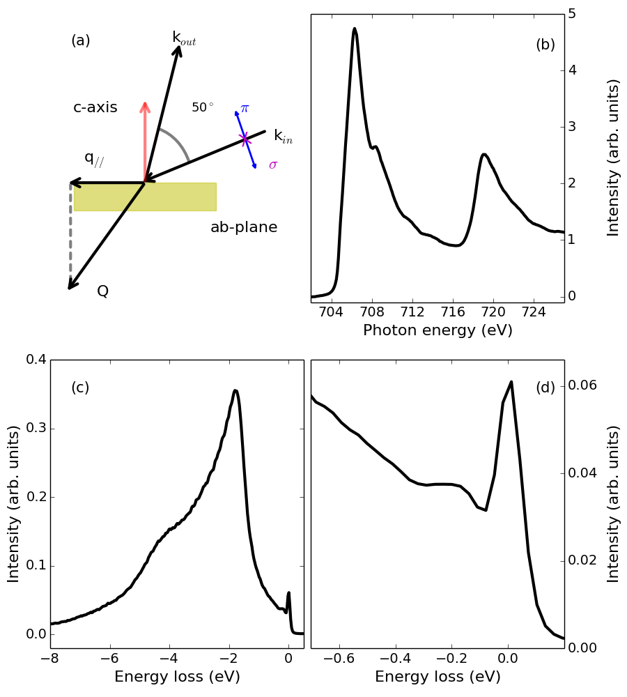
The samples were mounted with the ab plane perpendicular to the scattering plane and the c axis lying in it (sketch in Fig. 1(a) and post-cleaved in situ at a pressure better than 2.0x10⁻¹⁰ mbar. The directions studied are (1, 0) and (1, 1) according to the orthorhombic unfolded crystallographic notation Park et al. (2010). We use the convention of 1 Fe per unit cell. All the measurements were carried out at 10 K. X-ray Absorption Spectra (XAS) and RIXS experiments were performed at the ADRESS beamline of the Swiss Light Source, Paul Scherrer Institute, Villigen, Switzerland Strocov et al. (2010); Ghiringhelli et al. (2006). XAS spectra were measured in Total Fluorescence Yield (TFY). The RIXS spectrometer was set to a scattering angle of 130∘ and the incidence angle on the samples surface was varied to change the in plane momentum transferred (q) from (0, 0) to (0.47, 0 r.l.u.) (relative lattice units expressed in qa/2) and from (0, 0) to (0.34, 0.34 r.l.u.) as shown in Fig. 1(a). All measurements are recorded in grazing incidence configuration. The total energy resolution was 110 meV, measured by means of elastic scattering from a carbon-filled acrylic tape.
We measured Fe L2,3 XAS spectra for the two crystallographic orientations at 15∘ incidence angle and polarization. Fig. 1(b) displays the XAS spectrum of the (1, 0) orientation. The XAS along (0, 0)(1, 1) (not displayed) is analogue to the (0, 0)(1,0) direction. The spectrum is composed of a broad peak centered at 707 eV, typical of metallic systems containing Fe Zhou et al. (2013); Kurmaev et al. (2009); Yang et al. (2009); Pelliciari et al. (2016). The incident energy for RIXS was tuned at the main Fe-L3 peak. In Fig. 1(c) an exemplary RIXS spectrum of at (0.47, 0) is shown. The main line in this spectrum resembles emission from metallic systems with a broad asymmetric peak displaying a maximum at around -2 eV in energy loss (-) Zhou et al. (2013); Kurmaev et al. (2009); Hancock et al. (2010); Pelliciari et al. (2016), arising from resonant emission of itinerant electrons. In contrast to doped 1111 Nomura et al. (2016), but in agreement with other Fe pnictides, the emission line is not showing any sharp features ascribable to dd-excitations Zhou et al. (2013); Kurmaev et al. (2009); Yang et al. (2009); Pelliciari et al. (2016). This confirms the moderate electronic correlations of Fe pnictides contrasting with Fe chalcogenides where dd-excitations have been observed employing hard X-ray RIXS Gretarsson et al. (2015).
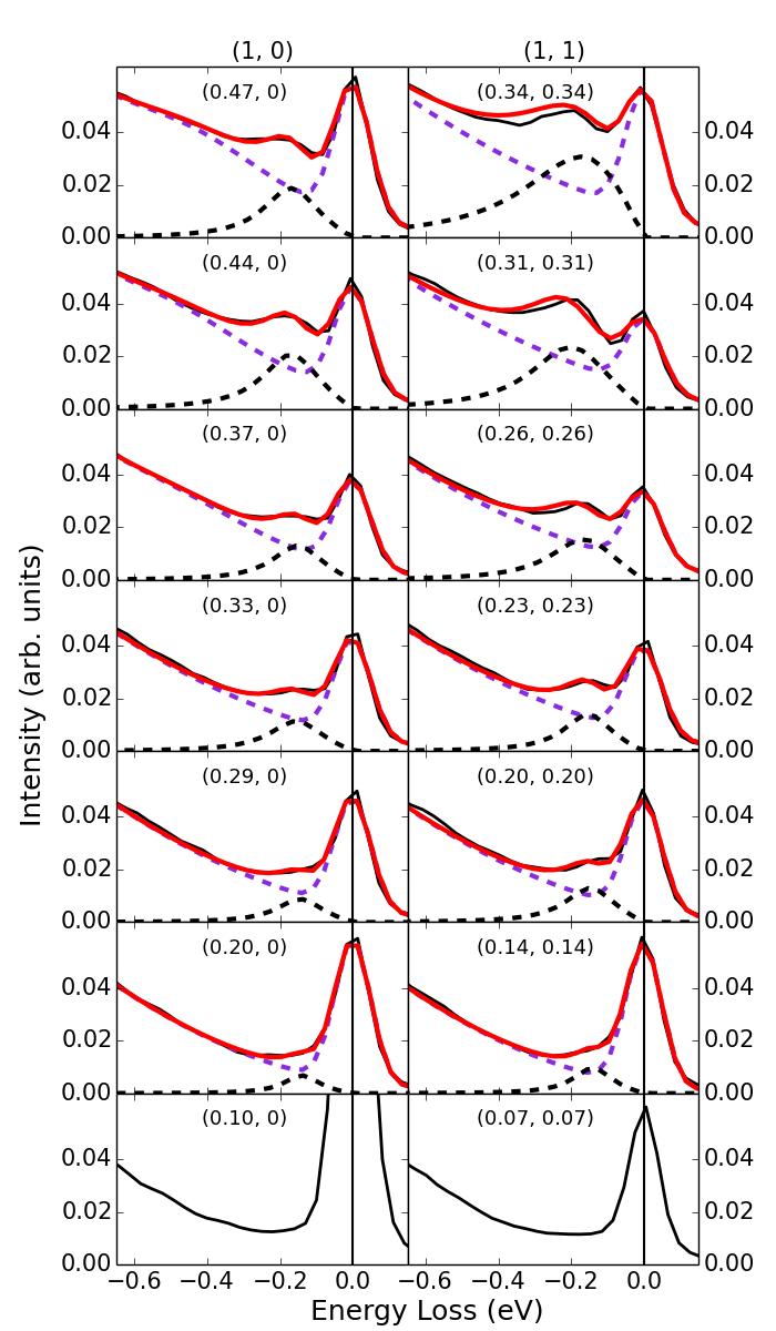
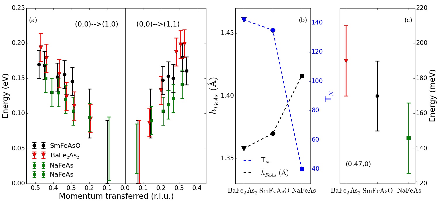
On the low energy side of the RIXS spectra at (0.47, 0) and (0.34, 0.34), we observe a peak emerging from the background having an energy of 170 meV as illustrated in Fig. 1(d) and 2. This peak moves towards the elastic line when the in-plane momentum transferred is decreased and at low q// it merges into the elastic line. The energy range and the dispersive nature of this mode resemble the behavior of spin excitations detected by RIXS in Zhou et al. (2013) and (measurements shown in Supplemental Material). We fit the background, the elastic and the magnetic line in agreement to Pelliciari et al. (2016); Zhou et al. (2013); Hancock et al. (2010) and plot the results in Fig. 2. At low values of q//, we do not attempt any fitting procedure because of the high overlap between the elastic and the magnetic peak. However, we believe that an estimation of a rough energy range is still possible. In Fig. 3(a) we show the dispersion curve arising from this fitting procedure as black dots with error bars together with the dispersion curves extracted from Zhou et al. (2013) and measured in the same experimental conditions (see the raw data and the fittings of in the Supplemental Material).
Figure 3(a) illustrates the dispersion relation of magnetic excitations for (black dots with error bars), (green dots with error bars), and (red dots with error bars) along (0, 0)(1, 0) and (0, 0)(1, 1). Clearly, there is a renormalization of the magnetic bandwidth between , that shows the highest energy (190 meV 20 meV), displaying an intermediate energy (170 meV 20 meV), and having the lowest bandwidth (150 meV 20 meV) as summarized in Fig. 3(c). This trend is in qualitative agreement with the decreasing values of magnetic moment from to and Stewart (2011); Johnson et al. (2015); Johnston (2010); Zhang et al. (2014); Dai (2015). However a quantitative comparison between ordered magnetic moment and spin excitations is not straightforward and is beyond the goal of this paper. The relevance of the structural parameters for SC and magnetism has been widely discussed in Refs. Stewart (2011); Johnston (2010); Zhang et al. (2014), with hFeAs and the angle of the tetrahedron formed by FeAs4 as the main possible parameters affecting TC and TN. Since there is no SC in these parent compounds, in Fig. 3(b) we show only the values of TN and how they correlate with hFeAs Stewart (2011); Johnston (2010); Zhang et al. (2014). In details, hFeAs increases from 1.358 Å in to 1.37 Å in and then to 1.416 Å in , whereas TN decreases Stewart (2011); Johnson et al. (2015); Johnston (2010); Zhang et al. (2014); Dai (2015) as well as the magnetic bandwidth measured in our experiments and outlined in Fig. 3(c). This highlights the importance of the structure for the spin excitations in Fe pnictides as well as the expected role of structural deformations due to doping, pressure or even defects in phase transitions like nematicity and / or SC.

In conclusion, we measured the spin excitations spectrum of , parent compound of the 1111 series, bypassing the polar cleaving problem. Comparison with other parent Fe pnictides, measured in the same experimental configuration, show that the bandwidth of spin waves is slightly renormalized to lower values in compared to but higher than . Here, we have illustrated how the structure, in particular hFeAs, influences the magnetic properties of Fe pnictides possibly triggering most of the modifications. We claim that, if such structural modifications can affect the magnetism and bandwidth, then the perturbations provided by doping and / or pressure might lead to instabilities such as Cooper pairing and / or nematicity, not triggered by electronic effects alone but by structural effects as well.
Acknowledgements.
J.P. and T.S. acknowledge financial support through the Dysenos AG by Kabelwerke Brugg AG Holding, Fachhochschule Nordwestschweiz, and the Paul Scherrer Institut. Part of this research has been funded by the Swiss National Science Foundation through the D-A-CH programme (SNSF Research Grant 200021L 141325). Experiments have been performed at the ADRESS beamline of the Swiss Light Source at Paul Scherrer Institut. The work at IOP-CAS is supported by NSF and MOST through research projects.References
- Kamihara et al. (2008) Y. Kamihara, T. Watanabe, M. Hirano, and H. Hosono, Journal of the American Chemical Society 130, 3296 (2008).
- Stewart (2011) G. R. Stewart, Reviews of Modern Physics 83, 1589 (2011).
- Johnston (2010) D. C. Johnston, Advances in Physics 59, 803 (2010).
- Scalapino (2012) D. J. Scalapino, Reviews of Modern Physics 84, 1383 (2012).
- Chubukov (2012) A. Chubukov, Annual Review of Condensed Matter Physics 3, 57 (2012).
- Fernandes et al. (2014) R. M. Fernandes, A. V. Chubukov, and J. Schmalian, Nature Physics 10, 97 (2014).
- Richard et al. (2009) P. Richard, T. Sato, K. Nakayama, S. Souma, T. Takahashi, Y.-M. Xu, G. F. Chen, J. L. Luo, N. L. Wang, and H. Ding, Physical Review Letters 102, 047003 (2009).
- Liu et al. (2010a) H. Liu, G. F. Chen, W. Zhang, L. Zhao, G. Liu, T.-L. Xia, X. Jia, D. Mu, S. Liu, S. He, Y. Peng, J. He, Z. Chen, X. Dong, J. Zhang, G. Wang, Y. Zhu, Z. Xu, C. Chen, and X. J. Zhou, Physical Review Letters 105, 027001 (2010a).
- Yang et al. (2011) L. X. Yang, B. P. Xie, B. Zhou, Y. Zhang, Q. Q. Ge, F. Wu, X. F. Wang, X. H. Chen, and D. L. Feng, Journal of Physics and Chemistry of Solids Spectroscopies in Novel Superconductors 2010SNS 2010, 72, 460 (2011).
- Dai (2015) P. Dai, Reviews of Modern Physics 87, 855 (2015).
- Tranquada et al. (2014) J. M. Tranquada, G. Xu, and I. A. Zaliznyak, Journal of Magnetism and Magnetic Materials 350, 148 (2014).
- Fujita et al. (2011) M. Fujita, H. Hiraka, M. Matsuda, M. Matsuura, J. M. Tranquada, S. Wakimoto, G. Xu, and K. Yamada, Journal of the Physical Society of Japan 81, 011007 (2011).
- (13) D. S. Inosov, Comptes Rendus Physique 10.1016/j.crhy.2015.03.001.
- Zhang et al. (2014) C. Zhang, L. W. Harriger, Z. Yin, W. Lv, M. Wang, G. Tan, Y. Song, D. Abernathy, W. Tian, T. Egami, K. Haule, G. Kotliar, and P. Dai, Physical Review Letters 112, 217202 (2014).
- Zhou et al. (2013) K.-J. Zhou, Y.-B. Huang, C. Monney, X. Dai, V. N. Strocov, N.-L. Wang, Z.-G. Chen, C. Zhang, P. Dai, L. Patthey, J. van den Brink, H. Ding, and T. Schmitt, Nature Communications 4, 1470 (2013).
- Pelliciari et al. (2016) J. Pelliciari, Y. Huang, T. Das, M. Dantz, V. Bisogni, P. O. Velasco, V. N. Strocov, L. Xing, X. Wang, C. Jin, and T. Schmitt, Physical Review B 93, 134515 (2016).
- Luo et al. (2012) H. Luo, Z. Yamani, Y. Chen, X. Lu, M. Wang, S. Li, T. A. Maier, S. Danilkin, D. T. Adroja, and P. Dai, Physical Review B 86, 024508 (2012).
- Luo et al. (2013) H. Luo, X. Lu, R. Zhang, M. Wang, E. A. Goremychkin, D. T. Adroja, S. Danilkin, G. Deng, Z. Yamani, and P. Dai, Physical Review B 88, 144516 (2013).
- Wang et al. (2013) M. Wang, C. Zhang, X. Lu, G. Tan, H. Luo, Y. Song, M. Wang, X. Zhang, E. A. Goremychkin, T. G. Perring, T. A. Maier, Z. Yin, K. Haule, G. Kotliar, and P. Dai, Nature Communications 4 (2013), 10.1038/ncomms3874.
- de Jong et al. (2009) S. de Jong, Y. Huang, R. Huisman, F. Massee, S. Thirupathaiah, M. Gorgoi, F. Schaefers, R. Follath, J. B. Goedkoop, and M. S. Golden, Physical Review B 79, 115125 (2009).
- Eschrig et al. (2010) H. Eschrig, A. Lankau, and K. Koepernik, Physical Review B 81, 155447 (2010).
- Yang et al. (2010) L. X. Yang, B. P. Xie, Y. Zhang, C. He, Q. Q. Ge, X. F. Wang, X. H. Chen, M. Arita, J. Jiang, K. Shimada, M. Taniguchi, I. Vobornik, G. Rossi, J. P. Hu, D. H. Lu, Z. X. Shen, Z. Y. Lu, and D. L. Feng, Physical Review B 82, 104519 (2010).
- Liu et al. (2010b) C. Liu, Y. Lee, A. D. Palczewski, J.-Q. Yan, T. Kondo, B. N. Harmon, R. W. McCallum, T. A. Lograsso, and A. Kaminski, Physical Review B 82, 075135 (2010b).
- Ramazanoglu et al. (2013) M. Ramazanoglu, J. Lamsal, G. S. Tucker, J.-Q. Yan, S. Calder, T. Guidi, T. Perring, R. W. McCallum, T. A. Lograsso, A. Kreyssig, A. I. Goldman, and R. J. McQueeney, Physical Review B 87, 140509 (2013).
- Strocov et al. (2010) V. N. Strocov, T. Schmitt, U. Flechsig, T. Schmidt, A. Imhof, Q. Chen, J. Raabe, R. Betemps, D. Zimoch, J. Krempasky, X. Wang, M. Grioni, A. Piazzalunga, and L. Patthey, Journal of Synchrotron Radiation 17, 631 (2010).
- Dean et al. (2012) M. P. M. Dean, R. S. Springell, C. Monney, K. J. Zhou, J. Pereiro, I. Božović, B. Dalla Piazza, H. M. Rønnow, E. Morenzoni, J. van den Brink, T. Schmitt, and J. P. Hill, Nature Materials 11, 850 (2012).
- Park et al. (2010) J. T. Park, D. S. Inosov, A. Yaresko, S. Graser, D. L. Sun, P. Bourges, Y. Sidis, Y. Li, J.-H. Kim, D. Haug, A. Ivanov, K. Hradil, A. Schneidewind, P. Link, E. Faulhaber, I. Glavatskyy, C. T. Lin, B. Keimer, and V. Hinkov, Physical Review B 82, 134503 (2010).
- Ghiringhelli et al. (2006) G. Ghiringhelli, A. Piazzalunga, C. Dallera, G. Trezzi, L. Braicovich, T. Schmitt, V. N. Strocov, R. Betemps, L. Patthey, X. Wang, and M. Grioni, Review of Scientific Instruments 77, 113108 (2006).
- Kurmaev et al. (2009) E. Z. Kurmaev, J. A. McLeod, N. A. Skorikov, L. D. Finkelstein, A. Moewes, Y. A. Izyumov, and S. Clarke, Journal of Physics: Condensed Matter 21, 345701 (2009).
- Yang et al. (2009) W. L. Yang, A. P. Sorini, C.-C. Chen, B. Moritz, W.-S. Lee, F. Vernay, P. Olalde-Velasco, J. D. Denlinger, B. Delley, J.-H. Chu, J. G. Analytis, I. R. Fisher, Z. A. Ren, J. Yang, W. Lu, Z. X. Zhao, J. van den Brink, Z. Hussain, Z.-X. Shen, and T. P. Devereaux, Physical Review B 80, 014508 (2009).
- Hancock et al. (2010) J. N. Hancock, R. Viennois, D. van der Marel, H. M. Rønnow, M. Guarise, P.-H. Lin, M. Grioni, M. Moretti Sala, G. Ghiringhelli, V. N. Strocov, J. Schlappa, and T. Schmitt, Physical Review B 82, 020513 (2010).
- Nomura et al. (2016) T. Nomura, Y. Harada, H. Niwa, K. Ishii, M. Ishikado, S. Shamoto, and I. Jarrige, Physical Review B 94, 035134 (2016).
- Gretarsson et al. (2015) H. Gretarsson, T. Nomura, I. Jarrige, A. Lupascu, M. H. Upton, J. Kim, D. Casa, T. Gog, R. H. Yuan, Z. G. Chen, N.-L. Wang, and Y.-J. Kim, Physical Review B 91, 245118 (2015).
- Johnson et al. (2015) P. D. Johnson, G. Xu, and W.-G. Yin, eds., Iron-Based Superconductivity, Springer Series in Materials Science, Vol. 211 (Springer International Publishing, Cham, 2015).