The quartet ground state in CeB6: an inelastic x-ray scattering study
Abstract
We investigated the ground state symmetry of the cubic hidden order compound CeB6 by means of core level non-resonant inelastic x-ray scattering (NIXS). The information is obtained from the directional dependence of the scattering function that arises from higher than dipole transitions. Our new method confirms that the ground state is well described using a localized crystal-field model assuming a quartet ground state.
I Introduction
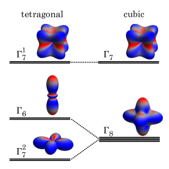
The material class of rare earth hexaborides has attracted considerable attention over the years. It comprises of a variety of different fascinating ground states (see Ref. Sun and Wu (2016) and references therein) which include exotic magnetically ordered phases, heavy fermion behavior, as well as Kondo insulating ground states. CeB6 is an important member of this material class, well known for its so-called hidden magnetic order. The very recent theoretical suggestion that SmB6 could be a strongly correlated topological insulator Dzero et al. (2010); Takimoto (2011) even caused a flurry of new investigations (see Ref. Dzero et al. (2016) and references therein), thereby raising speculations that also YbB6 under pressure could be topological. The standard and at the same time pressing question in all these studies concerns the symmetry of the ground state wave function of the crystal-electric field split 4 multiplet. Here we explore the feasibility of using a recently developed experimental method, namely core-level non-resonant x-ray scattering (NIXS), to determine the ground state wave function of CeB6, a system which crystallizes in the cubic CsCl structure. Fig. 1 displays how the crystal-electric field splits the sixfold degenerate multiplet state of the Ce into a quartet and doublet.
CeB6 is a heavy fermion compound that has been intensively studied for its rich magnetic phase diagram J.M. Effantin et al. (1985). Upon cooling CeB6 enters a hidden order phase at 3.2 K followed by an antiferromagnetic phase below 2.4 K. The hidden order parameter is not accessible with e.g. neutron or standard x-ray diffraction at zero field. The application of an external field, however, induces a dipole component with the wave vector of the quadrupolar ordering Erkelens et al. (1987). Theory suggests that the multipolar moments of the localized 4 electrons interact with each other via the itinerant 5 conduction electrons, breaking up the fourfold ground state degeneracy of the Ce 4 wave function in the cubic crystal field stabilizing an antiferro-quadrupolar (AFQ) order Shiina et al. (1997), a conjecture that by now has received credibility from a resonant x-ray diffraction study Matsumura et al. (2009); Lovesey (2002). The observation of a spin resonance in the inelastic neutron data of CeB6 Friemel et al. (2012); Portnichenko et al. (2016) shows the importance of itinerancy for the formation of the multipolar and magnetic order Akbari and Thalmeier (2012), the latter being supported by electronic structure investigations of CeB6 Neupane et al. (2015); Koitzsch et al. (2016). Inelastic neutron scattering finds in agreement with Raman scattering a crystal-field excitation at 46 meV and it is generally accepted that the intriguing magnetic properties of CeB6 evolve out of the fourfold degenerate ground state. The quartet ground state had been originally deduced from an unusual low temperature shift of the crystal-field excitation in Raman and inelastic neutron scattering data Zirngiebl et al. (1984); Loewenhaupt et al. (1985). The energy shift was interpreted as a splitting of the quartet ground state in the low temperature phase in accordance with electron paramagnetic resonance (EPR) measurements Terzioglu et al. (2001). A quartet ground state is also consistent with findings of the magnetic anisotropy Sato et al. (1984) and magnetic neutron form factor measurements Givord et al. (2003). We have now revisited the symmetry aspect of CeB6 in the paramagnetic phase using core level Ce N4,5 () NIXS, a spectroscopic technique that directly probes the charge distribution of the Ce electrons.
II Spectroscopic Technique
In the recent past we have shown that soft x-ray absorption spectroscopy (XAS) with linear polarized light is a very useful local probe for determining the anisotropy of wave functions in tetragonal Hansmann et al. (2008) or orthorhombic heavy fermion compounds Strigari et al. (2012), and for detecting small variations with unprecedented accuracy Willers et al. (2015). However, for cubic compounds XAS cannot be applied since it relies on dipole transitions which cannot distinguish between the quartet and doublet state (see Fig. 1). We have therefore performed an experiment that probes the symmetry with higher multipole transitions. This can be realized in a core level non-resonant inelastic x-ray scattering (NIXS) experiment with large momentum transfers . For large enough the expansion of the transition operator e in the scattering function S(q,) can no longer be truncated after the first term and as a result higher multipole terms contribute to S(q,). These extra multipole contributions then give information that is not accessible in a dipole experiment Larson et al. (2007); Haverkort et al. (2007); Gordon et al. (2008, 2009); Bradley et al. (2010); Caciuffo et al. (2010); Sen Gupta et al. (2011); Bradley et al. (2011); Gordon et al. (2011); Hiraoka et al. (2011); van der Laan (2012).
Bradley et al. Bradley et al. (2011) and Gordon et al. Gordon et al. (2011) were the first to observe higher multipole transitions in rare earth materials at the N4,5 core level excitation for large momentum transfers and the data were well described with a local many body approach by Haverkort et al. Haverkort et al. (2007). Already the early papers suggested that vector q dependent NIXS experiments on a single crystal should give insight into the ground state symmetry in analogy to an XAS experiment with linear polarized light Haverkort et al. (2007); Gordon et al. (2008, 2011); Bradley et al. (2011), and indeed, an experiment on cubic single crystals of MnO and CeO2 at the Mn M2,3 and Ce N4,5 edges revealed direction dependencies in the higher multipole scattering function Gordon et al. (2009). Very recently, NIXS has been successfully used to determine the ground state symmetry and/or determine the rotation of the orbitals in fourfold symmetry in Ce single crystals Willers et al. (2012); Rueff et al. (2015); Sundermann et al. (2015).
III Experimental
The single-crystal samples of CeB6 were grown by the Al-flux method. Typically 0.7g of CeB6 (as the elements) are heated with 60 g of high purity Al (59) to 1450 C, held there for 8 hr and then cooled to 1000 C at 2 C/hr, when the furnace is shut off. The crystals are leached from the Al in NaOH solution.
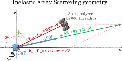
The NIXS measurements on the CeB6 Ce N4,5 core level were performed at the beamline P01 of PETRA-III. The incident energy was selected with a Si(311) double monochromator. The P01 NIXS end station has a vertical geometry with twelve Si(660) 1 m radius spherically bent crystal analyzers that are arranged in 3 x 4 array (see Fig. 2). The fixed final energy was 9690 eV. The analyzers were positioned at scattering angles of 2 150∘, 155∘, and 160∘ which corresponds at elastic scattering to an averaged momentum transfer of = (9.6 0.1) Å-1. The scattered beam was detected by a position sensitive custom-made Lambda detector, based on a Medipix3 chip detector. The elastic line was regularly measured and pixelwise calibration yields an instrumental energy resolution of FWHM 0.7 eV. A sketch of the scattering geometry, showing the incoming and outgoing photons as well as the transferred momentum , is given in Fig. 2 for a scan with q [100] in specular geometry. In order to realize another crystallographic direction, e.g. q [110], the sample can be turned with respect to the scattering triangle, or a different sample with another polished surface may be mounted in specular geometry.
Two crystals with (100) and (110) surfaces were mounted in a vacuum cryostat with Kapton windows. The measurements were performed with a pressure in the 10-6 mbar range. The two samples were oriented such that for q [100] and q [110] a specular scattering geometry was realized, i.e. with the surface normal parallel to the momentum transfer ( = = ). In order to check reliability, the q [110] measurement was repeated on the (100) crystal but with the surface normal being rotated 45∘ away from q ( = ∘ - 45∘). The data were fully consistent. The q [111] situation was realized by turning the (110) crystal to = ∘ - 35∘.
IV Results and Discussion

Fig. 3 shows the NIXS spectrum across the Ce N4,5 (), N2,3 (), and N1 () edges. The accompanying Compton contribution has its maximum at about 350 eV energy transfer. It is important to note that the Ce white lines are clearly discerned from the Compton scattering, and that especially the Ce N4,5 white lines stand out with an excellent signal to background ratio. This shows that N4,5 NIXS is an extremely suitable experimental method for the study of the local electronic structure of CeB6, and for that matter, the class of rare-earth hexaborides.
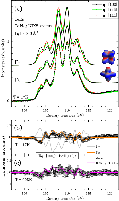
The top panel(a) of Fig. 4 shows the Ce N4,5 NIXS spectra of CeB6 (dots) taken at 17 K, for the three momentum directions q [100] (black dots), [110] (green dots), and [111] (red dots). The temperature of 17 K is low enough to assure that only the ground state is populated. We recall that the excited crystal-field state is 46 meV above the ground state Zirngiebl et al. (1984); Loewenhaupt et al. (1985). Here only a constant background has been subtracted to account for the (weak) Compton signal (about 12% of the signal peak) (see Fig. 3). The size of the dots resembles the statistical error bar.
There is a clear direction dependence that shows up strongest in the energy interval of 103 to 106 eV. Especially the q [100] direction differs from the q [110] and [111]. We can obtain a more detailed view at the directional dependence by constructing the difference spectra I - I that is displayed as dichroism in the bottom panel(b) of Fig. 4 (black dots).
The Ce N4,5 NIXS data are simulated by calculating the transition using the full multiplet code QuantyHaverkort (2016) which includes Coulomb as well as spin-orbit interactions. A Gaussian and a Lorentzian broadening of FWHM = 0.7 eV and 0.4 eV, respectively, are used to account for the instrumental resolution and life time effects. The atomic Hartree-Fock values were adjusted via the peak positions, resulting in reductions of 30 % and 22 % for the 4-4 and 4-4 Coulomb interactions, respectively. The reduction accounts for configuration interaction effects not included in the Hartree-Fock scheme Tanaka and Jo (1994). A momentum transfer of = 9.2 Å-1 has been used for the simulations (and not the experimental value of 9.6 0.1) Å-1) so that the experimental peak ratio of the two main features around 108 and 110 eV is reproduced best. This fine tuning optimizes the multipole contributions to the scattering functions to mimic for a minor adjustment of the calculated radial wave functions of the Ce3+ atomic wave function (see e.g. Ref. Willers et al. (2012)).
We now compare the measured spectra and the dichroism therein with the simulations for the two possible scenarios, namely one with the doublet as ground state and the other with the quartet. The results are plotted in Fig. 4 (a). The quartet scenario reproduces in great detail the experimental spectra for all three q directions. Actually, the match is excellent. In contrast, the simulation based on the doublet exhibits large discrepancies with respect to the experiment: the intensities of several features in the spectra are not correct. To make the difference between the two scenarios even more contrasting, we compare the experimental and calculated dichroic spectra, i.e. I - I, as displayed in bottom panel(b). There is an excellent match for the quartet ground state scenario but a large mismatch for the doublet. From these comparisons we can unambiguously conclude that the quartet forms the ground state in CeB6.
In addition, we have taken spectra at T = 295 K. The spectra look very similar to the low temperature data but the dichroism is reduced by about 20%, see bottom panel (c) of Fig. 4. This reduction in the dichroism is fully consistent with a partial population of the excited state at 46 meV. A simulation in which the Boltzmann weighted contributions of the and states are taken into account is represented by the magenta line in panel (c) of Fig. 4. The excellent agreement provides yet another evidence for the thorough understanding we have obtained using NIXS on the Ce symmetry and crystal-electric field effects in CeB6.
V Summary
Using Ce N4,5 non-resonant inelastic x-ray scattering (NIXS) we were able to establish that the ground state symmetry of the cubic hidden order compound CeB6 is the quartet. The high signal to background ratio of the N4,5 NIXS signal indicates that this bulk sensitive and element specific spectroscopic technique is a powerful method to study the local electronic structure of the rare-earth ions in rare-earth borides. With NIXS probing directly the charge distribution of the electrons, it complements nicely neutron scattering based techniques which provide direct information on the spin distribution.
Acknowledgements.
We thank P. Thalmeier for valuable discussions. We also thank C. Becker and T. Mende from MPI-CPfS, and F.-U. Dill, S. Mayer, and other members from beamline P01 for their skillful technical support, C.J. Sahle and M. Harder for their valuable contribution to the data processing. Part of this research was carried out at the light source PETRA III at DESY, a member of the Helmholtz Association (HGF). K.C., M.S. and A.S. benefited from the financial support of the Deutsche Forschungsgemeinschaft (DFG) under projects SE 1441/1-2 and SE 1441/1-3.References
- Sun and Wu (2016) L. Sun and Q. Wu, Rep. Prog. Phys. 79, 084503 (2016).
- Dzero et al. (2010) M. Dzero, K. Sun, V. Galitski, and P. Coleman, Phys. Rev. Lett. 104, 106408 (2010).
- Takimoto (2011) T. Takimoto, J. Phys. Soc. Jpn. 80, 123710 (2011).
- Dzero et al. (2016) M. Dzero, J. Xia, V. Galitski, and P. Coleman, Annual Rev. Cond. Matt. Phys. 7, 249 (2016).
- J.M. Effantin et al. (1985) J. J.M. Effantin, J. Rossat-Mignod, P. Burlet, H. Bartholin, S. Kunii, and T. Kasuya, J. Mag. Mag. Mat. 47-48, 145 (1985).
- Erkelens et al. (1987) W. Erkelens, L. P. egnault, P. Burlet, J. Rossat-Mignod, S. Kunii, and T. Kasuya, J. Mag. Mag. Mat. 64 & 64, 61 (1987).
- Shiina et al. (1997) R. Shiina, H. Shiba, and P. Thalmeier, J. Phys. Soc. Jpn. 66, 1741 (1997).
- Matsumura et al. (2009) T. Matsumura, T. Yonemura, K. Kunimori, M. Sera, and F. Iga, Phys. Rev. Lett. 103, 017203 (2009).
- Lovesey (2002) S. W. Lovesey, Journal of Physics: Condensed Matter 14, 4415 (2002).
- Friemel et al. (2012) G. Friemel, Y. Li, A. Dukhnenko, N. Shitsevalova, N. Sluchanko, A. Ivanov, V. Filipov, B. Keimer, and D. Inosov, Nat. Commun. 3, 830 (2012).
- Portnichenko et al. (2016) P. Y. Portnichenko, S. V. Demishev, A. V. Semeno, H. Ohta, A. S. Cameron, M. A. Surmach, H. Jang, G. Friemel, A. V. Dukhnenko, N. Y. Shitsevalova, V. B. Filipov, A. Schneidewind, J. Ollivier, A. Podlesnyak, and D. S. Inosov, Phys. Rev. B 94, 035114 (2016).
- Akbari and Thalmeier (2012) A. Akbari and P. Thalmeier, Phys. Rev. Lett. 108, 146403 (2012).
- Neupane et al. (2015) M. Neupane, N. Alidoust, I. Belopolski, G. Bian, S.-Y. Xu, D.-J. Kim, P. P. Shibayev, D. S. Sanchez, H. Zheng, T.-R. Chang, H.-T. Jeng, P. S. Riseborough, H. Lin, A. Bansil, T. Durakiewicz, Z. Fisk, and M. Z. Hasan, Phys. Rev. B 92, 104420 (2015).
- Koitzsch et al. (2016) A. Koitzsch, N. Heming, M. Knupfer, P. Y. Büchner, B. Portnichenko, A. V. Dukhnenko, N. Y. Shitsevalova, V. B. Filipov, L. L. Lev, V. N. Strocov, J. Ollivier, and D. S. Inosov, Nat. Commun. 7, 10876 (2016).
- Zirngiebl et al. (1984) E. Zirngiebl, B. Hillebrands, S. Blumenröder, G. Güntherodt, M. Loewenhaupt, J. M. Carpenter, K. Winzer, and Z. Fisk, Phys. Rev. B 30, 4052 (1984).
- Loewenhaupt et al. (1985) M. Loewenhaupt, J. M. Carpenter, and C.-K. Loong, J. Mag. Mag. Mat. 52, 245 (1985).
- Terzioglu et al. (2001) C. Terzioglu, D. A. Browne, R. G. Goodrich, A. Hassan, and Z. Fisk, Phys. Rev. B 63, 235110 (2001).
- Sato et al. (1984) N. Sato, S. Kunii, I. Oguro, T. Komatsubara, and T. Kasuya, J. Phys. Soc. Jpn 53, 3967 (1984).
- Givord et al. (2003) F. Givord, J.-X. Boucherle, P. Burlet, B. Gillon, and S. Kunii, J. Phys.: Cond. Matt. 15, 3095 (2003).
- Hansmann et al. (2008) P. Hansmann, A. Severing, Z. Hu, M. W. Haverkort, C. F. Chang, S. Klein, A. Tanaka, H. H. Hsieh, H.-J. Lin, C. T. Chen, B. Fåk, P. Lejay, and L. H. Tjeng, Phys. Rev. Lett. 100, 066405 (2008).
- Strigari et al. (2012) F. Strigari, T. Willers, Y. Muro, K. Yutani, T. Takabatake, Z. Hu, Y.-Y. Chin, S. Agrestini, H.-J. Lin, C. T. Chen, A. Tanaka, M. W. Haverkort, L. H. Tjeng, and A. Severing, Phys. Rev. B 86, 081105(R) (2012).
- Willers et al. (2015) T. Willers, F. Strigari, Z. Hu, V. Sessi, N. Brookes, E. Bauer, J. Sarrao, J. Thompson, A. Tanaka, S. Wirth, L. Tjeng, and A. Severing, PNAS 112, 2384 (2015).
- Larson et al. (2007) B. C. Larson, W. Ku, J. Z. Tischler, C.-C. Lee, O. D. Restrepo, A. G. Eguiluz, P. Zschack, and K. D. Finkelstein, Phys. Rev. Lett. 99, 026401 (2007).
- Haverkort et al. (2007) M. W. Haverkort, A. Tanaka, L. H. Tjeng, and G. A. Sawatzky, Phys. Rev. Lett. 99, 257401 (2007).
- Gordon et al. (2008) R. A. Gordon, G. T. Seidler, T. T. Fister, M. W. Haverkort, G. A. Sawatzky, A. Tanaka, and T. K. Sham, Europhys. Lett. 81, 26004 (2008).
- Gordon et al. (2009) R. A. Gordon, M. W. Haverkort, S. Sen Gupta, and G. A. Sawatzky, J. Phys. Conf. Series 190 (2009), 10.1088/1742-6596/190/1/012047.
- Bradley et al. (2010) J. A. Bradley, S. Sen Gupta, G. T. Seidler, K. T. Moore, M. W. Haverkort, G. A. Sawatzky, S. D. Conradson, D. L. Clark, S. A. Kozimor, and K. S. Boland, Phys. Rev. B 81, 193104 (2010).
- Caciuffo et al. (2010) R. Caciuffo, G. van der Laan, L. Simonelli, T. Vitova, C. Mazzoli, M. A. Denecke, and G. H. Lander, Phys. Rev. B 81, 195104 (2010).
- Sen Gupta et al. (2011) S. Sen Gupta, J. A. Bradley, M. W. Haverkort, G. T. Seidler, A. Tanaka, and G. A. Sawatzky, Phys. Rev. B 84, 075134 (2011).
- Bradley et al. (2011) J. A. Bradley, K. T. Moore, G. van der Laan, J. P. Bradley, and R. A. Gordon, Phys. Rev. B 84, 205105 (2011).
- Gordon et al. (2011) R. A. Gordon, G. T. Seidler, T. T. Fister, and K. P. Nagle, J. Electron Spectrosc. Relat. Phenom. 184, 220 (2011).
- Hiraoka et al. (2011) N. Hiraoka, M. Suzuki, K. D. Tsuei, Y. Q. Cai, M. W. Haverkort, C. C. Lee, and K. Ku, EPL 96 (2011), 10.1209/0295-5075/96/37007.
- van der Laan (2012) G. van der Laan, Phys. Rev. Lett. 108, 077401 (2012).
- Willers et al. (2012) T. Willers, F. Strigari, N. Hiraoka, Y. Q. Cai, M. W. Haverkort, K.-D. Tsuei, Y. F. Liao, S. Seiro, C. Geibel, F. Steglich, L. H. Tjeng, and A. Severing, Phys. Rev. Lett. 109, 046401 (2012).
- Rueff et al. (2015) J.-P. Rueff, J. M. Ablett, F. Strigari, M. Deppe, M. W. Haverkort, L. H. Tjeng, and A. Severing, Phys. Rev. B 91, 201108 (2015).
- Sundermann et al. (2015) M. Sundermann, F. Strigari, W. T., H. Winkler, A. Prokofiev, J. M. Ablett, J.-P. Rueff, D. Schmitz, E. Weschke, M. Moretti Sala, A. Al-Zein, A. Tanaka, M. W. Haverkort, D. Kasninathan, L. H. Tjeng, S. Paschen, and A. Severing, Sci. Rep. 5, 17937 (2015).
- Haverkort (2016) M. W. Haverkort, J. Phys.: Conf. Ser. 712, 012001 (2016).
- Tanaka and Jo (1994) A. Tanaka and T. Jo, J Phys. Soc. Jpn. 63, 2788 (1994).