Symmetry and Size of Membrane Protein Polyhedral Nanoparticles
Abstract
In recent experiments [T. Basta et al., Proc. Natl. Acad. Sci. U.S.A. 111, 670 (2014)] lipids and membrane proteins were observed to self-assemble into membrane protein polyhedral nanoparticles (MPPNs) with a well-defined polyhedral protein arrangement and characteristic size. We develop a model of MPPN self-assembly in which the preferred symmetry and size of MPPNs emerge from the interplay of protein-induced lipid bilayer deformations, topological defects in protein packing, and thermal effects. With all model parameters determined directly from experiments, our model correctly predicts the observed symmetry and size of MPPNs. Our model suggests how key lipid and protein properties can be modified to produce a range of MPPN symmetries and sizes in experiments.
pacs:
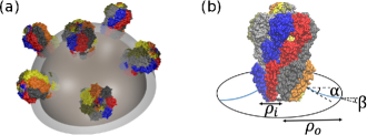
87.16.D-, 87.17.-dMembrane proteins play a central role in a variety of essential cellular processes McMahon and Gallop (2005); Engelman (2005) such as ion exchange, signaling, and membrane curvature regulation. The biologically relevant structures, and resulting functions, of many membrane proteins depend critically Engelman (2005); Phillips et al. (2009) on their lipid bilayer environment, and on chemical and voltage gradients across the cell membrane. Yet, determination of membrane protein structure in lipid bilayer environments and in the presence of physiologically relevant transmembrane gradients has largely remained elusive. Recent experiments on membrane protein polyhedral nanoparticles (MPPNs) Basta et al. (2014) present an exciting step towards overcoming this challenge. In these experiments, lipids and mechanosensitive channels of small conductance (MscS) Bass et al. (2002); Steinbacher et al. (2007) were observed to self-assemble Basta et al. (2014); Wu et al. (2013) into lipid bilayer vesicles with a polyhedral protein arrangement [see Fig. 1(a)]. The dominant polyhedral symmetry of MPPNs was found Basta et al. (2014) to be the snub cube (snub cuboctahedron) with MscS located at its 24 vertices and a characteristic MPPN radius nm. The well-defined symmetry and characteristic size of MPPNs may permit Basta et al. (2014) structural analysis of membrane proteins with the membrane proteins embedded in a lipid bilayer environment and the closed surfaces of MPPNs supporting physiologically relevant transmembrane gradients.
Utilization of MPPNs for high-resolution structural studies requires Basta et al. (2014) control over the symmetry and size of MPPNs. In this Letter we develop a physical description of MPPNs which establishes a quantitative link between the shape of MPPNs and key molecular properties of their constituents. We first describe a simple mean-field model of MPPNs inspired by previous work on membrane budding Góźdź and Gompper (2001); Auth and Gompper (2009); Müller and Deserno (2010) and viral capsid self-assembly Bruinsma et al. (2003). Our mean-field model of MPPNs accounts for the lipid bilayer bending deformations induced by MscS Bass et al. (2002); Steinbacher et al. (2007); Phillips et al. (2009) and the MscS packing defects resulting from the spherical topology of MPPNs, and yields the MPPN energy as a function of the number of MscS per MPPN without any free parameters. We confirm some of the key assumptions underlying our mean-field model by carrying out Monte Carlo simulations of a minimal molecular model of MPPN organization, which we formulate following previous work on viral capsid symmetry Zandi et al. (2004). Finally, we use our mean-field model of MPPNs to calculate Bruinsma et al. (2003); Safran (2003); Ben-Shaul and Gelbart (1994) the MPPN self-assembly phase diagram as a function of protein concentration, bilayer-protein contact angle, and protein size. We show that our model correctly predicts, with all model parameters determined directly from experiments, the observed Basta et al. (2014) symmetry and size of MPPNs formed from MscS. Our results suggest that the preferred symmetry and size of MPPNs emerge from the interplay of protein-induced lipid bilayer deformations, topological defects in protein packing, and thermal effects.
Mean-field model.—The membrane-spanning region of MscS Bass et al. (2002); Steinbacher et al. (2007) has an approximately conical shape Phillips et al. (2009); sm with radius nm in the lipid bilayer midplane and bilayer-protein contact angle – rad, yielding Phillips et al. (2009) protein-induced lipid bilayer bending deformations [see Fig. 1(b)]. The preferred MscS arrangement minimizing bilayer bending energy is expected Góźdź and Gompper (2001); Auth and Gompper (2009); Müller and Deserno (2010) to be a uniform hexagonal lattice. Our simple mean-field model of MPPNs therefore considers, on the one hand, contributions to the MPPN energy arising from MscS-induced bilayer bending deformations for hexagonal protein arrangements Dan et al. (1994); Góźdź and Gompper (2001); Auth and Gompper (2009); Müller and Deserno (2010). On the other hand, the spherical shape of MPPNs necessitates defects in the preferred hexagonal packing of MscS which, in analogy to viral capsids Bruinsma et al. (2003); Zandi et al. (2004), yields an energy penalty characteristic of the number of proteins per MPPN, . Thus, we allow in the total MPPN energy for contributions due to protein-induced bilayer bending, , and topological defects in protein packing, , respectively.
We estimate the MPPN bending energy using a formalism developed in the context of membrane budding Góźdź and Gompper (2001); Auth and Gompper (2009); Müller and Deserno (2010), which we summarize here for completeness. The unit cell associated with uniform hexagonal protein arrangements can be approximated Góźdź and Gompper (2001); Auth and Gompper (2009); Müller and Deserno (2010) by a circular membrane patch with the protein at its center and boundary conditions set by the spherical shape of MPPNs. We denote the projected radius of the circular membrane patch by [Fig. 1(b)], where is the bilayer midplane radius of MPPNs and is the patch boundary angle. We have so that the total area covered by membrane patches, , is equal to the total MPPN area, . Minimization of the Helfrich-Canham-Evans bending energy Canham (1970); Evans (1974); Helfrich (1973); Boal (2012) with respect to the bilayer midplane height field then yields Auth and Gompper (2009) the MPPN bending energy,
| (1) |
where the bilayer bending rigidity Rawicz et al. (2000) for the diC14:0 lipids used for MPPNs formed from MscS Basta et al. (2014); Wu et al. (2013), and the slopes at the bilayer-protein boundary and at the membrane patch boundary . To account for steric constraints on lipid and protein size we only allow sm in Eq. (1) for membrane patch sizes when calculating the MPPN energy, where the lipid radius nm for diC14:0 lipids Damodaran and Merz (1993). Equation (1) yields Auth and Gompper (2009) a preferred unit cell size of hexagonally packed proteins, with , which can be achieved if , corresponding to close-packed catenoidal bilayer deformation profiles.
Topological defects perturb the hexagonal packing of proteins in MPPNs. To estimate, for each and , the energy cost of the resulting deviations from the preferred protein arrangement in Eq. (1) we adopt a mean-field approach developed in the context of viral capsid self-assembly Bruinsma et al. (2003) and approximate the hexagonal spring network associated with Eq. (1) Auth and Gompper (2009) by a uniform elastic sheet Phillips et al. (2012). In the continuum limit, the minimum-energy protein arrangement in Eq. (1) satisfying then yields Kantor et al. (1987); sm the stretching modulus
| (2) |
which depends on via . At the mean-field level, the deviation from the preferred hexagonal packing of proteins in Eq. (1) due to the spherical shape of MPPNs can be quantified Bruinsma et al. (2003) through the fraction of the surface of a sphere enclosed by identical non-overlapping circles at closest packing Clare and Kepert (1991), , which yields in our model the protein arrangement at each . The local maxima of correspond to locally optimal protein packings [see Fig. 2(inset)]. We thus find sm the MPPN defect energy
| (3) |
where corresponds to the uniform hexagonal protein arrangements assumed in Eq. (1).
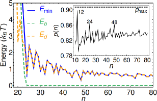
We minimize the total MPPN energy given by Eqs. (1)–(3), at each , with respect to , which yields the minimum MPPN energy with all parameters determined directly by the molecular properties of the lipids and proteins forming MPPNs (see Fig. 2). We find that MPPNs with , where for MPPNs formed from MscS Basta et al. (2014); Wu et al. (2013) with rad, are strongly penalized by the MPPN bending energy, which cannot be minimized to zero in this regime. Furthermore, MPPNs with also tend to be penalized by the MPPN defect energy because in Eq. (2) can be large for small sm . For , in which case also , we find a range of favorable corresponding to locally optimal protein packings. However, for the MPPN energies associated with distinct fall within just a few of each other and, as we discuss further below, thermal effects are therefore crucial in this regime. Finally we note that, for which allow in Fig. 2, the preferred protein separation (and, hence, MPPN size) is set, within , by in Eq. (1).
Minimal molecular model.—Our mean-field model of MPPNs assumes Góźdź and Gompper (2001); Auth and Gompper (2009); Müller and Deserno (2010); Bruinsma et al. (2003) that, for a given , the protein arrangement in MPPNs is determined by close packing of circular membrane patches, each with a protein at its center. Following previous work on viral capsid symmetry Zandi et al. (2004), we test these assumptions through Monte Carlo simulations of a minimal molecular (particle-based) model of MPPN organization, which focuses on short-range interactions between lipids and proteins. In this model, we represent sm the lipid bilayer and membrane proteins by disks lying on the surface of a sphere and assume that lipids interact with other lipids and proteins via Lennard-Jones potentials Zandi et al. (2004) with, for simplicity, hardcore steric repulsion between proteins. These interactions can be parametrized sm based on experiments and previous calculations Steinbacher et al. (2007); Damodaran and Merz (1993); Boal (2012); Ben-Tal et al. (1996); Choe et al. (2008) but our simulation results are not sensitive to the particular interactions used. We employed simulated annealing Monte Carlo simulations Kirkpatrick et al. (1983) with linear cooling to numerically determine the minimum-energy configuration of lipids and proteins in our minimal molecular model of MPPN organization. Following experiments on MPPNs formed from MscS Basta et al. (2014) we focused in our simulations on MPPNs with and a total of lipids sm .
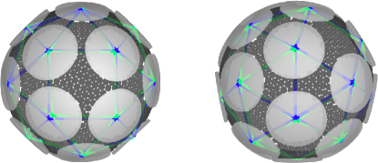
Figure 3 shows the minimum-energy MPPN configuration found in our simulations. The results in Fig. 3 suggest that, in the ground state of the system, MscS are arranged in the form of a snub cube, in agreement with the corresponding optimal protein packing assumed in the mean-field model of MPPNs in Fig. 2. To quantify the quality of the polyhedral fit in Fig. 3 we proceeded as in experiments on MPPNs Basta et al. (2014) and used least-square minimization sm to calculate the minimum fit error for 132 convex polyhedra Hart (2000): the Platonic, Archimedean, Catalan, and Johnson solids. We define Basta et al. (2014) the fit error as the sum over the squared distances between the simulated positions of protein centers and the closest fitted polyhedron vertices. We find sm that the snub cube (dextro) yields the best fit with a fit error Å2, while the second- and third-best fits are provided by the truncated cuboctahedron and pentagonal hexecontahedron (levo) with the substantially larger fit errors Å2 and Å2, respectively.
Phase diagram.—Based on our mean-field model of MPPNs we construct the MPPN phase diagram from Bruinsma et al. (2003) the statistical thermodynamics of amphiphile self-assembly in dilute aqueous solutions Safran (2003); Ben-Shaul and Gelbart (1994). Let denote the total number of proteins bound in MPPNs with proteins each and the total number of solvent molecules, which we take Basta et al. (2014); Wu et al. (2013) to be dominated by contributions due to water. For MPPNs formed from MscS the protein concentration mg/mL Basta et al. (2014), with the molecular mass g/mol for MscS Vásquez et al. (2007), yielding the protein number fraction . We assume here that all proteins in the system are incorporated into MPPNs, a point we return to below. In the dilute limit with no interactions between MPPNs we have Safran (2003); Ben-Shaul and Gelbart (1994) the mixing entropy , where the MPPN number fraction . This then allows Safran (2003); Ben-Shaul and Gelbart (1994) construction of the Helmholtz free energy with , in which the minimum MPPN energy is determined by our mean-field model of MPPNs via Eqs. (1)–(3). Minimization of with respect to Safran (2003); Ben-Shaul and Gelbart (1994) results in
| (4) |
where and the protein chemical potential is fixed by the constraint . As in Fig. 2, we restrict to the range for simplicity. Finally, we calculate the MPPN equilibrium distribution from Eq. (4) via .
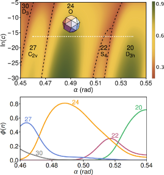
Figure 4 shows the MPPN self-assembly phase diagram as a function of protein number fraction and bilayer-protein contact angle for the region of parameter space relevant for MPPNs formed from MscS Basta et al. (2014); Wu et al. (2013). In agreement with experiments Basta et al. (2014); Wu et al. (2013) our model predicts that MPPNs with are dominant for MPPNs formed from MscS. As observed experimentally Basta et al. (2014) the MPPNs in Fig. 4 with have the symmetry of a snub cube with MscS located at the polyhedron vertices. We note that MPPNs with and icosahedral symmetry, which exhibit the closest packing of MscS for [Fig. 2(inset)], yield in Fig. 4 due to the relatively small Auth and Gompper (2009) of MscS Bass et al. (2002); Steinbacher et al. (2007); Phillips et al. (2009), which results in a large for . Our mean-field model of MPPNs predicts, with all parameters fixed directly by experiments Basta et al. (2014); Bass et al. (2002); Steinbacher et al. (2007); Phillips et al. (2009); Rawicz et al. (2000), that MPPNs with have a bilayer midplane radius nm for MPPNs formed from MscS Basta et al. (2014); Wu et al. (2013). Adjusting for the length of the MscS cytoplasmic region nm Steinbacher et al. (2007); sm (Fig. 1), the size of the dominant MPPNs predicted by our model is in quantitative agreement with the total MPPN radius nm measured in experiments Basta et al. (2014) for . Apart from the dominant MPPNs with , experiments also suggest Basta et al. (2014) that lipids and MscS can self-assemble into MPPNs with a smaller average radius, but the symmetry of these MPPNs is unclear. The observed sub-dominant MPPNs Basta et al. (2014) may correspond to the low-symmetry structures competing with MPPNs with in Fig. 4. In particular, Fig. 4 predicts that the most abundant low-symmetry MPPNs correspond to MPPNs with , symmetry, and a radius at the bilayer midplane which is reduced by nm compared to MPPNs with .
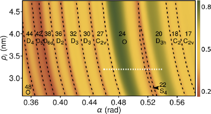
Figure 4 shows that the dominant MPPN symmetry and size only weakly depend on . This suggests that even if not all proteins in the system are incorporated into MPPNs, and the effective value of is smaller than Basta et al. (2014), the key model predictions discussed above remain unchanged—indeed, smaller tend to increase the dominance of MPPNs with and (Fig. 4) while leaving the MPPN radius unchanged. In contrast, Fig. 4 suggests that is a key parameter setting the preferred symmetry and size of MPPNs. Calculating the MPPN self-assembly phase diagram as a function of and the protein radius (see Fig. 5), we find that the dominant MPPN symmetry is more sensitive to variations in than . With the exception of , which is almost as closely packed as the locally optimal packing state [Fig. 2(inset)], all of the dominant MPPN symmetries in Fig. 5 correspond to locally optimal protein packings, with yielding the largest . Finally, we note that the bilayer bending rigidity Rawicz et al. (2000) of the lipids used for MPPNs Basta et al. (2014) is small compared to other lipids Rawicz et al. (2000); Phillips et al. (2009), and that it has also been suggested Partenskii and Jordan (2002); Kim et al. (2012); Lee et al. (2013) that may be increased in the vicinity of membrane proteins. We find sm that, as is being increased, the dominance of MPPNs with becomes increasingly pronounced for MPPNs formed from MscS Basta et al. (2014); Wu et al. (2013).
Conclusion.—To aid the utilization of MPPNs for high-resolution structural studies Basta et al. (2014); Wu et al. (2013) we have developed a simple physical description of MPPNs which connects the symmetry and size of MPPNs to key molecular properties of the lipids and proteins forming MPPNs. Our model accounts for the energy cost of protein-induced lipid bilayer bending deformations Góźdź and Gompper (2001); Auth and Gompper (2009); Müller and Deserno (2010) and topological defects in protein packing in MPPNs Bruinsma et al. (2003); Zandi et al. (2004), and the statistical thermodynamics Bruinsma et al. (2003); Zandi et al. (2004); Safran (2003); Ben-Shaul and Gelbart (1994) of MPPN self-assembly. With all model parameters determined directly from experiments, our model correctly predicts the observed Basta et al. (2014) symmetry and size of MPPNs formed from MscS. Our results suggest that the MPPN bending and defect energies determine a lower cutoff on the number of proteins per MPPN, with the MPPN defect energy and thermal effects yielding MPPNs with locally optimal protein packings close to this cutoff as the dominant MPPN symmetry and size. Our model suggests how, through suitable choices of key lipid and protein properties, a range of well-defined MPPN symmetries and sizes can be produced in experiments.
Acknowledgements.
This work was supported by NSF award numbers DMR-1554716 and DMR-1206332, an Alfred P. Sloan Research Fellowship in Physics, the James H. Zumberge Faculty Research and Innovation Fund at the University of Southern California, and the USC Center for High-Performance Computing. We also acknowledge support through the Kavli Institute for Theoretical Physics, Santa Barbara, via NSF award number PHY-1125915. We thank W. S. Klug, R. Phillips, D. C. Rees, M. H. B. Stowell, and H. Yin for helpful comments.References
- McMahon and Gallop (2005) H. T. McMahon and J. L. Gallop, Nature 438, 590 (2005).
- Engelman (2005) D. M. Engelman, Nature 438, 578 (2005).
- Phillips et al. (2009) R. Phillips, T. Ursell, P. Wiggins, and P. Sens, Nature 459, 379 (2009).
- Basta et al. (2014) T. Basta, H.-J. Wu, M. K. Morphew, J. Lee, N. Ghosh, J. Lai, J. M. Heumann, K. Wang, Y. C. Lee, D. C. Rees, et al., Proc. Natl. Acad. Sci. U.S.A. 111, 670 (2014).
- Bass et al. (2002) R. B. Bass, P. Strop, M. Barclay, and D. C. Rees, Science 298, 1582 (2002).
- Steinbacher et al. (2007) S. Steinbacher, R. B. Bass, P. Strop, and D. C. Rees, Structures of the Prokaryotic Mechanosensitive Channels MscL and MscS, vol. 58 of Curr. Top. Membr. (Academic Press, 2007).
- Wu et al. (2013) H.-J. Wu, T. Basta, M. Morphew, D. Rees, M. Stowell, and Y. Lee, Micro Nano Lett. 8, 672 (2013).
- Humphrey et al. (1996) W. Humphrey, A. Dalke, and K. Schulten, J. Mol. Graphics 14, 33 (1996).
- Góźdź and Gompper (2001) W. T. Góźdź and G. Gompper, Europhys. Lett. 55, 587 (2001).
- Auth and Gompper (2009) T. Auth and G. Gompper, Phys. Rev. E 80, 031901 (2009).
- Müller and Deserno (2010) M. M. Müller and M. Deserno, Prog. Theor. Phys. Supp. 184, 351 (2010).
- Bruinsma et al. (2003) R. F. Bruinsma, W. M. Gelbart, D. Reguera, J. Rudnick, and R. Zandi, Phys. Rev. Lett. 90, 248101 (2003).
- Zandi et al. (2004) R. Zandi, D. Reguera, R. F. Bruinsma, W. M. Gelbart, and J. Rudnick, Proc. Natl. Acad. Sci. U.S.A. 101, 15556 (2004).
- Safran (2003) S. A. Safran, Statistical Thermodynamics of Surfaces, Interfaces, and Membranes (Westview Press, Boulder, 2003).
- Ben-Shaul and Gelbart (1994) A. Ben-Shaul and W. M. Gelbart, in Micelles, Membranes, Microemulsions, and Monolayers (Springer New York, 1994), pp. 1–104.
- (16) See Supplemental Material for futher details.
- Dan et al. (1994) N. Dan, A. Berman, P. Pincus, and S. A. Safran, J. Phys. II 4, 1713 (1994).
- Canham (1970) P. Canham, J. Theor. Biol. 26, 61 (1970).
- Evans (1974) E. Evans, Biophys. J. 14, 923 (1974).
- Helfrich (1973) W. Helfrich, Z. Naturforsch. C 28, 693 (1973).
- Boal (2012) D. H. Boal, Mechanics of the Cell (Cambridge University Press, Cambridge, 2012), 2nd ed.
- Rawicz et al. (2000) W. Rawicz, K. Olbrich, T. McIntosh, D. Needham, and E. Evans, Biophys. J. 79, 328 (2000).
- Damodaran and Merz (1993) K. V. Damodaran and K. M. Merz, Langmuir 9, 1179 (1993).
- Phillips et al. (2012) R. Phillips, J. Kondev, J. Theriot, and H. Garcia, Physical Biology of the Cell (Garland Science, London and New York, 2012).
- Kantor et al. (1987) Y. Kantor, M. Kardar, and D. R. Nelson, Phys. Rev. A 35, 3056 (1987).
- Clare and Kepert (1991) B. Clare and D. Kepert, J. Math. Chem. 6, 325 (1991).
- Ben-Tal et al. (1996) N. Ben-Tal, A. Ben-Shaul, A. Nicholls, and B. Honig, Biophys. J. 70, 1803 (1996).
- Choe et al. (2008) S. Choe, K. A. Hecht, and M. Grabe, J. Gen. Physiol. 131, 563 (2008).
- Kirkpatrick et al. (1983) S. Kirkpatrick, C. D. Gelatt, and M. P. Vecchi, Science 220, 671 (1983).
- Hart (2000) G. Hart, The encyclopedia of polyhedra, Available at www.georgehart.com/virtual-polyhedra/vp.html. Accessed Nov. 12, 2015 (2000).
- Vásquez et al. (2007) V. Vásquez, D. M. Cortes, H. Furukawa, and E. Perozo, Biochemistry 46, 6766 (2007).
- Partenskii and Jordan (2002) M. B. Partenskii and P. C. Jordan, J. Chem. Phys. 117, 10768 (2002).
- Kim et al. (2012) T. Kim, K. I. Lee, P. Morris, R. W. Pastor, O. S. Andersen, and W. Im, Biophys. J. 102, 1551 (2012).
- Lee et al. (2013) K. I. Lee, R. W. Pastor, O. S. Andersen, and W. Im, Chem. Phys. Lipids 169, 19 (2013).
- Monera et al. (1995) O. D. Monera, T. J. Sereda, N. E. Zhou, C. M. Kay, and R. S. Hodges, J. Pept. Sci. 1, 319 (1995).
- Goulian et al. (1993) M. Goulian, R. Bruinsma, and P. Pincus, Europhys. Lett. 22, 145 (1993).
- Weikl et al. (1998) T. R. Weikl, M. M. Kozlov, and W. Helfrich, Phys. Rev. E 57, 6988 (1998).
- Kim et al. (1998) K. S. Kim, J. Neu, and G. Oster, Biophys. J. 75, 2274 (1998).
- Fournier (1999) J.-B. Fournier, Eur. Phys. J. B. 11, 261 (1999).
- Weitz and Destainville (2013) S. Weitz and N. Destainville, Soft Matter 9, 7804 (2013).
- Dommersnes and Fournier (1999) P. Dommersnes and J.-B. Fournier, Eur. Phys. J. B 12, 9 (1999).
- Kim et al. (1999) K. S. Kim, J. C. Neu, and G. F. Oster, Europhys. Lett. 48, 99 (1999).
- Kim et al. (2008) K. S. Kim, T. Chou, and J. Rudnick, Phys. Rev. E 78, 011401 (2008).
- Atılgan and Sun (2004) E. Atılgan and S. X. Sun, J. Chem. Phys. 121, 10392 (2004).
- Reynwar et al. (2007) B. J. Reynwar, G. Illya, V. A. Harmandaris, M. M. Müller, K. Kremer, and M. Deserno, Nature 447, 461 (2007).
- Haselwandter and Phillips (2013) C. A. Haselwandter and R. Phillips, Europhys. Lett. 101, 68002 (2013).
- Haselwandter and Wingreen (2014) C. A. Haselwandter and N. S. Wingreen, PLoS Comput. Biol. 10, e1003932 (2014).
- De Kruijff et al. (1975) B. De Kruijff, P. Cullis, and G. Radda, BBA-Biomembranes 406, 6 (1975).
- Parsegian (1966) V. A. Parsegian, T. Faraday Soc. 62, 848 (1966).
- Sukharev (2002) S. Sukharev, Biophys. J. 83, 290 (2002).