Self-aligned local electrolyte gating of 2D materials with nanoscale resolution
Abstract
In the effort to make 2D materials-based devices smaller, faster, and more efficient, it is important to control charge carrier at lengths approaching the nanometer scale. Traditional gating techniques based on capacitive coupling through a gate dielectric cannot generate strong and uniform electric fields at this scale due to divergence of the fields in dielectrics. This field divergence limits the gating strength, boundary sharpness, and pitch size of periodic structures, and restricts possible geometries of local gates (due to wire packaging), precluding certain device concepts, such as plasmonics and transformation optics based on metamaterials. Here we present a new gating concept based on a dielectric-free self-aligned electrolyte technique that allows spatially modulating charges with nanometer resolution. We employ a combination of a solid-polymer electrolyte gate and an ion-impenetrable e-beam-defined resist mask to locally create excess charges on top of the gated surface. Electrostatic simulations indicate high carrier density variations of across a length of at the mask boundaries on the surface of a 2D conductor, resulting in a sharp depletion region and a strong in-plane electric field of across the so-created junction. We apply this technique to the 2D material graphene to demonstrate the creation of tunable p-n junctions for optoelectronic applications. We also demonstrate the spatial versatility and self-aligned properties of this technique by introducing a novel graphene thermopile photodetector.
Modulation of charge carrier concentration of semiconductors lies at the heart of many electronic and optoelectronic device operation principles Neamen (2003); Chuang (2012). This modulation is especially essential for two-dimensional (2D) van der Waals materials Ferrari et al. (2015); Bao and Loh (2012); Avouris and Freitag (2014); Koppens et al. (2014); Wang et al. (2012); Britnell et al. (2013); Liu et al. (2014); Sassi et al. (2016) where it is usually much stronger (from 0.15 electrons/cell to 15 electrons/cell) compared to bulk materials and can be dynamically tuned with electrostatic gating methods. In recent years, rapidly developing device concepts and applications impose stronger and stronger requirements on the spatial resolution and highest-achievable carrier concentration of gating techniques. For example, a spatially sharp () p-n junction and a high carrier density contrast across the junction is the key to the realization of concepts such as tunnel diodes Roy et al. (2015) and negative electron refractive index Cheianov, Fal’ko, and Altshuler (2007). A strong in-plane electric field across the junction as a result of the junction sharpness facilitates electron-hole pair separation in the photovoltaic (PV) effectShang et al. (2011) and thus can improve the quantum efficiency of PV-based solar cells and photodetectors Konstantatos and Sargent (2010). Many novel device concepts also rely on the ability to create metamaterials with spatial carrier density variations down to the nanometer scale, including for instance graphene with periodically doped nanodisk or nanoribbon arrays for complete optical absorption in the visible and near-infrared Thongrattanasiri, Koppens, and de Abajo (2012); Ye et al. (2015), graphene with doped waveguide, bend and resonator patterns for a plasmon-based nanophotonic network Vakil and Engheta (2011), and superlattices based on graphene and other 2D materials for concepts such as electron beam supercollimation Yankowitz et al. (2012); Kang et al. (2013); Park et al. (2008a, b). Implementing these concepts calls for a gating method that allows for sharp p-n junctions with narrow depletion regions (), large carrier density contrasts (), strong in-plane electric fields (), and the versatility to generate complex spatial doping profiles with a nanoscale resolution.
The state-of-the-art electrostatic gating technique for modulating charge carrier concentration and creating p-n junctions is the metal-dielectric split gate technique Baugher et al. (2014); Pospischil, Furchi, and Mueller (2014); Ross et al. (2014). This method is based on the electric field effect Novoselov et al. (2004) in which electric voltages are applied across a gate dielectric to induce extra charges on the 2D material surface. A p-n junction can be created by applying opposing electric potentials to the two sides of a boundary to induce charges with opposite polarities. Although this technique is convenient, several limitations restrict its use when more extreme requirements are desirable: In terms of carrier density contrast, dielectric-based gating can only induce a carrier concentration variation of less than for typical dielectrics such as \ceSiO2, \ceHfO2, \ceSiN, and hexagonal-\ceBN, due to a maximal applicable voltage across the dielectrics before the dielectric breakdown (molecular bond breakage and defects) McPherson et al. (2003); Sire et al. (2007); Hattori et al. (2014). In terms of junction sharpness, the carrier density has a slowly varying profile across the junction due to electric field divergence in dielectrics, with a characteristic length similar to the thickness of the dielectric, making it hard to create sharp junctions at nanoscale unless with extremely thin (a few nanometers) dielectrics which typically has undesirable leakage and tunneling currents. Furthermore, due to wire-packaging difficulties and fabrication limitation of the electrodes, complex gating patterns and device geometries with large numbers of gating electrodes at the nanoscale is practically challenging.
In this paper we present a self-aligned gating concept with a spatial resolution down to sub- based on electrolytic gating. In contrast to dielectric-based gating, electrolyte gating can concentrate excess charges directly on the surface of the 2D material and reach a capacitance of (250 times higher than a typical \ceSiO2 gate) Efetov and Kim (2010); Ueno et al. (2011); Ohno et al. (2009), which enables carrier density modification up to . Since patterning of electrolyte is challenging, a general method for local electrolyte gates at nanoscale has not been demonstrated so far. To achieve this goal we introduce a lithographical masking technique based on e-beam over-exposed Poly(methyl methacrylate) (PMMA) that can screen ions in electrolyte. This e-beam patterned mask can prevent the mask-protected areas from being in contact with, and thus modulated by, the electrolyte gate. The mask hence effectively creates lithographically-defined local electrolyte gates with versatile geometries and feature sizes down to several nanometers.
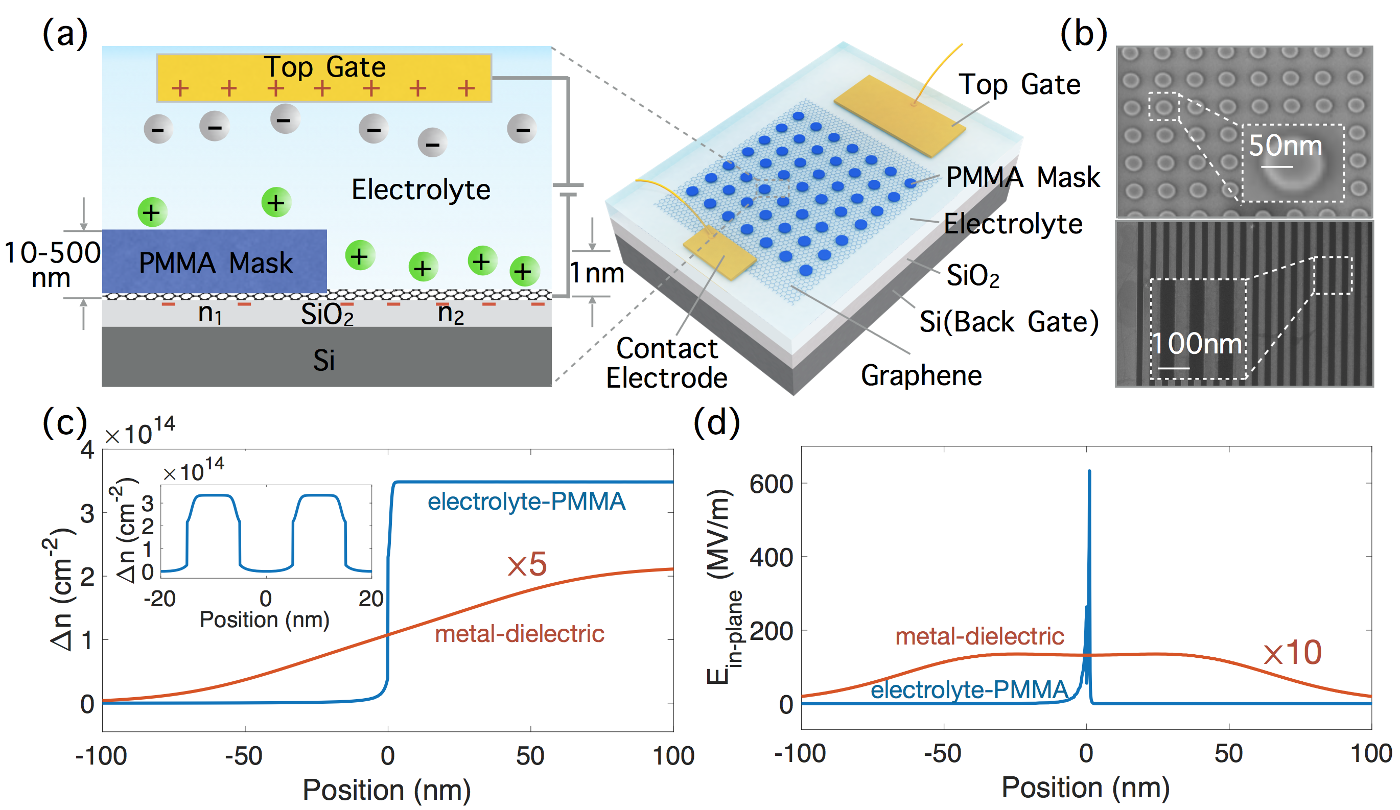
Figure 1(a) illustrates the technique for a graphene sheet. When a voltage is applied between the electrolyte top gate and the graphene, ions in the electrolyte accumulate on the graphene surface, only in regions that are uncovered by the PMMA mask, creating a self-aligned electrolyte gating pattern defined by the shape of the mask. An additional \ceSiO2 back gate allows weak p or n doping of the regions covered by the mask.

For an electrolyte gate voltage of , a carrier density contrast of can be created at the PMMA mask boundary across only a few nanometers, as shown in the blue curve in Figure 1(c), produced by finite element simulations with COMSOL-Multiphysics Comsol (2015). Plotted in Figure 1(d) in the blue curve is the calculated in-plane electric field intensity across the junction, showing a maximum magnitude as high as at the close vicinity of the mask boundary. The simulation assumes a Stern-Gouy-Chapman electrical double layer model Bard et al. (1980) of the electrolyte ions and calculates the electric potential and the flux of ions under the influence of both ion diffusion due to the ionic concentration gradient and ion migration due to the electric field. This process is governed by the Poisson-Nernst-Planck equations. Additional details about the double layer model and parameters used in the simulation are in the Supplementary Information.
For comparison, the simulated doping contrast and the in-plane electric field are much lower in a metal-dielectric split gate, as indicated in the red curves in Figure 1(c) and (d), rescaled for better visibility with factors of 5 and 10, respectively. A carrier density contrast of at most (1 order of magnitude lower than that with the electrolyte-PMMA-mask technique) across a length scale of is induced when a voltage of is applied, corresponding to an in-plane electric field of ( times lower than that with the electrolyte-PMMA-mask technique). This simulation assumes a dielectric constant of 3.9 (\ceSiO2) and a thickness of for the gate dielectric, and a gap of between the two gate electrodes, which are typical values in the literatureBaugher et al. (2014); Pospischil, Furchi, and Mueller (2014). The dielectric thickness and the gap width between the gate electrodes are the limiting factors for the junction sharpness. To achieve a sharpness of only a few nanometers, both the dielectric and the gap width have to be only a few nanometers in size too (see Supplementary Information). The former would result in undesirable leaking and tunneling currents and the latter is currently challenging from a fabrication standpoint.
In summary, our self-aligned electrolyte gating technique can enable nanometer-sharp junctions, and carrier density contrast and in-plane electric field orders of magnitude higher than split metal gate structures.
To implement this self-aligned electrolyte gating technique with the screening mask, the choice of material for the mask is essential. Two requirements need to be met: (1) To ensure high spatial resolution of the self-aligned local electrolyte gates, the lithographyical resolution of the mask has to be high; (2) To ensure reliable spatial selectivity and doping level control, the mask must be impenetrable to ions in the electrolyte with no leaks.
One candidate for the mask is the e-beam resist PMMA. Commonly used as a high-resolution positive-tone e-beam resist Chen and Ahmed (1993); Beaumont et al. (1981), PMMA becomes a negative-tone resist when exposed at a much higher dose (), where it is cross-linked and transformed into graphitic nanostructures from a polymeric resist carbonization process Duan et al. (2010); Duan, Xie, and Han (2008). Cross-linked PMMA allows sub- e-beam lithography resolution Duan et al. (2010). In separate experiments, we measured negligible current between a graphene sheet covered by cross-linked PMMA ( thickness) and the electrolyte gate, verifying that the mask is essentially impermeable to ions in solid polymer electrolyte \cePEO-LiClO4. These cyclic voltammetry (CV) results are in the Supplementary Information.
The spatial patterning resolution of the self-aligned gates is determined by two factors: the e-beam lithography resolution of the screening mask and the Debye length of the electrolyte. As mentioned above, the e-beam resolution is and the Debye length is Efetov (2014), so the spatial resolution for patterning local electrolyte gates using this technique is dominated by the e-beam resolution which is . The simulation in the inset of Figure 1(c) indicates a well-defined carrier density modulation profile resulting from periodic local electrolyte gates with a half-pitch of . Figure 1(b) shows scanning-electron-micrographs (SEMs) of two examples of PMMA mask on graphene with nanometer feature size and different geometries, including disks and ribbons.
As proof-of-principle studies, we will now apply this technique to demonstrate two different device concepts: a graphene p-n junction and a graphene compact thermopile.
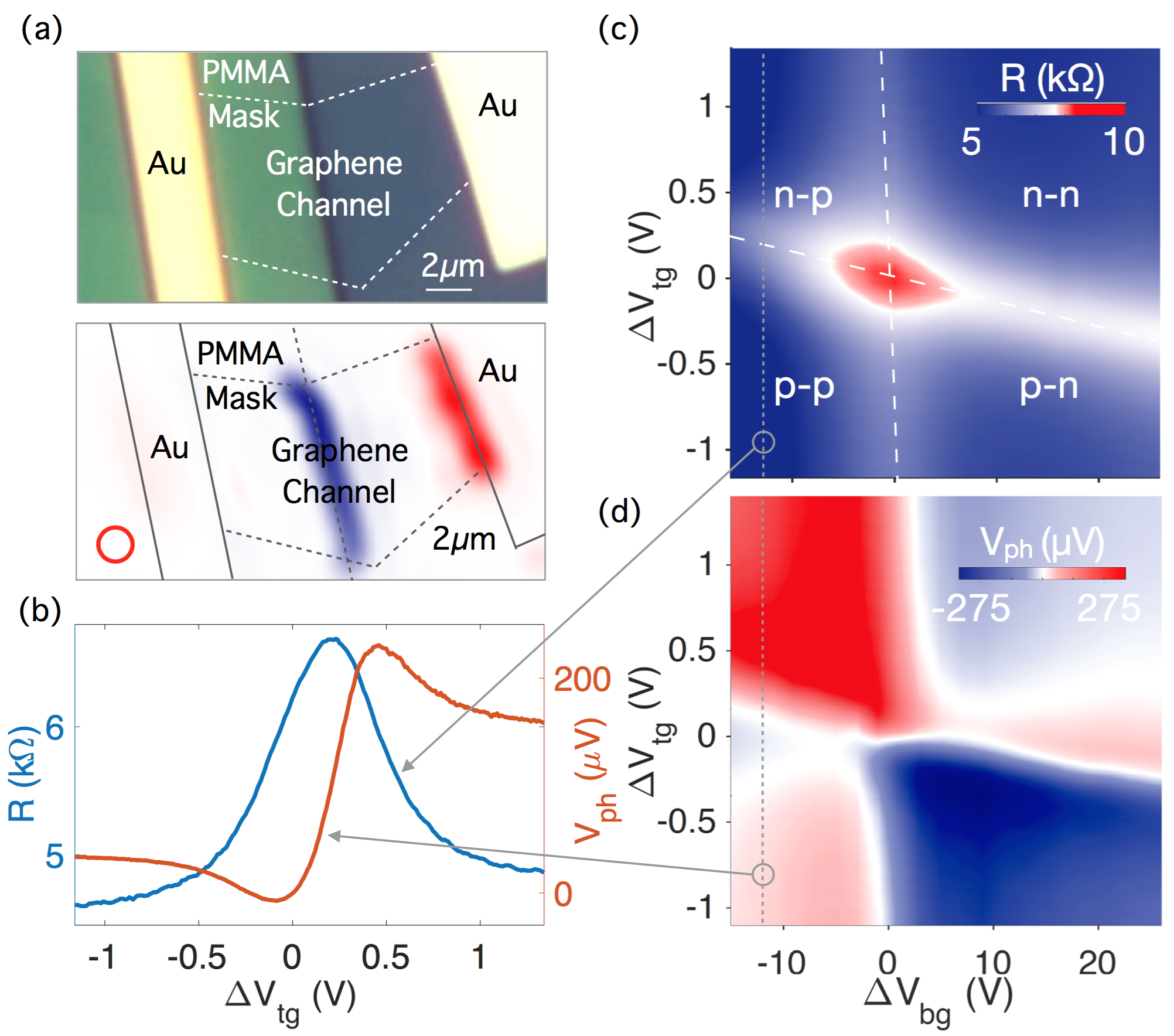
Figure 2 shows a graphene p-n junction and the dynamical tuning of its doping level and photoresponse. The structure of the p-n junction device, shown in Figure 2(a) (top panel), consists of a graphene channel covered in half by a PMMA mask. The graphene is exfoliated onto a \ceSiO2 substrate thermally grown on doped \ceSi. It is then patterned by e-beam lithography and reactive ion etching (RIE) into a channel roughly in length and in width. A pair of \ceCr/\ceAu contacts and a PMMA mask are then defined by e-beam lithography. Solid polymer electrolyte \cePEO-LiClO4 is then drop-casted to cover the entire device.

Large-range doping level control of the two regions in a p-n junction can be achieved by tuning electrolyte top gate and \ceSiO2 back gate voltages, where controls the mask-uncovered region and mostly controls the mask-covered region. Figure 2(c) shows the channel resistance versus and , showing four distinct characteristic regions that indicate gate-voltage-tunable charge density at a p-n interface Williams, DiCarlo, and Marcus (2007). Two intersecting lines of high resistance (white dashed), representing charge neutrality points of the two regions respectively, divide the resistance map into four low-resistance regions: p-n, p-p, n-p, and n-n. A vertical line trace of the 2D resistance map along the dotted gray line, shown in the blue curve of Figure 2(b), exhibits distinct Dirac peak indicating modulation of graphene’s Fermi level across the charge neutrality point.
Photoresponse observed at the graphene p-n junction can also be dynamically tuned by the gate voltages. Figure 2(a) (bottom panel) shows the spatially-resolved open-circuit photovoltage map of the device under zero bias voltage across the channel, conducted on a near-infrared () confocal scanning microscopy setup at room temperature. As the laser excitation is scanned over the device, a large photovoltage is observed at the self-aligned electrolyte gate defined junction. This photovoltage at the junction as a function of and , plotted in Figure 2(d), exhibits a distinct six-fold pattern with alternating photovoltage signs, showing a strong dependence of the photoresponse on the relative doping level of the graphene junction. This six-fold pattern indicates a photo-induced hot carrier-assisted photoresponse process at the graphene p-n junction known as the photo-thermoelectric (PTE) effect Song et al. (2011). A vertical line trace of the 2D photovoltage map along the same dotted gray line, plotted in the red curve of Figure 2(b), shows typical non-monotonic gate voltage dependence as a result of the PTE effect.

Next we demonstrate a compact graphene thermopile in the mid-infrared that takes advantage of the flexible gating geometries enabled by this self-aligned technique. In this design a complex doping pattern of graphene is created to enhance the photodetector’s photovoltage responsivity. For PTE effect, the photovoltage generated can be expressed as , where is the Seebeck coefficient of graphene, a function of charge carrier density, and is the increase in electron temperature from the environment. For free-space incident light that typically has a spherical Gaussian profile, the temperature gradient points in the radial direction, so the photovoltage is maximized when it is collected radially.
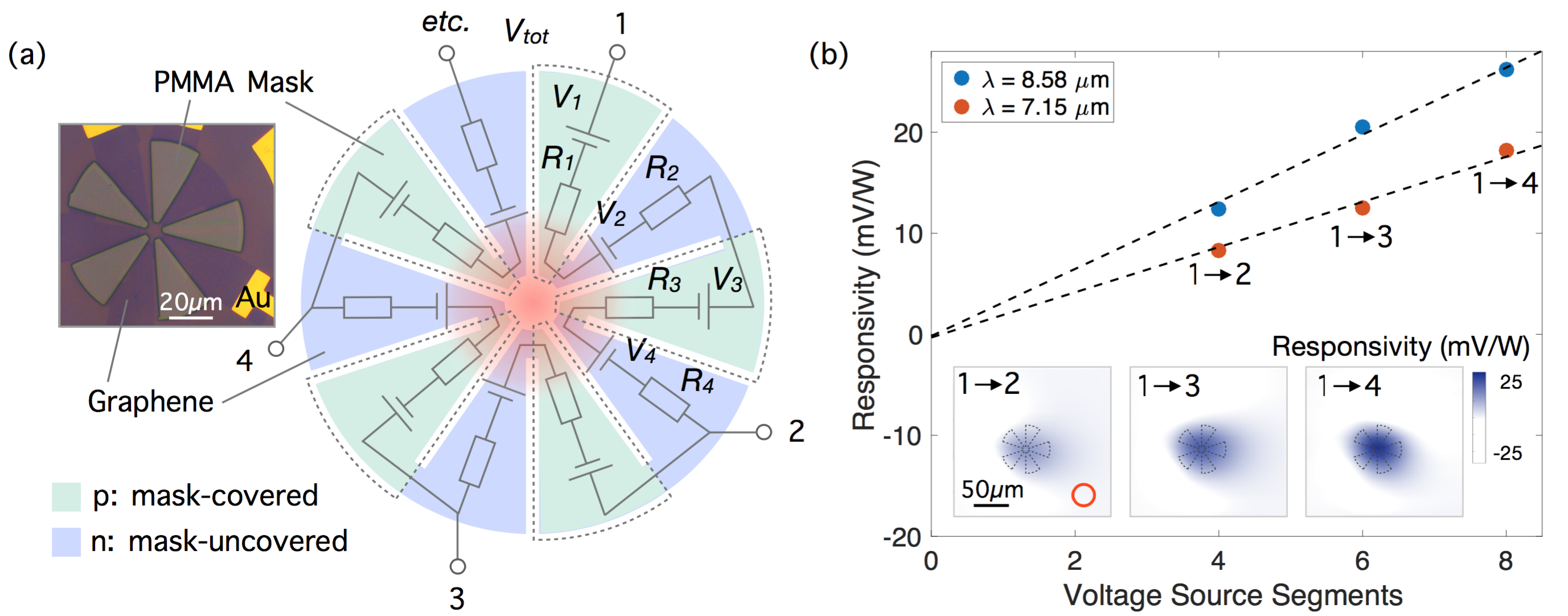
The designed thermopile geometry, whose equivalent circuit diagram is illustrated in Figure 3(a), consists of several thermocouples connected in series whose photovoltages are all collected in the radial direction. Each graphene segment is considered a voltage source with a resistance. For the photovoltage to sum up along the meandering graphene channel, each segment is or doped in an alternating fashion so that neighboring photovoltages point in opposite directions (Seebeck coefficient has opposite signs). The alternating doping is achieved using our gating technique by covering every other segments with the PMMA mask and applying positive and negative respectively. Compared to existing graphene thermopiles such as that in ref. Hsu et al. (2015), this approach eliminates the need for embedded gates and external wiring of thermocouples, enabling a compact thermopile based solely on graphene. The achievable nanoscale dimensions and the more complex geometries could lead to more efficient photovoltage collection.
Spatially-resolved open-circuit photovoltage mapping of this thermopile is conducted on a confocal scanning microscopy setup with a mid-infrared laser source at two wavelengths and . The photovoltage between several sets of terminals (indicated with numbers in Figure 3(a)) are measured to study the individual contributions of thermocouples. As the laser spot is scanned over the device, a maximal photovoltage is observed at the center of the thermopile, as shown in the photovoltage spatial maps in the inset of Figure 3(b). The spatial maximum for responsivity is then plotted as a function of the number of voltage source segments between the terminals. The well-fitted linear relation at both wavelengths confirms the summation of photovoltages from each individual segment. The maximal responsivity of this device is . Compared to a previous graphene thermocouple with only one single p-n junction, studied in similar conditions Herring et al. (2014), the carefully designed doping pattern of graphene results in an about 7 times enhancement in photovoltage responsivity. Further optimization of parameters including device dimension and the number of voltage segments can be done to achieve higher responsivity.
The flexibility to tailor the dimension and geometry of electrolyte gates on 2D materials at the nanoscale with strong doping ability expands the possibility of 2D-material-based tunable optoelectronic devices. Also, the non-destructive fabrication procedure maintains the high quality of the 2D material samples since there is no need for nanopatterning of graphene itself in harsh environments, and the cross-linked PMMA has previously been shown to have insignificant effect on the mobility of graphene Henriksen, Nandi, and Eisenstein (2012). Moreover, the broadband optical transparency of the PMMA mask also ensures non-interfered optical spectroscopy on the fabricated device. These are important practical considerations that can be crucial for the experimental implementation of many novel device concepts such as the compact thermopile we have demonstrated above. Plasmonics in graphene would be another example. To achieve a plasmonic resonant wavelength of or less, graphene nanostructures need to have a feature size as small as . This typically requires direct patterning of the graphene sheet, which on this small scale would create significant edge scattering and reduce carrier mobility, limiting the quality factor of the graphene plasmonic resonances Low and Avouris (2014). The nanoscale electrolytic gating scheme proposed here would be an alternative and promising way of generating tunable graphene plasmons with an improved quality factor and a broader wavelength range.
To conclude, we have proposed and numerically simulated a self-aligned local electrolyte gating method of 2D materials that allows for a carrier density contrast of more than across a length of and an in-plane electric field of . We have also developed an experimental implementation of this technique and demonstrated two different device concepts based on graphene, including a single p-n junction with tunable doping level and photoresponse and a novel compact thermopile with enhanced photovoltage responsivity in the mid-infrared. This novel nanoscale electrolytic gating scheme is a promising and versatile experimental approach to numerous 2D material-based device concepts in tunable nanophotonics and optoelectronics, and can potentially be used for other low-dimensional material classes too.
Acknowledgement
The research leading to these results has received funding from the U.S. Office of Naval Research (Award N00014-14-1-0349), the European Commission H2020 Programme (no. 604391, “Graphene Flagship”), the European Research Council starting grant (307806, CarbonLight) and project GRASP (FP7-ICT-2013-613024-GRASP). C.P. was supported in part by the Stata Family Presidential Fellowship of MIT and in part by the U.S. Office of Naval Research (Award N00014-14-1-0349). D.K.E. was supported in part by an Advanced Concept Committee (ACC) program from MIT Lincoln Laboratory. S.N. was supported by the European Commission (FP7-ICT-2013-613024-GRASP) and thanks M. Batzer and R. Parret for their help in exploratory work on this subject. R.-J.S. was supported in part by the Center for Excitonics, an Energy Frontier Research Center funded by the U.S. Department of Energy, Office of Science, Office of Basic Energy Sciences under award no. DE-SC0001088. G.G. was supported by the Swiss National Science Foundation (SNSF). Device fabrication was performed at the NanoStructures Laboratory at MIT and the Center for Nanoscale Systems, a member of the National Nanotechnology Infrastructure Network supported by the National Science Foundation (NSF).
References
- Neamen (2003) D. A. Neamen, Semiconductor physics and devices (McGraw-Hill Higher Education, 2003).
- Chuang (2012) S. L. Chuang, Physics of photonic devices, Vol. 80 (John Wiley & Sons, 2012).
- Ferrari et al. (2015) A. C. Ferrari, F. Bonaccorso, V. Falko, K. S. Novoselov, S. Roche, P. Bøggild, S. Borini, F. Koppens, V. Palermo, N. Pugno, J. a. Garrido, R. Sordan, A. Bianco, L. Ballerini, M. Prato, E. Lidorikis, J. Kivioja, C. Marinelli, T. Ryhänen, A. Morpurgo, J. N. Coleman, V. Nicolosi, L. Colombo, A. Fert, M. Garcia-Hernandez, A. Bachtold, G. F. Schneider, F. Guinea, C. Dekker, M. Barbone, C. Galiotis, A. Grigorenko, G. Konstantatos, A. Kis, M. Katsnelson, C. W. J. Beenakker, L. Vandersypen, A. Loiseau, V. Morandi, D. Neumaier, E. Treossi, V. Pellegrini, M. Polini, A. Tredicucci, G. M. Williams, B. H. Hong, J. H. Ahn, J. M. Kim, H. Zirath, B. J. van Wees, H. van der Zant, L. Occhipinti, A. Di Matteo, I. a. Kinloch, T. Seyller, E. Quesnel, X. Feng, K. Teo, N. Rupesinghe, P. Hakonen, S. R. T. Neil, Q. Tannock, T. Löfwander, and J. Kinaret, “Science and technology roadmap for graphene, related two-dimensional crystals, and hybrid systems,” Nanoscale 7, 4598–4810 (2015).
- Bao and Loh (2012) Q. Bao and K. P. Loh, “Graphene photonics, plasmonics, and broadband optoelectronic devices,” ACS nano 6, 3677–3694 (2012).
- Avouris and Freitag (2014) P. Avouris and M. Freitag, “Graphene photonics, plasmonics, and optoelectronics,” IEEE Journal of selected topics in quantum electronics 1, 6000112 (2014).
- Koppens et al. (2014) F. Koppens, T. Mueller, P. Avouris, A. Ferrari, M. Vitiello, and M. Polini, “Photodetectors based on graphene, other two-dimensional materials and hybrid systems,” Nature nanotechnology 9, 780–793 (2014).
- Wang et al. (2012) Q. H. Wang, K. Kalantar-Zadeh, A. Kis, J. N. Coleman, and M. S. Strano, “Electronics and optoelectronics of two-dimensional transition metal dichalcogenides,” Nature nanotechnology 7, 699–712 (2012).
- Britnell et al. (2013) L. Britnell, R. M. Ribeiro, A. Eckmann, R. Jalil, B. D. Belle, A. Mishchenko, Y.-J. Kim, R. V. Gorbachev, T. Georgiou, S. V. Morozov, A. N. Grigorenko, A. K. Geim, C. Casiraghi, A. H. C. Neto, and K. S. Novoselov, “Strong light-matter interactions in heterostructures of atomically thin films,” Science 340, 1311–1314 (2013).
- Liu et al. (2014) C.-H. Liu, Y.-C. Chang, T. B. Norris, and Z. Zhong, “Graphene photodetectors with ultra-broadband and high responsivity at room temperature,” Nature nanotechnology 9, 273–278 (2014).
- Sassi et al. (2016) U. Sassi, R. Parret, S. Nanot, M. Bruna, S. Borini, S. Milana, D. De Fazio, Z. Zhuang, E. Lidorikis, F. Koppens, et al., “Graphene-based, mid-infrared, room-temperature pyroelectric bolometers with ultrahigh temperature coefficient of resistance,” arXiv preprint arXiv:1608.00569 (2016).
- Roy et al. (2015) T. Roy, M. Tosun, X. Cao, H. Fang, D.-H. Lien, P. Zhao, Y.-Z. Chen, Y.-L. Chueh, J. Guo, and A. Javey, “Dual-gated mos2/wse2 van der waals tunnel diodes and transistors,” Acs Nano 9, 2071–2079 (2015).
- Cheianov, Fal’ko, and Altshuler (2007) V. V. Cheianov, V. Fal’ko, and B. Altshuler, “The focusing of electron flow and a veselago lens in graphene pn junctions,” Science 315, 1252–1255 (2007).
- Shang et al. (2011) X. Shang, J. He, H. Wang, M. Li, Y. Zhu, Z. Niu, and Y. Fu, “Effect of built-in electric field in photovoltaic inas quantum dot embedded gaas solar cell,” Applied Physics A 103, 335–341 (2011).
- Konstantatos and Sargent (2010) G. Konstantatos and E. H. Sargent, “Nanostructured materials for photon detection,” Nature nanotechnology 5, 391–400 (2010).
- Thongrattanasiri, Koppens, and de Abajo (2012) S. Thongrattanasiri, F. H. Koppens, and F. J. G. de Abajo, “Complete optical absorption in periodically patterned graphene,” Physical review letters 108, 047401 (2012).
- Ye et al. (2015) C. Ye, Z. Zhu, W. Xu, X. Yuan, and S. Qin, “Electrically tunable absorber based on nonstructured graphene,” Journal of Optics 17, 125009 (2015).
- Vakil and Engheta (2011) A. Vakil and N. Engheta, “Transformation optics using graphene,” Science 332, 1291–1294 (2011).
- Yankowitz et al. (2012) M. Yankowitz, J. Xue, D. Cormode, J. D. Sanchez-Yamagishi, K. Watanabe, T. Taniguchi, P. Jarillo-Herrero, P. Jacquod, and B. J. LeRoy, “Emergence of superlattice dirac points in graphene on hexagonal boron nitride,” Nature Physics 8, 382–386 (2012).
- Kang et al. (2013) J. Kang, J. Li, S.-S. Li, J.-B. Xia, and L.-W. Wang, “Electronic structural moire pattern effects on mos2/mose2 2d heterostructures,” Nano letters 13, 5485–5490 (2013).
- Park et al. (2008a) C.-H. Park, L. Yang, Y.-W. Son, M. L. Cohen, and S. G. Louie, “Anisotropic behaviours of massless dirac fermions in graphene under periodic potentials,” Nature Physics 4, 213–217 (2008a).
- Park et al. (2008b) C.-H. Park, Y.-W. Son, L. Yang, M. L. Cohen, and S. G. Louie, “Electron beam supercollimation in graphene superlattices,” Nano letters 8, 2920–2924 (2008b).
- Baugher et al. (2014) B. W. Baugher, H. O. Churchill, Y. Yang, and P. Jarillo-Herrero, “Optoelectronic devices based on electrically tunable pn diodes in a monolayer dichalcogenide,” Nature nanotechnology 9, 262–267 (2014).
- Pospischil, Furchi, and Mueller (2014) A. Pospischil, M. M. Furchi, and T. Mueller, “Solar-energy conversion and light emission in an atomic monolayer pn diode,” Nature nanotechnology 9, 257–261 (2014).
- Ross et al. (2014) J. S. Ross, P. Klement, A. M. Jones, N. J. Ghimire, J. Yan, D. G. Mandrus, T. Taniguchi, K. Watanabe, K. Kitamura, W. Yao, D. H. Cobden, and X. Xu, “Electrically tunable excitonic light-emitting diodes based on monolayer WSe2 p–n junctions,” Nature Nanotechnology 9, 268–272 (2014).
- Novoselov et al. (2004) K. S. Novoselov, A. K. Geim, S. V. Morozov, D. Jiang, Y. Zhang, S. V. Dubonos, I. V. Grigorieva, and A. A. Firsov, “Electric field effect in atomically thin carbon films,” science 306, 666–669 (2004).
- McPherson et al. (2003) J. McPherson, J.-Y. Kim, A. Shanware, and H. Mogul, “Thermochemical description of dielectric breakdown in high dielectric constant materials,” Applied Physics Letters 82, 2121 (2003).
- Sire et al. (2007) C. Sire, S. Blonkowski, M. J. Gordon, and T. Baron, “Statistics of electrical breakdown field in hfo2 and sio2 films from millimeter to nanometer length scales,” Applied Physics Letters 91, 2905 (2007).
- Hattori et al. (2014) Y. Hattori, T. Taniguchi, K. Watanabe, and K. Nagashio, “Layer-by-layer dielectric breakdown of hexagonal boron nitride,” ACS nano 9, 916–921 (2014).
- Efetov and Kim (2010) D. K. Efetov and P. Kim, “Controlling electron-phonon interactions in graphene at ultrahigh carrier densities,” Physical review letters 105, 256805 (2010).
- Ueno et al. (2011) K. Ueno, S. Nakamura, H. Shimotani, H. Yuan, N. Kimura, T. Nojima, H. Aoki, Y. Iwasa, and M. Kawasaki, “Discovery of superconductivity in ktao3 by electrostatic carrier doping,” Nature nanotechnology 6, 408–412 (2011).
- Ohno et al. (2009) Y. Ohno, K. Maehashi, Y. Yamashiro, and K. Matsumoto, “Electrolyte-gated graphene field-effect transistors for detecting ph and protein adsorption,” Nano Letters 9, 3318–3322 (2009).
- Comsol (2015) Comsol, COMSOL Multiphysics: Version 5.0 (Comsol, 2015).
- Bard et al. (1980) A. J. Bard, L. R. Faulkner, J. Leddy, and C. G. Zoski, Electrochemical methods: fundamentals and applications, Vol. 2 (Wiley New York, 1980).
- Chen and Ahmed (1993) W. Chen and H. Ahmed, “Fabrication of 5–7 nm wide etched lines in silicon using 100 kev electron-beam lithography and polymethylmethacrylate resist,” Applied physics letters 62, 1499–1501 (1993).
- Beaumont et al. (1981) S. Beaumont, P. Bower, T. Tamamura, and C. Wilkinson, “Sub-20-nm-wide metal lines by electron-beam exposure of thin poly (methyl methacrylate) films and liftoff,” Applied Physics Letters 38, 436–439 (1981).
- Duan et al. (2010) H. Duan, D. Winston, J. K. Yang, B. M. Cord, V. R. Manfrinato, and K. K. Berggren, “Sub-10-nm half-pitch electron-beam lithography by using poly (methyl methacrylate) as a negative resist,” Journal of Vacuum Science & Technology B 28, C6C58–C6C62 (2010).
- Duan, Xie, and Han (2008) H. Duan, E. Xie, and L. Han, “Turning electrospun poly (methyl methacrylate) nanofibers into graphitic nanostructures by in situ electron beam irradiation,” Journal of Applied Physics 103, 046105 (2008).
- Efetov (2014) D. K. Efetov, “Towards inducing superconductivity into graphene,” (2014).
- Williams, DiCarlo, and Marcus (2007) J. Williams, L. DiCarlo, and C. Marcus, “Quantum hall effect in a gate-controlled pn junction of graphene,” Science 317, 638–641 (2007).
- Song et al. (2011) J. C. Song, M. S. Rudner, C. M. Marcus, and L. S. Levitov, “Hot carrier transport and photocurrent response in graphene,” Nano letters 11, 4688–4692 (2011).
- Hsu et al. (2015) A. L. Hsu, P. Herring, N. Gabor, S. Ha, Y. C. Shin, Y. Song, M. Chin, M. Dubey, A. Chandrakasan, J. Kong, P. Jarillo-Herrero, and T. Palacios, “Graphene-based thermopile for thermal imaging applications,” Nano letters 15, 7211–7216 (2015).
- Herring et al. (2014) P. K. Herring, A. L. Hsu, N. M. Gabor, Y. C. Shin, J. Kong, T. Palacios, and P. Jarillo-Herrero, “Photoresponse of an electrically tunable ambipolar graphene infrared thermocouple,” Nano letters 14, 901–907 (2014).
- Henriksen, Nandi, and Eisenstein (2012) E. Henriksen, D. Nandi, and J. Eisenstein, “Quantum hall effect and semimetallic behavior of dual-gated aba-stacked trilayer graphene,” Physical Review X 2, 011004 (2012).
- Low and Avouris (2014) T. Low and P. Avouris, “Graphene plasmonics for terahertz to mid-infrared applications,” ACS nano 8, 1086–1101 (2014).