Scroll-wave dynamics in an anatomically… \sodtitleScroll-wave dynamics in the presence of ionic and conduction inhomogeneities in an anatomically realistic mathematical model for the pig heart \rauthorR. Majumder, Rahul Pandit, A. V. Panfilov \sodauthorMajumder, Pandit, Panfilov \dates22 August 2016
Scroll-wave dynamics in the presence of ionic and conduction inhomogeneities in an anatomically realistic mathematical model for the pig heart
Abstract
Nonlinear waves of the reaction-diffusion (RD) type occur in many biophysical systems, including the heart, where they initiate cardiac contraction. Such waves can form vortices called scroll waves, which result in the onset of life-threatening cardiac arrhythmias. The dynamics of scroll waves is affected by the presence of inhomogeneities, which, in a very general way, can be of (i) ionic type, i.e., they affect the reaction part, or (ii) conduction type, i.e., they affect the diffusion part of an RD equation. We demostrate, for the first time, by using a state-of-the-art, anatomically realistic model of the pig heart, how differences in the geometrical and biophysical nature of such inhomogeneities can influence scroll-wave dynamics in different ways. Our study reveals that conduction-type inhomogeneities become increasingly important at small length scales, i.e., in the case of multiple, randomly distributed, obstacles in space at the cellular scale (). Such configurations can lead to scroll-wave break up. In contrast, ionic inhomogeneities, affect scroll-wave dynamics significantly at large length scales, when these inhomogeneities are localized in space at the tissue level ( mm). In such configurations, these inhomogeneities can (a) attract scroll waves, by pinning them to the heterogeneity, or (b) lead to scroll-wave breakup.
74.50.+r, 74.80.Fp
Introduction: Nonlinear waves occur in excitable media of physical, chemical, and biological origin. Such waves can form vortices in two and three dimensions; these are called spiral and scroll waves, respectively, and they are involved in the spatiotemporal organization of wave dynamics in various complex systems. Therefore, the study of such waves is a subject of interest in a broad area of research. One of the most important applications of such studies is the formation of vortices in cardiac tissue, which is associated with the onset and development of lethal cardiac arrhythmias [1, 2, 3, 4, 5, 6, 7]. Thus, understanding the factors that determine the dynamics of scroll waves is a topic of great interest. Cardiac arrhythmias, such as ventricular tachycardias (VT) are generally associated with stationary, meandering, or drifting, periodic or quasiperiodic scroll waves; whereas, ventricular fibrillation (VF) is associated with scroll-wave break up. The dynamical behaviour of scroll waves in cardiac tissue is affected significantly by the presence of inhomogeneities [8, 9, 10, 11, 2, 3, 12, 13, 14, 15, 16], which can occur in the heart in many forms. However, biophysically, they can be grouped into two major classes: (i) Ionic-type, i.e., inhomogeneities in the properties of different cells that constitute the system; and (ii) conduction-type, i.e., inexcitable obstacles. An in-depth knowledge of the role of these inhomogeneities is essential for understanding the mechanisms that underlie most cardiac arrhythmias.
In experiments, it is often difficult to study systematically the role of
these inhomogeneities in the development of arrhythmias, with regard to the
nature, position, and distribution of these inhomogeneities within the heart.
Thus, it is important to search for alternative methods of investigation.
Mathematical modelling provides an important tool here; it has been used
extensively, with outstanding success, in interdisciplinary
science. From a mathematical point of view, the excitable, cardiac-tissue
medium is described by a reaction-diffusion (RD) equation of the type:
| (1) |
with the reaction part accounting for properties of cardiac cells and the diffusion part , for the connection of cells to tissue. In this setting, an ionic inhomogeneity represents a modification of , whereas a conduciton inhomogeneity involves a modification of .
In this Letter, we present an extensive numerical study of scroll-wave dynamics
in the presence of inhomogeneities in an anatomically realistic model of the
pig heart. We have used the single-cell, modified, Luo-Rudy I (mLRI)
model [17] to construct our cardiac-tissue model and the anatomically
realistic geometry obtained in [18]. We have studied the effects
on scroll-wave dynamics of (i) large-length-scale, solitary
inhomogeneity (old infarction) and (ii) small-length-scale, multiple,
conduction inhomogeneities (fibrosis) and compared our results from these
studies with those we have obtained from similar (i) large- and
(ii) small-length-scale ionic inhomogeneities. Our results illustrate,
for the first time, that conduction inhomogeneities influence scroll-wave
dynamics significantly, when they occur at small length scales (sub-millimeter)
in distributed patterns; by contrast, ionic inhomogeneities play a significant
role in influencing such dynamics at large length scales (millimeters), when
they are localized in space.
Methods: A modified version of the original Luo-Rudy I model [19], namely, the mLRI [17], was used to model the electrophysiological properties of the pig cardiac cell. The original parameters of the mLRI model, including the effects of Eqs.4-7 of Qu, et al. [17] were used to simulate the pig heart electrophysiology in our studies. In two dimensions (2D), this parameter set yielded a spiral wave rotating at a frequency , the approximate frequency of spiral waves [20, 21] in the pig heart.
Here the transmembrane potential () of a cardiac cell depends on the sum of ionic currents () and the applied current stimulus () according to the following partial differential equation:
| (2) |
where is the specific membrane capacitance of the cell. The diffusion tensor is a matrix [22, 23] with elements
| (3) |
Diffusion coefficients for longitudinal () and transverse () propagation are chosen as , respectively, to obtain conduction velocities and , respectively, in the longitudinal and transverse directions; these are consistent with the normally accepted values for pig cardiac tissue [24]. The vector specifies the local, muscle-fiber orientation.
To construct an anatomically realistic simulation domain, processed DTMRI data points, have been embedded into a cubical simulation domain [22, 23], with vertices. Each node in this cubical domain are labeled as a heart point (HP), if the node coincides with one of the points from the processed data set, or as a non-heart point (NHP) otherwise. The temporal part of Eq. 2 is solved by using Euler’s method; we use a centered, finite-difference scheme with cm to solve Eq. 2 in space. Zero-flux boundary conditions are incorporated on the boundaries of the anatomically realistic heart by adopting a phase-field approach [25].
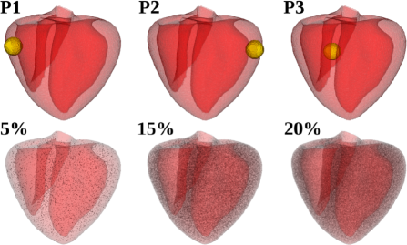
To model large-scale inhomogeneities, spheres of radius were
embedded in different positions of the simulated heart wall (, ,
and ), with the possibility of protrusion into the inner cavities, or out
of the exterior surface of the heart. Small-scale inhomogeneities were modeled
as randomly distributed cubical cells of side [23],
that contained node each ( , and by number).
To model conduction-type inhomogeneities, was set to
inside the inhomogeneity. To model ionic inhomogeneities, only the value of the
slow, inward conductance was set to [3]
at the sites covered by the inhomogeneity, without adjusting the elements of
the diffusion tensor. Figure 1 shows the positions and
distributions of inhomogeneities considered.
Results: No inhomogeneities: In the absence of inhomogeneities, we obtain a single stable periodically
rotating scroll, with an average frequency . We then study
scroll-wave dynamics in the presence of large- and small-scale conduction and
ionic inhomogeneities. Thus, in total, we consider different cases with
different inhomogeneities. Our main findings from these cases are listed
in Tables I & II. The details of our results are also discussed below with
figures to illustrate the most important types of dynamical behaviours.
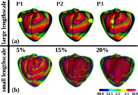
Conduction Inhomogeneities: Figure 2 shows the effects of various conduction heterogeneities
on scroll-wave dynamics (cases 1-2). We find that solitary, large-scale
conduction inhomogeneities do not have any pronounced effect on scroll-wave
dynamics. Indeed, at all positions of the inhomogeneity and ,
the scroll wave remains insensitive to the presence of the obstacle
(Figures 2 (a)). However, small-scale conduction inhomogeneities
affect scroll-wave dynamics substantially by changing the characteristics of
the scroll wave and causing its breakup. At all distributions of small-scale
conduction inhomogeneities that we have considered, namely, and
inhomogeneity (Case 2), we observe the following: (i) a
shortening of the spatial wavelength of the scroll, and (ii)
scroll-wave breakup at inhomogeneities 15% (Figure 2
(b)).
| Table I: Conduction inhomogeneity | ||
|---|---|---|
| Case no. | Inhomogeneity type | Dynamics |
| 1 | large () | Scroll wave remains passive |
| large () | towards the presence of the | |
| large () | inhomogeneity. | |
| 2 | small () | Scroll wavelength reduces. |
| small () | Scroll wavelength reduces. | |
| small () | Unstable breakup. | |
| small () | Stable breakup. | |
Ionic Inhomogeneities: Figure 3 illustrates the effects of various ionic
heterogeneities on scroll-wave dynamics (Cases 3-4). We see that solitary,
large-scale ionic inhomogeneities have a substantial effect on scroll-wave
dynamics. We observe interesting dynamical behaviour, such as, scroll-wave
breakup (Figure 3(a):P1) and anchoring P3)
(Case 3). On the contrary, small-scale ionic inhomogeneities do not lead to
qualitatively interesting dynamics: for all the inhomogeneities we have
considered, namely, and (Case 4)
(Figure 3 (b)) we do not find a pronounced change in
scroll-wave dynamics.
| Table II: Ionic inhomogeneity | ||
|---|---|---|
| Case no. | Inhomogeneity type | Dynamics |
| 3 | large () | Stable breakup. |
| large () | No change. | |
| large () | Stable anchoring. | |
| 4 | small () | No significant qualitative |
| small () | change. Dynamics is | |
| small () | insensitive to the presence | |
| small () | of the inhomogeneity. | |
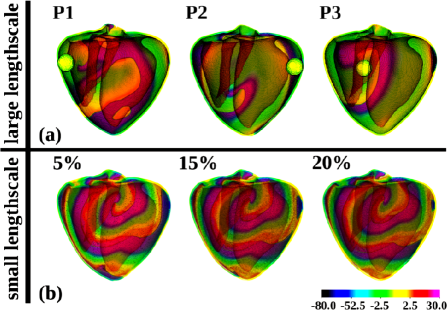
Taken together, our results demonstrate that large-scale conduction
inhomogeneities do not affect scroll-wave dynamics in the pig heart. However,
if the inhomogeneity is of ionic type, it can lead to scroll-wave breakup.
On the contrary, small-scale conduction inhomogeneities have a significant
influence on the dynamics of scroll waves; such inhomogeneities generally
lead to some decrease in the spatial wavelength of the scroll wave and
initiate scroll-wave breakup. Small-scale ionic inhomogeneities, however,
prove to be protective against breakup.
Discussion:We have carried out a comprehensive numerical study
of scroll-wave dynamics in an ionic model for pig cardiac tissue; and
we have compared, in the same conditions, the effects of conduction
and ionic heterogeneities, both for small and large length scales [3, 12, 13], on such scroll-wave dynamics.
Our principal, qualitative result that small-scale inhomogeneities are
important in the diffusion part is a consequence of the effect of the
diffusion processes on the reaction part (called the electrotonic effect
in electrophysiology) [26]. However, we have also found that
small-scale conduction inhomogeneities are not averaged out by the diffusion.
Therefore, their mean-field consideration, e.g., by using homogenization
techniques, should be done with caution. For large-scale heterogeneities,
our results are in line with findings for human cardiac-tissue
simulations [27]; however, these have been
performed on a completely different cardiac geometry, different cell models,
and for substantially different values of scroll wavelengths. In addition
to dynamical anchoring (via the transient-breakup phase) described
in Ref. [27] we have also observed anchoring of the other type
resulting from a drift of the scroll for qualitatively different positions of
the heterogeneity: in particular, we have placed heterogeneity inside the
septum and have found that it can attract scroll waves
and thus lead to interesting new dynamics.
The work of RM and RP was supported by DST, UGC, and CSIR (India). RM and RP would like to thank SERC (IISc) for computational resources. The work of AVP was supported by the Research Foundation-Flanders (FWO Vlaanderen).
References
- [1] Clayton RH, Bernus O, Cherry EM, Dierckx H, Fenton FH, et al. Prog Biophys Mol Biol. 104, 1–3:22–48 (2011).
- [2] Shajahan TK, Sinha S, and Pandit R, Phys. Rev. E. 75:011929-1 - 011929-8 (2007).
- [3] Shajahan TK, Nayak AR, and Pandit R, PLoS ONE. 4(3):e4738 (2009).
- [4] Jalife J, Gray RA, Morley GE, and Davidenko JM, Chaos. 8, 1:79-93 (1998).
- [5] Mann DL, Circ. Res. 91:988-998 (2002).
- [6] Kléber AG, Rudy Y, Physiol. Rev. 84:431-488 (2004).
- [7] Vigmond EJ, Weber dos Santos R, Prassl AJ, Deo M, Plank G, Prog Biophys Mol Biol. 96:3-18 (2008).
- [8] Ikeda T, Yashima M, Uchida T, Hough D, Fishbein MC, et al. Circ. Res. 81:753 (1997).
- [9] Valderr/’abano M, Kim YH, Yashima M, Wu TJ, Karagueuzian HS, et al. J Am Coll Cardiol. 36: 2000 (2000).
- [10] Lim ZY, Maskara B, Aguel F, Emokpae Jr. R, Tung L, Circulation. 114:2113-2121 (2006).
- [11] Davidenko JM, Pertsov AV, Salomonsz R, Baxter W, and Jalife J, Nature. 355:349 (1992).
- [12] Majumder R, Nayak AR, Pandit R, PLoS ONE. 6(4):e18052 (2011).
- [13] Majumder R, Nayak AR, Pandit R, PLoS ONE. 7(10):e45040 (2012).
- [14] Nayak AR, Shajahan TK, Panfilov AV, and Pandit R, PLoS ONE. 8(9):e72950 (2013).
- [15] Rudenko AN, Panfilov AV, Studia Biophysica. 98:183-188 (1983).
- [16] Panfilov AV, Vasiev BN, Physica D.49:107-113, (1991).
- [17] Qu Z, Weiss JN, and Garfinkel A, Am J Physiol Heart Circ Physiol. 276:269-283 (1999).
- [18] Stevens C, Remme E, LeGrice IJ, Hunter PJ, J Biomech. 36:737-748 (2003).
- [19] Luo CH, Rudy Y, Circ. Res. 68:1501-1526 (1991).
- [20] Panfilov AV, Heart Rhythm. 3(7):862-864 (2006).
- [21] Newton JC, Smith WM, Ideker RE, Circ. Res. 94:836-842 (2004).
- [22] Clayton RH, and Panfilov AV, Prog Biophys Mol Biol. 96:19-43 (2008).
- [23] Ten Tusscher KHWJ, Panfilov AV, Europace. 9:v138-v145, (2007).
- [24] Kléber A, Janse MJ, Wilms-Schopmann FJG, Wilde AAM, and Coronel R, Circulation 73(1):189-198 (1986).
- [25] Fenton FH, Cherry EM, Karma A, and Rappel WJ, Chaos 15, 013502 (2005).
- [26] Defauw A, Kazbanov IV, Dierckx H, Dawyndt P, Panfilov AV, PLoS One. 8(11):e79607/1-e79607/12, (2013).
- [27] Defauw A, Vandersickel N, Dawyndt P, Panfilov AV, Am J Physiol Heart Circ Physiol. 307:H1456-68, (2014).