11email: teppo. valtonen @ttl. fi,
POBox 40, 00250, Helsinki, Finland
A short review and primer on using video for psychophysiological observations in human-computer interaction applications
Abstract
The application of psychophysiological measures in human-computer interaction is a growing field with significant potential for future smart personalised systems. Working in this emerging field requires comprehension of an array of physiological signals and analysis techniques.
An important aspect in measuring psychophysiological variables in real-world settings is the invasiveness of the measurement setup. Video is a signal which can be captured from a distance without interrupting the subject. Furthermore, the advancements in camera technologies enable detecting a growing variety of psychophysiological phenomena from a video signal with an increasing accuracy.
This paper aims to serve as a primer for the novice, enabling rapid familiarisation with the latest core concepts. We put special emphasis on everyday human-computer interface applications to distinguish from the more common clinical or sports uses of psychophysiology.
This paper is an extract from a comprehensive review of the entire field of ambulatory psychophysiology, including 12 similar chapters, plus application guidelines and systematic review. Thus any citation should be made using the following reference:
B. Cowley, M. Filetti, K. Lukander, J. Torniainen, A. Henelius, L. Ahonen, O. Barral, I. Kosunen, T. Valtonen, M. Huotilainen, N. Ravaja, G. Jacucci. The Psychophysiology Primer: a guide to methods and a broad review with a focus on human-computer interaction. Foundations and Trends in Human-Computer Interaction, vol. 9, no. 3-4, pp. 150–307, 2016.
Keywords:
video, psychophysiology, human-computer interaction, primer, review, chapter1 Introduction
Advances in camera technologies make video an attractive possibility for measuring a variety of physiological phenomena, especially in environments such as the workplace, where contextual factors (e.g., ambient light) can be accounted for. Ever smaller and more accurate camera systems enable unobtrusive observations with sufficiently high precision for many interesting applications, and as long as there is a line of sight between the camera and the object of interest, a video signal may reveal, for example, a person’s cognitive state without interrupting the work that is being done.
Here we consider digital video-based systems aimed at assessing some aspect of physiology or behaviour from a distance, in order to augment human–computer interaction. We exclude the ocular system from discussion, since it is dealt with in Cowley et al. (2016).
2 Background
Typically, a video signal comprises measurements of the intensity of electromagnetic radiation in the spectra of visible (wavelengths of about 390 to 700 nm) and infrared (wavelengths from about 700 to 1000 nm) light, on a plane (for example, the image sensor in digital cameras). Changes in intensity arise mainly from a change in the original light source or the various points of reflection along the path of the ray of light from the source to the sensor. Accordingly, any movement within the measurement space (for instance, the eyebrows rising when the subject is surprised or expansion of the lungs when one is inhaling) or changes in the reflective properties of the reflection points (such as a change in skin colour due to increased blood flow) may be detected via the sensor. There is great variety in the video technologies and systems available today.
High-speed cameras
A typical video camera captures 24 to 30 frames per second, depending on the encoding. While this is sufficient to make a video stream seem smooth for the human visual system, systems with higher frame rates have been developed too. One of the fastest methods, known as compressed ultra-fast photography (CUP), can capture non-repetitive time-evolving events at up to 1011 frames per second (Gao et al., 2014). For many physiological phenomena, a frame rate on the magnitude of 100 frames per second is adequate, and 200 frames a second may already allow, for example, the use of video-based photoplethysmography in clinical settings (Sun et al., 2012a).
Webcams
The first system to feature a video camera that streamed an image in real time through a computer network came about in 1991 (Stafford-Fraser, 1995). The camera was pointed at a coffee pot in the Cambridge University Computer Lab. Since then, video cameras have become a basic feature in laptop computers and the screens of desktop computers, and they have been used mainly for video calls.
Cameras in hand-held devices
While cameras forming part of traditional computers are widespread, probably the most ubiquitous camera systems today are those embedded in hand-held devices, since almost all modern mobile phones and tablet computers have one or more cameras on their faces. The main camera typically points away from the user and is intended for photography. There is often another, however, intended for video calls and points in the same direction as the screen. In addition to conveying a video image to a caller, the front-facing camera can be used to detect, for example, whether or not there is a face in front of the screen. Once a face is detected, it may reveal various attributes of the user, such as emotional engagement as assessed from facial expressions (Kang et al., 2008). Also, however, as their name suggests, hand-held devices are often held in the user’s hand. It has been demonstrated that the optical sensor of a mobile phone can detect, for example, the following elements from touch: breathing rate, heart rate, blood oxygen saturation, and even atrial fibrillation or blood loss (Scully et al., 2012).
3D camera systems
Whereas a single-sensor camera system typically is limited to collecting emitted visual information on a two-dimensional plane, adding sensors and possibly projectors to the system may enable the observation of three-dimensional structures. A system with two appropriately placed cameras (i.e., a stereo camera system) functions in the same way as human binocular vision and can produce three-dimensional images. In another commonly used method, the system utilises projections of structured light patterns onto a three-dimensional surface, whereby the sensor’s detection of distortions in the patterns received may reveal the precise three-dimensional co-ordinates of the surface (Ma et al., 2009). A third method, one that is quite new, uses a time-of-flight (ToF) camera system, in which distances from the source of a light pulse to a camera via each point in the visual field can be resolved from the time of flight of each light pulse on the basis of the known speed of light (Gokturk et al., 2004). Systems of this type have been used, for example, to monitor sleep (Lee et al., 2015).
3 Methods
With the above foundations laid, we now describe methods that have been used to extract information about human psychophysiology from a video signal. While there are diverse methods, we concentrate on three main categories here: light intensity analysis, 2D morphological analysis, and 3D morphological analysis. All of these areas are showing rapid development, and some solutions are still experimental. For practical methods, therefore, more research might be required.
Light intensity analysis
is the basis for video signal analysis and enables most higher-level interpretations. Even on its own, however, simply detecting changes in the intensity of the light in a fixed area in a video image may illuminate interesting psychophysiological variables. For example, while a plethysmograph reveals changes in volume in a body, typically due to changes in the amount of blood or air contained in that part of the body, photoplethysmography is an optical technique that can be used to detect variations in the intensity of light reflected from tissue that arise from changes in blood flow (Allen, 2007). For an optimal result, various light intensity parameters should be considered. These depend on the application. For example, pulses in line with heart rate seem the most apparent in the green colour channel of a colour camera feed (Sun et al., 2012b).
2D morphological analysis
is the analysis of interesting areas or shapes in a 2D image, and it is based on the detection of edges between areas that differ in light intensity. For HCI purposes, the most interesting part of the body is the human face. Face detection and recognition are established research topics, and there are free tools available for these (for example, OpenCV FaceRecognizer111 See http://docs.opencv.org/2.4/modules/contrib/doc/facerec/facerec_tutorial.html0.). Samal and Iyengar (1992) provide a good description of the process, from face detection all the way to the analysis of facial expressions and the classification of faces. More recently, Zhao et al. (2003) undertook an extensive review of face recognition. In a recent, thorough review, Martinez and Valstar (2016) concentrates on automatic recognition of facial expressions.
3D morphological analysis
is a broad category of analysis methods that rely on different optical sensor systems producing data on 3D structures within a sensor’s field of view. For example, in addition to using intensity analysis, one can collect plethysmographic data from a distance by measuring the movement of a body in three-dimensional space with an optical sensor. Even consumer-grade 3D sensors used in gaming may be utilised to measure heart and respiration rate (Bernacchia et al., 2014).
4 Applications
Novel video technologies and methods of signal processing give rise to interesting applications for observing psychophysiological phenomena. Here we describe two of the most interesting video-based applications for HCI: photoplethysmography and the recognition of facial expressions.
Plethysmographic data can provide basic information on psychophysiology. More thorough description is given, for example, in Cowley et al. (2016). Here we consider using video cameras for PPG, an optical technique that can be utilised to detect changes in blood flow (Allen, 2007). For example, Sun and colleagues extracted a PPG signal from a video and analysed pulse rate variability (a possible surrogate measure for HRV; see, for example, Gil et al. (2010)) from the palm of the subject’s hand, using a monochrome CMOS camera running at 200 frames per second in 10-bit greyscale (Sun et al., 2012a). Tarassenko et al. (2014) measured both heart and respiration rate in a clinical set-up from a five-megapixel face video with eight bits per pixel, recorded at 12 frames per second. For broader applicability, even low-cost webcams have been demonstrated to function as photoplethysmographic sensors. In another of their studies, Sun et al. (2012b) compared a high-performance camera and a low-cost webcam in normal office lighting. They fixed a high-speed colour CMOS camera and a colour webcam in front of the face, along with a gold-standard pulse oximetry contact sensor on the index finger of the user’s left hand. To ensure that they were measuring light reflected from the skin, the authors manually determined the region of interest (ROI) in each frame of the video signals. For HR detection, they used the green colour channel to analyse changes in the average intensity of the pixels within the ROI, since, especially in the webcam signal, pulsations were most apparent in this particular channel. They concluded that both imaging PPG systems can successfully measure important physiological variables (in their case, HR).
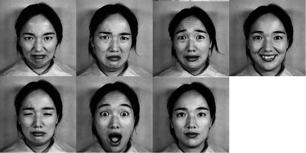
Mental state is high-level information, and identifying and conveying that information is gaining burgeoning interest in HCI research. Knowledge of the user’s mental state could augment not only user interfaces but also remote collaboration, telecommuting, and video conferencing. Facial expressions are an obvious signal as to the mental state, and the recognition of facial expressions is natural (and automatic) for humans; it is an important part of our communication. Both voluntary and involuntary facial actions convey, in particular, emotional information that is otherwise difficult to express – and difficult to conceal in face-to-face interaction (Ekman, 2003). Facial expressions can be categorised as reflecting six canonical emotions in addition to a neutral expression: anger, disgust, fear, happiness, surprise, and sadness (Dalgleish and Power, 1999). For an excellent review of automatic facial expression recognition, we recommend the work of Martinez and Valstar (2016). Traditionally, the process has begun with databases of portraits of actors mimicking the emotions (for example, as shown in Figure 1). A neutral expression is used as a reference for training the algorithm.
The core challenges with such a category-based approach are that the emotions each appear relatively rarely and that some expressions may differ in meaning on the basis of context. For example, someone might smile when embarrassed, not just when happy. Another method involves looking at the basic units of muscle activity in the human face, termed ‘action units’, in keeping with the Facial Action Coding System (FACS; see (Ekman and Friesen, 1976)). With the FACS approach, interpretation of mental state can be done at a later stage in the analysis pipeline, with the aid of additional information on the context. A third approach is to represent the mental state on two or more dimensions of affect, such as continua for arousal (ranging from relaxed to aroused) and valence (from pleasant to unpleasant). However, neither action units nor values on the affective dimensions are always detected with the current methods, especially in real-world settings.
5 Conclusion
The recent progress in video and signal processing methods renders video an interesting alternative to many traditional means of obtaining psychophysiological measurements, in areas such as plethysmography. In addition, video may enable new HCI applications, such as the remote and automatic identification of the mental state. Many of these methods are still in the early phases of development and require more research before they can achieve greater feasibility; however, there is already inexpensive hardware available, and, for many areas of study, excellent-quality free, open-source software tools and libraries exist. It is clear that the use of video in HCI is only just beginning.
References
- Allen [2007] John Allen. Photoplethysmography and its application in clinical physiological measurement. Physiological measurement, 28(3):R1, 2007.
- Bernacchia et al. [2014] Natascia Bernacchia, Lorenzo Scalise, Luigi Casacanditella, Ilaria Ercoli, Paolo Marchionni, and Enrico Primo Tomasini. Non contact measurement of heart and respiration rates based on kinect™. In Medical Measurements and Applications (MeMeA), 2014 IEEE International Symposium on, pages 1–5. IEEE, 2014.
- Cowley et al. [2016] Benjamin Cowley, Marco Filetti, Kristian Lukander, Jari Torniainen, Andreas Henelius, Lauri Ahonen, Oswald Barral, Ilkka Kosunen, Teppo Valtonen, Minna Huotilainen, Niklas Ravaja, and Giulio Jaccuci. The Psychophysiology Primer: a guide to methods and a broad review with a focus on human computer interaction. Foundations and Trends in HCI, 9(3-4):150–307, 2016.
- Dalgleish and Power [1999] Tim Dalgleish and Michael J Power. Handbook of cognition and emotion. Wiley Online Library, 1999.
- Ekman [2003] Paul Ekman. Darwin, deception, and facial expression. Annals of the New York Academy of Sciences, 1000(1):205–221, 2003.
- Ekman and Friesen [1976] Paul Ekman and Wallace V Friesen. Measuring facial movement. Environmental psychology and nonverbal behavior, 1(1):56–75, 1976.
- Gao et al. [2014] Liang Gao, Jinyang Liang, Chiye Li, and Lihong V Wang. Single-shot compressed ultrafast photography at one hundred billion frames per second. Nature, 516:74–77, 2014.
- Gil et al. [2010] E Gil, M Orini, R Bailón, J M Vergara, L Mainardi, and P Laguna. Photoplethysmography pulse rate variability as a surrogate measurement of heart rate variability during non-stationary conditions. Physiological Measurement, 31(9):1271, 2010.
- Gokturk et al. [2004] S.B. Gokturk, H. Yalcin, and C. Bamji. A time-of-flight depth sensor - system description, issues and solutions. In Computer Vision and Pattern Recognition Workshop, 2004. CVPRW ’04. Conference on, pages 35–35, June 2004.
- Kang et al. [2008] Sin-Hwa Kang, James H. Watt, and Sasi Kanth Ala. Social copresence in anonymous social interactions using a mobile video telephone. In Proceedings of the SIGCHI Conference on Human Factors in Computing Systems, CHI ’08, pages 1535–1544, New York, NY, USA, 2008. ACM. ISBN 978-1-60558-011-1.
- Lee et al. [2015] Jaehoon Lee, Min Hong, and Sungyong Ryu. Sleep monitoring system using kinect sensor. International Journal of Distributed Sensor Networks, 2015, 2015.
- Lyons et al. [1998] M. Lyons, S. Akamatsu, M. Kamachi, and J. Gyoba. Coding facial expressions with Gabor wavelets. In Proceedings Third IEEE International Conference on Automatic Face and Gesture Recognition, pages 200–205, Nara, Japan, 1998. IEEE Comput. Soc. ISBN 0-8186-8344-9.
- Ma et al. [2009] Lili Ma, Tianmin Xu, and Jiuxiang Lin. Validation of a three-dimensional facial scanning system based on structured light techniques. Computer Methods and Programs in Biomedicine, 94(3):290 – 298, 2009. ISSN 0169-2607.
- Martinez and Valstar [2016] Brais Martinez and Michel F Valstar. Advances, challenges, and opportunities in automatic facial expression recognition. In Advances in Face Detection and Facial Image Analysis, pages 63–100. Springer, 2016.
- Samal and Iyengar [1992] Ashok Samal and Prasana A Iyengar. Automatic recognition and analysis of human faces and facial expressions: A survey. Pattern recognition, 25(1):65–77, 1992.
- Scully et al. [2012] C. Scully, Jinseok Lee, J. Meyer, A.M. Gorbach, D. Granquist-Fraser, Y. Mendelson, and K.H. Chon. Physiological parameter monitoring from optical recordings with a mobile phone. Biomedical Engineering, IEEE Transactions on, 59(2):303–306, Feb. 2012. ISSN 0018-9294.
- Stafford-Fraser [1995] Quentin Stafford-Fraser. The trojan room coffee pot, 1995. [Online; accessed 19-July-2016].
- Sun et al. [2012a] Yu Sun, Sijung Hu, Vicente Azorin-Peris, Roy Kalawsky, and Stephen Greenwald. Noncontact imaging photoplethysmography to effectively access pulse rate variability. Journal of Biomedical Optics, 18(6):061205–061205, 2012a.
- Sun et al. [2012b] Yu Sun, Charlotte Papin, Vicente Azorin-Peris, Roy Kalawsky, Stephen Greenwald, and Sijung Hu. Use of ambient light in remote photoplethysmographic systems: Comparison between a high-performance camera and a low-cost webcam. Journal of Biomedical Optics, 17(3):037005–1–037005–10, 2012b.
- Tarassenko et al. [2014] L Tarassenko, M Villarroel, A Guazzi, J Jorge, D A Clifton, and C Pugh. Non-contact video-based vital sign monitoring using ambient light and auto-regressive models. Physiological Measurement, 35(5):807, 2014.
- Zhao et al. [2003] Wenyi Zhao, Rama Chellappa, P Jonathon Phillips, and Azriel Rosenfeld. Face recognition: A literature survey. ACM computing surveys (CSUR), 35(4):399–458, 2003.