Chimera-like in a neuronal network model of the cat brain
Abstract
Neuronal systems have been modelled by complex networks in different description levels. Recently, it has been verified that networks can simultaneously exhibit one coherent and other incoherent domain, known as chimera states. In this work, we study the existence of chimera-like states in a network considering the connectivity matrix based on the cat cerebral cortex. The cerebral cortex of the cat can be separated in 65 cortical areas organised into the four cognitive regions: visual, auditory, somatosensory-motor and frontolimbic. We consider a network where the local dynamics is given by the Hindmarsh-Rose model. The Hindmarsh-Rose equations are a well known model of neuronal activity that has been considered to simulate membrane potential in neuron. Here, we analyse under which conditions chimera-like states are present, as well as the affects induced by intensity of coupling on them. We identify two different kinds of chimera-like states: spiking chimera-like with desynchronised spikes, and bursting chimera-like with desynchronised bursts. Moreover, we find that chimera-like states with desynchronised bursts are more robust to neuronal noise than with desynchronised spikes.
keywords:
chimera-like , neuronal network , noisePACS:
05.45.Pq , 87.19.lj1 Introduction
The mammalian brain has neuronal mechanisms that give support to various anatomically and functionally distinct structures [1]. Mammals have the most complex brains of all vertebrates, which vary in size by a factor of [2]. Such a brain is arranged according to not only interacting elements on different levels, but also of different interconnections and functions [3]. For instance, the cat has approximately neurons in the brain and synapses [4, 5], while the human brain has approximately neurons and synapses [6].
One of the mammalian brain connectivity studies that has received considerable attention is the connectivity in the cat cerebral cortex [8, 7]. Scannell and Young [9] reported the connectional organisation of neuronal systems in the cat cerebral cortex. They arranged the cortex in four cognitive regions or connectional groups of areas: visual, auditory, somatosensory-motor, and frontolimbic. Regarding this realistic neuronal network, it was studied the relationship between structural and functional connectivity at different levels of synchronisation [10]. Lameu et al. analysed bursting synchronisation [11] and suppression of phase synchronisation [12] in network based on cat’s brain. They verified that the delayed feedback control can be an efficient method to have suppress synchronisation.
We focus on the existence of chimera-like states in a neuronal network model based on the cat cerebral cortex. The chimera states are spatiotemporal patterns in which coherent and incoherent domains mutually coexist [13, 14]. There are many studies about these patterns [15, 16]. Omel’chenko et al. observed chaotic motion of the chimera’s position along arrays of nonlocally coupled phase oscillators, where the chimera states have no artificially imposed symmetry [17]. Chimera and phase-cluster states in populations of coupled chemical oscillators were studied by Tinsley et al. [18]. Considering coupled Belousov-Zhabotinsky oscillators, they verified that chimera lifetime grows approximately exponentially with system size. There are also experimental investigations about chimera states, e.g., in [19] demonstrated the existence of these patterns in an open chain of electronic circuits with neuron-like spiking dynamics. In addition, Martens et al. [20] showed the appearance of chimera states in experiments with mechanical oscillators coupled in a hierarchical network.
Coexistence of states was reported in several animals that exhibit conflict between sleep and wakefulness, where one cerebral hemisphere sleeps and the other stays in an awake condition [21]. Recently, Andrzejak et al. [22] demonstrated analogies between chimera state collapses and epileptic seizures. In neuronal systems, chimera states were found in a network of coupled Hodgkin-Huxley equations [23], and also in the C. Elegans brain network by coupling Hindmarsh-Rose equations [24, 25].
We build a neuronal network according to the matrix of corticocortical connections in the cat [8, 9]. This topological connectivity is more complex than the matrix description from C. Elegans due to the fact that it is composed of cortical areas and axonal projections between them. In each cortical area we consider the neuron model proposed by Hindmarsh and Rose (HR) [26], and the axonal densities are considered as the connections between the areas. This neuronal network is able to reproduce EEG-like oscillations [27]. The HR neuron model can reproduce neuronal activities such as regular or chaotic spikes, and regular or chaotic bursting [28]. Baptista et al. [29] analysed a HR neuronal network on the rate of information and synchronous behaviour. Hizanidis et al. [30] observed chimera-like states in nonlocally coupled HR neuron models, where each neuron is connected with its nearest neighbours on both sides. In this work, we consider unidirectional connections both inside each connectional group of area (intra) and between groups of areas (inter). Unidirectional connections can be related to chemical synapses [31]. As a result, we verify the existence of two kinds of chimera-like: spiking chimera-like (SC) with desynchronised spikes, and bursting chimera-like (BC) with desynchronised bursts. Moreover, we include a neuronal noise by adding a random term in the external current. This way, we demonstrate that BC is more robust to noise than SC.
Firstly, we introduce the neuronal network described by a coupled HR neuronal model. Then, we discuss our results about the existence of two kinds of chimera-like states and noise robustness. Finally, we draw our conclusions.
2 Neuronal network
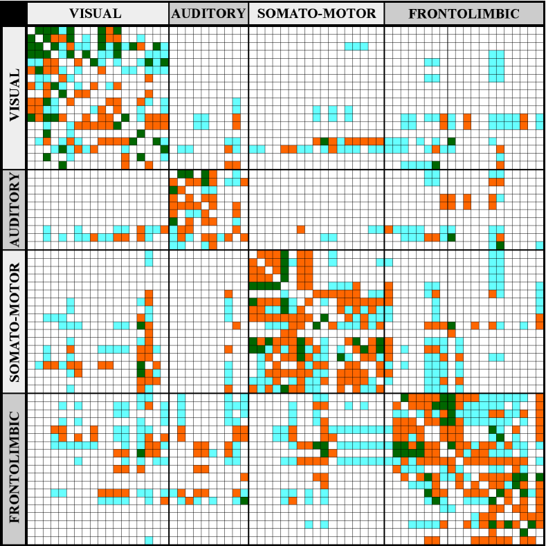
We consider a neuronal network composed for coupled HR according to the matrix that describes the corticocortical connectivity of the cat brain, obtained by Scannel et. al [8]. Fig. 1 shows the matrix of corticocortical connections in the cat brain with 1139 connections between 65 cortical areas which is organised into four cognitive regions: visual, auditory, somatosensory-motor and frontolimbic. In Ref. [8], the matrix was constructed by means of connections weighted , , , or , where corresponds to absent of connections (white), are sparse or weak (cyan), are intermediate (orange), and connections weighted are dense or strong (green).
The dynamic behaviour of the HR network if governed by the following equations
| (1) |
where , is the membrane potential, is related to the fast current (Na+ or K+), is associated with the slow current (Ca2+), controls the spiking frequency, corresponds to membrane input current (), is the reversal potential, and are sigmoidal function parameters, is responsible for the speed of variation of , governs adaptation, is the resting potential, is the intra connection strength, is the connection matrix of connections inside cortical areas (intra), is the inter connection strength, and is the connection matrix of connections between cortical areas (inter). We fix , , cortical areas, , , , , , and [25, 28]. The elements of the matrices and are (absent of connection), (weak), (intermediate), or (strong) according to the cat matrix (Fig. 1).
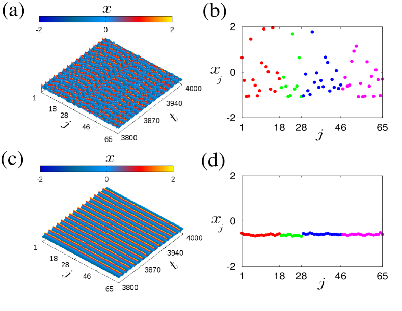
Fig. 2 shows space-time plots (left) and snapshot of the variable (right). We use, in the snapshot, a colour for each cortical areas: visual in red, auditory in green, somatosensory-motor in blue, and frontolimbic in magenta. In Fig. 2a for and , the network has a desynchronous behaviour, namely it is not possible to observe a synchronised firing pattern. Consequently, the dynamics is spatially incoherent, as shown in Fig. 2b. For and , there is a clear synchronised firing pattern (Fig. 2c) and the network displays a spatially coherent dynamics (Fig. 2d).
3 Chimera-Like states
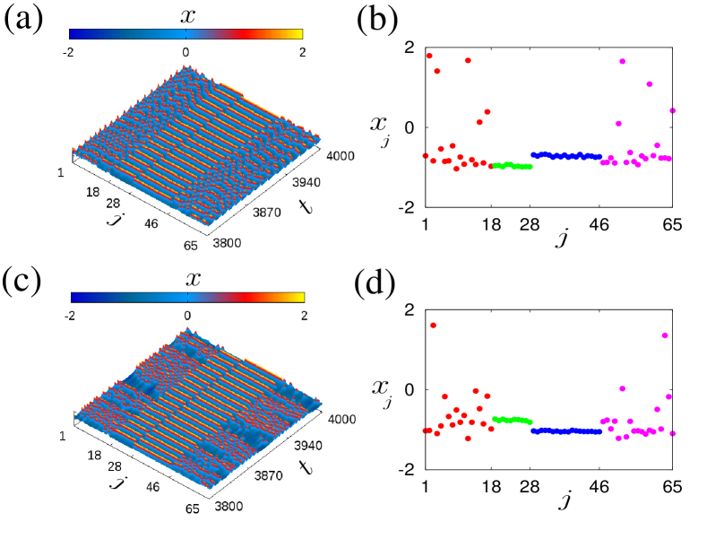
One fact that has been verified is the coexistence of coherence and incoherence structures in networks [34]. This phenomenon in spatiotemporal dynamical systems is so-called chimera states [13, 14]. Fig. 3 shows space-time plots (left) and snapshots (right) for different values of and in that it is possible to identify chimera-like states. In Figs. 3a and 3b for and , the auditory (green) and the somatosensory-motor area (blue) have synchronous behaviours, namely they exhibit spatially coherent dynamics, while the visual (red) and frontolimbic (magenta) areas are spatially incoherent with spikes patterns. This chimera-like is SC because the incoherent intervals are characterized by desynchronised spikes. For and (Figs. 3c and 3d), the auditory (green) and somatosensory-motor (blue) areas display synchronised patterns, whereas the other areas show incoherent structures. In this case, we verify BC, that is when desynchronised bursts are observed in the incoherent intervals. The transition between bursting and spiking resembles a continuous interior crisis [32, 33]. The interior crisis in the HR is a type of chaos-chaos transition, where it is observed changes of the size of the chaotic attractor due to collision between an unstable periodic orbit and a chaotic attractor.
In this work, we propose to use the recurrence plot as diagnostic tool to identify chimera-like in HR neurons coupled according to the cat matrix. To obtain this diagnostic, it is necessary to calculate the neuronal phase that is defined as
| (2) |
where is the firing time of the -th neuron. Then, the recurrence plot is given by [34]
| (3) |
where . Chimera-like states are found when there are blocks in the main diagonal and sparse points in the . We consider that chimera states occur when the sizes of the blocks are larger than of the total number of areas in each region. When there are only blocks and no sparse points, the network exhibits synchronized states (SI). Whereas, the incoherent states (IN) are identified when the blocks sizes are less than .
As a criterium to differentiate SC and BC we use the spiking time variance, that is given by
| (4) |
where , if the first spike occurs at time then the next one at time , and the symbol refers to the mean. For the network exhibits SC and for it presents BC.
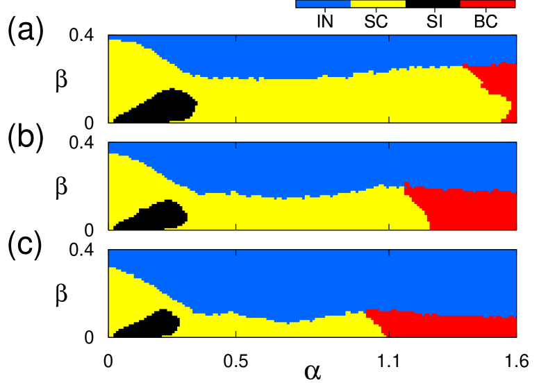
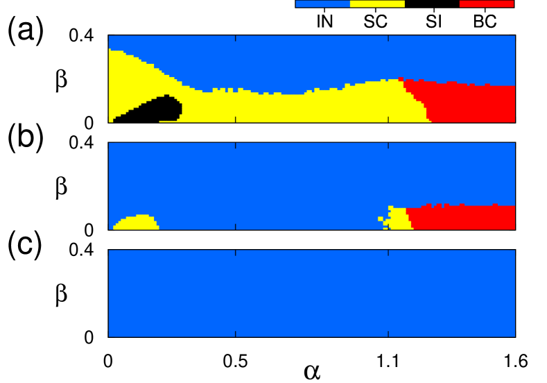
Figs. 4a, 4b and 4c show in the parameter space the regions for SC, BC, SI, and IN. In the parameter space , each point is computed by means of the average of initial conditions, where the initial conditions are randomly distributed in the intervals , and . In these intervals, the individual Hindmarsh-Rose neuron model exhibits spiking behaviour. This way we can identify the regions where chimera-like states appear by examining the recurrence plot. The yellow and red regions correspond to SC and BC, respectively, that are characterised by means of the spiking time variance. In the parameter space, the SC region is larger than the BC region. The black region exhibits SI and in the blue region we see IN. In our simulations, we observe that the IN region increases when decreases from to and, as a consequence, the SC and BC regions have a significant decrease in their sizes. In addition, for small values we verify that SC changes to BC.
4 Matching index
Some papers showed that the cat array has a high hierarchical level [10, 35]. This enables the formation of hierarchical synchronization [36, 37, 38]. We verify that the coherent regions in the chimera-like states are related to synchronisation between the neurons of the same connectional group of areas, mainly in auditory and somatosensory-motor.
Aiming to understand the formation of chimera-like states, we calculate the matching index (MI) of each two nodes and of the matrix showed in Fig. 1. The matching index of two nodes and is the overlap of their neighborhoods and is given by [39]
| (5) |
where is the adjacency matrix with elements equal to or according to whether and are connected or not. The MI is normalised dividing each element of the matrix by , where is the degree of the node . is equal to or if all inputs to and come from entirely different areas or and receive input completly from the same cortical areas, respectively.
Fig. 5 exhibits the normalised MI matrix from the anatomical connectivity. We see high MI values for the areas within the connectional group of visual, auditory, somatosensory-motor, and frontolimbic (internal MI). Areas in different group of areas (external) have heterogeneous MI values [10].
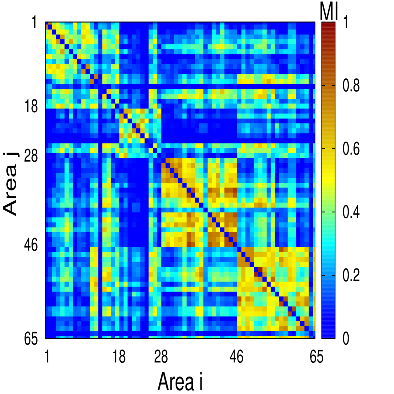
The average of the normalised MI is for the whole matrix. The internal MI values are , , , and for the connectional visual, auditory, somatosensory-motor, and frontolimbic group of areas, respectively. Furthermore, the auditory region has a higher value of MI () when it is only considered the areas between . In Refs. [10, 37] was showed that high values of MI can cause cluster synchronisation.
In our simulations, we verify that cluster synchronisation helps the appearance of coherent regions in the network, and as a consequence if incoherent regions mutually coexist, the network exhibits chimera-like states.
5 Neuronal noise
A relevant brain feature is noise, that can affect the transmission of signals and neuronal function [40]. There are many sources of noise in the brain, such as from genetic processes, thermal noise, ionic channel fluctuations, and synaptic events [41]. Serletis et al. [42] studied phase synchronisation of neuronal noise in mouse hippocampal. They reported from multi-spatial recordings that noise activity has a great influence on neurodynamic transitions in the healthy and epileptic brain.
We study the influence of neuronal noise on the chimera-like states in the cat brain. To do that, we consider an input current in Eq. 1 that is given by
| (6) |
where is the amplitude and is a normal distribution with mean and variance (Gaussian noise). For the isolated neuron exhibits spiking pattern. There are many studies that consider neurons under the influence of Gaussian noise [43]. Loos et al. [44] investigated chimera states under the influence of Gaussian noise in ring networks of Stuart-Landau oscillators. They showed that chimera death patterns persist under the impact of noise, and they also verified that the range of coupling parameters where chimera states occur is reduced with increasing noise amplitude.
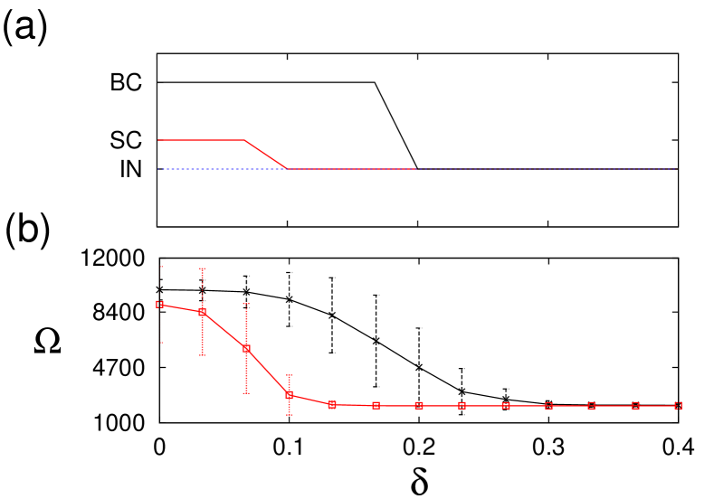
Fig. 6 shows the parameter spaces under an external current with noise (Eq. 6). For a small noise amplitude we do not observe significant alteration in the regions, as shown in Fig. 6a for . However, when is increased to it is possible to verify the destruction not only of synchronous behaviour, but also of chimera-like states (Fig. 6b). In Figs. 6c, where we consider , synchronisation and chimera-like states are completely suppressed.
We examine the robustness to noise of chimera-like states with spiking and bursting structures. To do that, we vary the amplitude and calculate the recurrence plot and the spiking variance. The results in Fig. 7 correspond to and into SC region (red line), and and into BC region (black line). In Fig. 7a, we see that for and the noise suppresses the SC and BC, respectively. Therefore, chimera-like states with desynchronised bursts are more robust to noise than with desynchronised spikes. We also calculate the average chimera-like lifetime , as shown in Fig. 7b, considering different initial conditions. The noise decreases the average chimera-like lifetime of both SC and BC.
6 Conclusions
We study a neuronal network composed by coupled HR model according to the connectivity matrix of the cat cerebral cortex. We consider unidirectional connections between neurons in the same area (intra) and between neurons in different areas (inter). This kind of connections can be associated with chemical synapses. The isolated HR neuron model has a regular spiking, but HR neurons in the network can exhibit bursting behaviour due to the coupling.
Varying the strength couplings, the network can exhibit synchronous and desynchronous behaviour. We also observe spatiotemporal patterns in that coherent and incoherent structures coexist, so-called chimera-like states. As diagnostic tool we propose to use the recurrence plot to identify chimera-like states in the neuronal network model of the cat brain. As a result, we verify that the network displays chimera-like states where the incoherent structures can be composed by desynchronised spikes or desynchronised bursts. SC occurs for small intra coupling strength, while BC appears for large intra coupling strength, and both for small inter coupling strength.
There are many sources of noise in the brain. With this in mind, we consider a noise in the external current to analyse effects on the chimera-like states. For small noise there is not significant changes in the parameter space () related to chimera-like, however, when the noise amplitude increases, chimera-like suppression is observed with the neuronal network exhibiting a desynchronous behaviour. In addition, we verify that BC is more robust to noise than SC, and the noise decreases the average chimera-like lifetime.
Acknowledgments
This study was possible by partial financial support from the following Brazilian government agencies: CNPq (154705/2016-0), CAPES, Fundação Araucária, and FAPESP (2016/16148-5, 2015/07311-7, and 2011/19296-1), and IRTG 1740/TRP 2011/50151-0 funded by the DFG/ FAPESP.
References
- [1] Barton RA, Harvey PH. Mosaic evolution of brain structure in mammals. Nature 2000;405:1055-1058.
- [2] Willemet R. Understanding the evolution of mammalian brain structures; the need for a (new) cerebrotype approach. Brain Sci 2012;2:203-224.
- [3] Zemanová L, Zhou C., Kurths J. Structural and functional clusters of complex brain networks. Physica D 2006;224:202-212.
- [4] Ananthanarayanan R, Esser SK, Simon HD, Modha DS. The cat is out of the bag: cortical simulations with 109 neurons and 1013 synapses. Proceedings IEEE/ACM Conference High Performance Networking Computing 2009;12:1-12.
- [5] Binzegger T, Douglas RJ, Martin KAC. A quantitative map of the circuit of cat primary visual cortex. J Neurosci 2004;29:8441-8453.
- [6] Williams RW, Herrup K. The control of neuron number. Ann Rev Neurosci 1988;11:423-453.
- [7] Beul SF, Grant S, Hilgetag CC. Brain Struct Funct 2015;220:3167-3184.
- [8] Scannell JW, Blakemore C, Young MP. J Neurosci 1995;15:1463-1483.
- [9] Scannell JW, Young MP. Curr Biol 1993;3:191-200.
- [10] Zhou C, Zemanová L, Zamora-López G, Hilgetag CC, Kurths J. New J Phys 2007;9:178.
- [11] Lameu EL, Borges FS, Borges RR, Batista AM, Baptista MS, Viana RL. Commun Nonlinear Sci Numer Simul 2016;34:45-54.
- [12] Lameu EL, Borges FS, Borges RR, Iarosz KC, Caldas IL, Batista AM, Viana RL, Kurths J. Chaos 2016;26:043107.
- [13] Kuramoto Y, Battogtokh D. Nonl Phen Compl Sys 2002;5:380-385.
- [14] Abrams DM, Strogatz SH. Phys Rev Lett 2004;93:174102.
- [15] Abrams DM, Mirollo R, Strogatz SH, Wiley DA, Phys Rev Lett 2008;101:084103.
- [16] Ujjwal SR, Punetha N, Ramaswamy R. . Phys Rev E 2016;93:012207.
- [17] Omel’chenko OE, Wolfrum M, Maistrenko YL. Phys Rev E 2010;81:065201.
- [18] Tinsley MR, Nkomo S, Showalter K. Nature Phys 2012;8:662-665.
- [19] Gambuzza LV, Buscarino A, Chessari S, Fortuna L, Meucci R, Frasca M. Phys Rev E 2014;90:032905.
- [20] Martens EA, Thutupalli S, Fourrière A, Hallatschek O. Proc Natl Acad Sci USA 2013;110:10563-10567.
- [21] Rattenborg NC, Amlaner CJ, Lima SL. Neurosci Biobehav Rev 2000;24:817-842.
- [22] Andrzejak RG, Rummel C, Mormann F, Schindler K. Sci Rep 2016;6:23000.
- [23] Sakaguchi H. Phys Rev E 2006;73:031907.
- [24] Hizanidis J, Kouvaris NE, Antonopoulos CG. Cybern Phys 2015;4:17-20.
- [25] Hizanidis J, Kouvaris NE, Zamora-López G, Díaz-Guilera A, Antonopoulos CG. Sci Rep. 2016;6:19845.
- [26] HINDMARSH J. L. and ROSE R. M., Proc. Roy. Soc. B, 221 (1984) 87.
- [27] SCHMIDT G., ZAMORA-LÓPEZ G. and KURTHS J., Int. J. Bifurcat. Chaos, 20 (2010) 859.
- [28] STORACE M., LINARO D. and LANGE D., Chaos, 18 (2008) 033128.
- [29] BAPTISTA M. S., KAKMENI MOUKAM F. M. and GREBOGI C., Phys. Rev. E, 82 (2010) 036203.
- [30] HIZANIDIZ J., KANAS V. G., BEZERIANOS A. and BOUNTIS T., Int. J. Bifurcat. Chaos, 24 (2014) 1450030.
- [31] ECCLES J. C., Ann. Rev. Neurosci., 5 (1982) 325.
- [32] GONZÁLEZ-MIRANDA J. M., Chaos, 13 (2003) 845.
- [33] INNOCENTI G., MORELLI A., GENESIO R. and TORCINI A., Chaos, 17 (2007) 043128.
- [34] SANTOS M. S., SZEZECH Jr J. D., BATISTA A. M., CALDAS I. L., VIANA R. L. and LOPES S. R., Phys. Lett. A, 379 (2015) 2188.
- [35] SPORNS O., HONEY C. J. and KÖTTER R., PLoS ONE, 2(10) (2007) e1049.
- [36] ZAMORA-LÓPEZ G., ZHOU C. and KURTHS J., Front. Neuroinform., 4 (2010) 1.
- [37] GOÓMEZ-GARDEÑES J., ZAMORA-LÓPES G., MORENO Y. and ARENAS A., PLoS ONE, 5(8) (2010) e12313.
- [38] STROUD J., BARAHONA M. and PEREIRA, T. Dynamics of Cluster Synchronisation in Modular Networks: Implications for Structural and Functional Networks in Applications of Chaos and Nonlinear Dynamics in Science and Engineering - Vol. 4, (Cham: Springer International Publishing) 2015.
- [39] HILGETAG C. C., KÖTTER R, STEPHAN K. E. and SPORNS O. Computational Neuroanatomy, (Totowa, NJ: Humana Press) 2002.
- [40] FAISAL A. A., SELEN L. P. J. and WOLPERT D. M., Nat. Rev. Neurosci., 9 (2008) 292.
- [41] DESTEXHE A. and RUDOLPH-LILITH M., Neuronal Noise, (Springer Series in Computational Neuroscience, New York) 2012.
- [42] SERLETICS D., CARLEN P. L., VALIANTE T. A. and BARDAKJIAN B. L., Int. J. Neural Syst., 23 (2013) 1250033.
- [43] LINDNER B., LONGTIN A. and BULSARA A., Neural Comput., 15 (2003) 1761.
- [44] LOOS S. A. M., CLAUSSEN J. C., SCHÖLL E. and ZAKHAROVA A., Phys. Rev. E, 93 (2016) 012209.
- [45] BARAHONA M. and PECORA L. M., Phys. Rev. Lett., 89 (2002) 054101.