*.pdf,.PDF
Recent advances in coarse-grained modeling of virus assembly
Abstract
In many virus families, tens to thousands of proteins assemble spontaneously into a capsid (protein shell) while packaging the genomic nucleic acid. This review summarizes recent advances in computational modeling of these dynamical processes. We present an overview of recent technological and algorithmic developments, which are enabling simulations to describe the large ranges of length-and time-scales relevant to assembly, under conditions more closely matched to experiments than in earlier work. We then describe two examples in which computational modeling has recently provided an important complement to experiments.
Capsid assembly and packaging of the genome are essential steps in the formation of an infectious virus. Thus, elucidating the mechanisms by which assembly proceeds could identify important targets for antiviral drugs and would advance our fundamental understanding of the viral lifecycle. However, assembly and packaging pathways remain incompletely understood for many viruses because most intermediates are transient and therefore undetectable or characterized only with low resolution. Computer simulations of virus assembly have overcome this limitation by revealing features of assembly processes that are not accessible to experiments alone. However, the large size of a virus (15-1000 nm) and the timescales required for assembly (ms-hours) prohibit simulating capsid formation with atomic-resolution, except for specific steps Jiang2015 . To this end, researchers have relied on simplified models, which aim to coarse-grain over atomic-scale details while accurately describing the essential physical features that control assembly.
This review describes recent advances in coarse-grained models of capsid assembly. We begin with a brief overview of models and simulation methodologies, followed by recent applications of these approaches. To accommodate space limitations, we limit our discussion of applications to two areas which have recently been the subject of intense modeling activity: the role of nucleic acids in the assembly of icosahedral viruses, and assembly of the mature HIV capsid.
Coarse-grained models for capsid assembly
One approach to model development seeks to describe a specific physical system with the greatest accuracy allowed by computational constraints, and by systematically coarse-graining from atomistic simulations (e.g. Davtyan2015 ; Dama2013 ). However, the conformational dynamics of capsid proteins restricts the accuracy of such techniques, and the complexity of the resulting coarse-grained models has limited their application to assembly. Therefore, capsid assembly models have relied on a combination of atomistic simulations, structural data, and fitting model parameters to kinetics and thermodynamic data. Often, the aim has been to construct the simplest model consistent with experimental data, to discover general, fundamental insights about capsid assembly.
Models for virus assembly can be separated into three classes. In the first, the time evolution of concentrations of capsid intermediates is represented by a system of rate equations Zlotnick1999 ; Endres2002 . Formulation of the model requires specifying the state space (the set of all possible assembly intermediates) and transition rates between each pair of intermediates. The rate equations can be numerically integrated Zlotnick1999 ; Endres2002 ; Moisant2010 ; Zandi2006 ; Schoot2007 , or trajectories consistent with the rate equations can be stochastically sampled using Gillespie-type algorithms Gillespie1977 ; Dykeman2013a ; Zhang2006a ; Xie2012 ; Smith2014 ; Dykeman2014 , and transition rates can be fit against experimental data Kumar2010 ; Xie2012 ; Zlotnick1999 ; Tsiang2012 . Despite their simplicity, such models reproduce many experimental observations on capsid assembly.
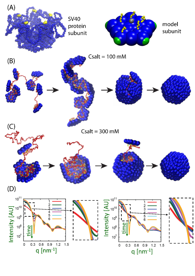
In the next class of models (particle-based simulations), subunits interact through pair potentials that drive assembly toward an ordered low-energy structure (e.g. an icosahedral shell Schwartz1998 ; Hagan2006 ; Nguyen2007 ; Rapaport1999 ; Rapaport2012 ; Baschek2012 ; Perlmutter2014 ; Perlmutter2015b ; Boettcher2015 , Fig. 1). Subunit motions are explicitly tracked by numerically integrating equations of motion (e.g. using molecular dynamics, Brownian dynamics (BD), or discontinuous molecular dynamics Rapaport2012 ; Perlmutter2014 ; Nguyen2007 . The third approach combines aspects of Gillespie-type and particle-based models, modeling assembly through the irreversible addition of triangular subunits to growing edges of an incomplete shell Hicks2006 ; Levandovsky2009 ; Wagner2015a . The shell is treated as an elastic sheet that relaxes to its minimum energy configuration after each accretion.
Of these approaches, particle-based simulations enable (at least in principle) the fewest assumptions about intermediate geometries and the highest resolution description of proteins. However, their high computational cost has limited model resolution, simulation timescales, and system sizes Hagan2014 ; Perilla2015 .
Recent algorithmic advances are beginning to overcome this limitation. For example, Grime and Voth Grime2014 designed an efficient parallelization scheme for spatially heterogeneous particle concentrations (which occur during assembly simulations with implicit solvent). Algorithms performing rigid body dynamics on GPUs show significant speedup in comparison to conventional CPUs. For example, the package HOOMD Nguyen2011 has been used to simulate virus assembly around nucleic acids and on membranes Perlmutter2013 ; Perlmutter2014 ; Perlmutter2015b ; Ruiz-Herrero2015 . Zuckerman and coworkers Spiriti2015 have shown that calculation of interparticle potentials between rigid bodies containing many interaction sites (such as high resolution models of proteins) can be speeded up by tabulation in distance and orientation space. Finally, enhanced sampling methods can focus computational time on critical but rare events, such as crossing nucleation barriers. Methods well-suited for the diverse pathways typical of assembly systems include multiple state transition path sampling Newton2015 , Markov state models Perkett2014 , weighted ensemble dynamics Spiriti2015 , and diffusion maps Long2014 .
Applications of coarse-grained modeling
There have been extensive applications of coarse-grained models to understand the assembly of small icosahedral capsids (reviewed in Hagan2014 ). Initial studies focused on modeling in vitro experiments in which pure proteins assemble into empty capsids. Recent works have included effects of crowding Smith2014 , assembly on membranes Matthews2013a ; Ruiz-Herrero2015 , and how assembly changes when subunits are generated during the course of a reaction, either by protein translation or advective transport Hagan2011 ; Boettcher2015 ; Castelnovo2014 . As noted above, we focus here on the role of nucleic acids (NAs) and other negatively charged cargos in the assembly of icosahedral viruses, and assembly of the mature HIV shell.
Assembly around nucleic acids and other cargoes. While there are several mechanisms of genome packaging Chelikani2014 , in many virus families with single-stranded (ss) RNA or DNA genomes, the capsid assembles spontaneously around the genomic NA. Electrostatics provides an important driving force for assembly around NAs due to the presence of positive charges, located on the inner surface of capsid proteins or on flexible terminal domains that dangle into the capsid interior.
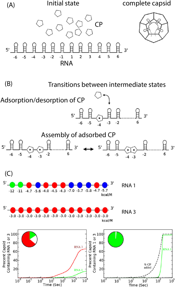
The dynamics of assembly around a NA (or other cargo) has been modeled by Gillespie-type simulations which implicitly build the NA into intermediate states Dykeman2013a ; Dykeman2014 (Fig. 2), and with particle-based simulations in which coarse-grained subunits assemble around flexible polyelectrolytes Perlmutter2013 ; Perlmutter2014 ; Perlmutter2015b ; Zhang2013 ; Zhang2013a , semiflexible polyelectrolytes Zhang2013a , or model NAs Perlmutter2013 . The latter simulations find that assembly around a cargo can proceed through two different mechanisms, see Fig. 1.
Most viruses with ssRNA or ssDNA genomes are overcharged, meaning that the negative charge on the encapsidated RNA significantly exceeds the positive charge on the interior of the capsid Belyi2006 ; Hu2008d . To explain this observation, the length of RNA which optimizes capsid thermostability has been investigated using scaling methods, continuum-models and BD simulations (e.g. Belyi2006 ; Zandi2009 ; Erdemci-Tandogan2014 ; Schoot2013 ; Perlmutter2013 ). These calculations suggest that overcharging arises because only a fraction of RNA charges can reside within the electrostatic screening distance of capsid charges, and that intra-molecular RNA base-pairing increases overcharging Perlmutter2013 ; Erdemci-Tandogan2014 ; Schoot2013 .
Despite the ability of nonspecific electrostatics to promote assembly around heterologous RNA, viruses package their genomic RNA with remarkable selectivity in vivo (e.g. 99% Routh2012 ). Several factors have been proposed to explain selective packaging. Experiments and simulations suggest that the physical features of viral RNAs (e.g. charge, and size due to tertiary structure) are optimized for assembly of their capsid Tubiana2015 ; Garmann2015 ; Perlmutter2013 ; Singaram2015 ; Comas-Garcia2012 ; Erdemci-Tandogan2014 ; Gopal2014 . Secondly, interactions between capsid proteins and specific sequences within the genome called packaging sites (PSs) have been identified for a number of viruses (e.g. Rao2006 ; Stockley2013a ; Dykeman2011 ). Experiments find that viral genomes contain many PSs (of order 30-60) with binding affinities to capsid proteins ranging from nanomolar to micromolar Stockley2013a .
Gillespie-type Dykeman2013a ; Dykeman2014 and BD simulations Perlmutter2015b of assembly around RNAs containing PSs predicted preferential packaging over uniform RNAs (without PSs) for certain parameter ranges, but predicted poor selectivity under conditions which are optimal for assembly around uniform RNAs. However, selectivity was significantly enhanced when considering the steadily increasing concentration of capsid proteins which occurs within a bacterial host (Fig. 2) Dykeman2014 .
Knowledge of the locations of PSs within genomes and models for how PS binding couples to the capsid geometry have been used as constraints for analyzing electron tomography data of RNA within the MS2 capsids Geraets2015 . The analysis suggested that the encapsidated RNA is highly organized, with similar conformations in most viruses. A tomography study of HBV virions led to a similar conclusion (e.g. Wang2014 ).
Additional factors have been proposed to enable selective RNA packaging in vivo, including subcellular localization of viral components and coordinated translation and assembly (reviewed in Rao2014 ). These factors have yet to be incorporated into assembly models.
Assembly of HIV. In contrast to the viruses described above, the capsid of the human immunodeficiency virus (HIV) lacks icosahedral symmetry. The virus initially assembles as an ‘immature’ spherical particle, constructed of a disordered lattice of uncleaved Gag proteins and surrounded by a lipid bilayer. The latter is derived from the plasma membrane of the host cell during budding (exiting) of the virus from the cell. Upon maturation, Gag polyproteins are cleaved into three distinct portions, the matrix (MA), capsid (CA), and nucleocapsid (NC) proteins, which constitute different components of the fully infectious virions Sundquist2012 ; Ganser-Pornillos2008 . The MA proteins are bound to the interface of the bilayer envelope, forming an outer shell. Inside, about 1500 of the CA proteins assemble into an unusual shell around a condensed complex of the RNA and NC proteins Sundquist2012 . In HIV-1, the CA shell forms as a “fullerene cone”: a hexagonal lattice containing 12 pentamers, usually with 5 pentamers at the cone tip and 7 at the base Ganser1999 . Molecular dynamics flexible fitting (MDFF), a technique in which MD is performed using constraints based on cryo-EM data, recently enabled atomic-resolution models for structures of the mature HIV capsid Zhao2013 . All-atom MD simulations on the resulting structure were used to examine contacts between hexamers, pentamers and various assembly units, which showed that the presence of pentamers gives rise to closer trimer contacts and higher surface curvature.
Despite cryo-electron tomographic studies Briggs2006 ; Sundquist2012 ; Woodward2015 , the structural details of the immature particle and the maturation pathway remain unknown. Two models have been proposed for how HIV CA proteins assemble into the mature conical shells. In the “displacive” model, the CA lattice from the immature shell does not completely disassemble, but undergoes conformational changes to build the mature capsid Frank2015 ; Meng2012 ; Keller2013 . In the other model, the immature spherical shell completely disassembles after Gag cleavage, followed by “de novo” reassembly of CA into the cone. Even among researchers who favor the de novo model, there is currently no consensus on the order of assembly of the cone. According to Briggs et al. Briggs2006 , the capsid nucleates from a narrow tip and then grows until hitting the opposite side of the enclosing membrane, at which point the cone base forms. Alternatively, the experiments of Benjamin et al. Benjamin2005 reveal a small hole (defect) on cone tips, suggesting an opposite pathway, in which the cone base assembles first, followed by formation of the body and then the narrow tip.
While there have been no modeling studies corresponding to the displacive model, several coarse-grained models have been employed to study the de novo assembly of conical capsids. Since experiments show that the CA protein can assemble to form conical and cylindrical shells in vitro in the absence of genome Ganser1999 ; Meng2012 ; Zhao2013 , thus far simulations have focused on assembly of conical shells in the absence of a membrane or genome. As the curvature of cone varies constantly along its axis of symmetry, a question naturally arises: how do the proteins adjust to sit in very different environments, and what are the asssembly pathways?
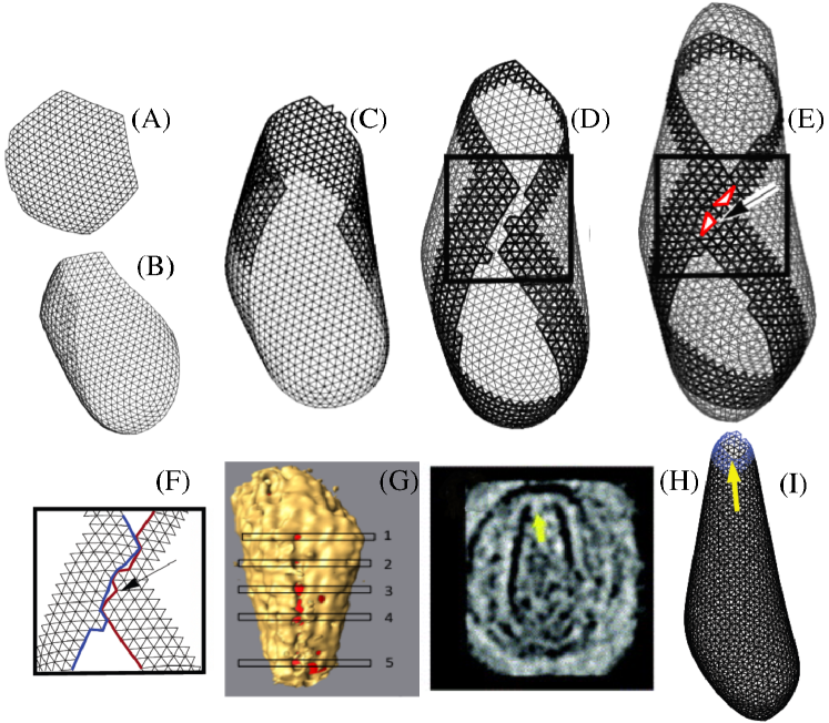
The growth of retrovirus shells was studied using the growing elastic sheet model described above Hicks2006 . While most of their simulated shells had irregular structures, addition of an attractive interaction between nearby edges of the growing shell (mimicking hydrophobic interactions) led to conical shells for large values of the Foppl von Karman number (meaning that the protein shell ˜resists stretching more strongly than bending) Levandovsky2009 ; Yu2013 . These simulations also explained the ‘seams’ observed in EM structures of mature capsids. Combining the model with experiments suggested that the HIV capsid can start to grow as a hexagonal lattice that eventually forms the body of the shell, with the tip and base of the cone closing toward the end of the assembly (Fig. 3) Yu2013 .
Several particle-based models for CA dimers have been developed, in which subunit shapes and short-ranged interactions between specific sites on subunits were based on solution NMR structures or all-atom MD simulations Qiao2015 ; Grime2014 ; Zhao2013 . Because solution NMR studies identify flexibility between the N-terminal and C-terminal domains within a dimer, simulations included flexible dimers Grime2012 or ensembles of dimers with different configurations based on an all-atom MD simulation Qiao2015 . Simulations of the assembly process (reaching up to 20-40 % of a complete capsid) found that associations between trimer-of-dimers intermediates play a key role in the assembly process. Relatedly, fitting of a rate equation model against in vitro experiments in which CA proteins assemble into tubes identified a trimer-of-dimers as the critical nucleus Tsiang2012 .
Outlook
The coarse-grained models presented in this review have elucidated key aspects of the virus formation process, which are not accessible to experiments or all-atom simulations. Looking ahead, an important area which is only beginning to be addressed by coarse-grained models is how the environment of a host cell contributes to viral assembly. In conjunction with quantitative experiments studying the effects of specific host factors, such models can advance our understanding of how viruses propagate, and pave the way for discovering new approaches to combat viral infections.
Acknowledgements
The acknowledge support from NIH grant R01GM108021 (MFH), the NSF Brandeis MRSEC, DMR-1420382 (MFH) and NSF grant DMR-1310687 (RZ).
References
-
(1)
J. J. Jiang, J. Yang, Y. V. Sereda, P. J. Ortoleva, Early stage p22 viral capsid self-assembly
mediated by scaffolding protein: Atom-resolved model and molecular dynamics
simulation, J. Phys. Chem. B 119 (16) (2015) 5156–5162.
URL <GotoISI>://WOS:000353604800006 -
(2)
A. Davtyan, J. F. Dama, G. A. Voth, H. C. Andersen,
Dynamic
force matching: A method for constructing dynamical coarse-grained models
with realistic time dependence, J. Chem. Phys. 142 (15) (2015) 154104.
doi:doi:http://dx.doi.org/10.1063/1.4917454.
URL http://scitation.aip.org/content/aip/journal/jcp/142/15/10.1063/1.4917454 -
(3)
J. F. Dama, A. V. Sinitskiy, M. McCullagh, J. Weare, B. Roux, A. R. Dinner,
G. A. Voth, The theory of
ultra-coarse-graining. 1. general principles, J. Chem. Theory Comput. 9 (5)
(2013) 2466–2480.
doi:10.1021/ct4000444.
URL http://dx.doi.org/10.1021/ct4000444 - (4) A. Zlotnick, J. M. Johnson, P. W. Wingfield, S. J. Stahl, D. Endres, A theoretical model successfully identifies features of hepatitis B virus capsid assembly, Biochemistry 38 (44) (1999) 14644–14652.
- (5) D. Endres, A. Zlotnick, Model-based analysis of assembly kinetics for virus capsids or other spherical polymers, Biophys. J. 83 (2) (2002) 1217–1230, * A comprehensive presentation of the rate equation approach to describing assembly of polyhedral shells.
- (6) P. Moisant, H. Neeman, A. Zlotnick, Exploring the Paths of (Virus) Assembly, Biophys. J. 99 (5) (2010) 1350–1357.
- (7) R. Zandi, P. van der Schoot, D. Reguera, W. Kegel, H. Reiss, Classical nucleation theory of virus capsids, Biophys. J. 90 (6) (2006) 1939–1948.
- (8) P. van der Schoot, R. Zandi, Kinetic theory of virus capsid assembly, Phys. Biol. 4 (4) (2007) 296–304.
- (9) D. T. Gillespie, Exact Stochastic Simulation of Coupled Chemical Reactions, J. Phys. Chem. 81 (25) (1977) 2340–2361.
- (10) E. C. Dykeman, P. G. Stockley, R. Twarock, Building a viral capsid in the presence of genomic RNA, Phys Rev E 87 (2) (2013) 022717. doi:10.1103/PhysRevE.87.022717.
- (11) T. Q. Zhang, R. Schwartz, Simulation study of the contribution of oligomer/oligomer binding to capsid assembly kinetics, Biophys. J. 90 (1) (2006) 57–64.
- (12) L. Xie, G. Smith, X. Feng, R. Schwartz, Surveying Capsid Assembly Pathways through Simulation-Based Data Fitting, Biophys. J. 103 (7) (2012) 1545–1554.
- (13) G. R. Smith, L. Xie, B. Lee, R. Schwartz, Applying Molecular Crowding Models to Simulations of Virus Capsid Assembly In Vitro, Biophys. J. 106 (1) (2014) 310–320.
- (14) E. C. Dykeman, P. G. Stockley, R. Twarock, Solving a Levinthal’s paradox for virus assembly identifies a unique antiviral strategy, Proc. Natl. Acad. Sci. U. S. A. 111 (14) (2014) 5361–5366, **This article shows that a time-varying protein concentration, such as occurs during some viral infections, can dramatically enhance specificity for viral RNA. doi:10.1073/pnas.1319479111.
- (15) M. S. Kumar, R. Schwartz, A parameter estimation technique for stochastic self-assembly systems and its application to human papillomavirus self-assembly, Phys. Biol. 7 (4) (2010) 045005.
- (16) M. Tsiang, A. Niedziela-Majka, M. Hung, D. Jin, E. Hu, S. Yant, D. Samuel, X. Liu, R. Sakowicz, A Trimer of Dimers Is the Basic Building Block for Human Immunodeficiency Virus-1 Capsid Assembly, Biochemistry 51 (22) (2012) 4416–4428.
- (17) R. Schwartz, P. W. Shor, P. E. Prevelige, B. Berger, Local rules simulation of the kinetics of virus capsid self-assembly, Biophys. J. 75 (6) (1998) 2626–2636.
- (18) M. F. Hagan, D. Chandler, Dynamic pathways for viral capsid assembly, Biophys. J. 91 (1) (2006) 42–54.
- (19) H. D. Nguyen, V. S. Reddy, C. L. Brooks, Deciphering the kinetic mechanism of spontaneous self-assembly of icosahedral capsids, Nano Lett. 7 (2) (2007) 338–344.
- (20) D. C. Rapaport, J. E. Johnson, J. Skolnick, Supramolecular self-assembly: molecular dynamics modeling of polyhedral shell formation, Comput. Phys. Commun. 122 (1999) 231–235.
-
(21)
D. C. Rapaport,
Molecular dynamics
simulation of reversibly self-assembling shells in solution using trapezoidal
particles, Phys. Rev. E 86 (2012) 051917.
doi:10.1103/PhysRevE.86.051917.
URL http://link.aps.org/doi/10.1103/PhysRevE.86.051917 -
(22)
J. E. Baschek, H. C. R. Klein, U. S. Schwarz, Stochastic dynamics of virus capsid formation:
direct versus hierarchical self-assembly, Bmc Biophysics 5, baschek, Johanna
E. Klein, Heinrich C. R. Schwarz, Ulrich S. Schwarz, Ulrich/K-4111-2014
Schwarz, Ulrich/0000-0003-1483-640X.
doi:10.1186/2046-1682-5-22.
URL <GotoISI>://WOS:000314583100001 - (23) J. D. Perlmutter, M. R. Perkett, M. F. Hagan, Pathways for virus assembly around nucleic acids, J. Mol. Biol. * Simulations demonstrate that capsid assembly around RNA can proceed by two different mechanisms, which can be tuned by solution conditions and capsid protein sequence, and can be experimentally distinguished. doi:10.1016/j.jmb.2014.07.004.
- (24) J. D. Perlmutter, M. F. Hagan, The Role of Packaging Sites in Efficient and Specific Virus Assembly., J. Mol. Biol.doi:10.1016/j.jmb.2015.05.008.
-
(25)
M. A. Boettcher, H. C. R. Klein, U. S. Schwarz, Role of dynamic capsomere supply for viral capsid
self-assembly, Phys. Biol. 12 (1).
doi:10.1088/1478-3975/12/1/016014.
URL <GotoISI>://WOS:000349948000014 - (26) S. D. Hicks, C. L. Henley, Irreversible growth model for virus capsid assembly, Phys. Rev. E 74 (3) (2006) 031912.
- (27) A. Levandovsky, R. Zandi, Nonequilibirum assembly, retroviruses, and conical structures, Phys. Rev. Lett. 102 (19) (2009) 198102–198102.
- (28) J. Wagner, R. Zandi, The robust assembly of small symmetric nanoshells, Biophys. J. 109 (2015) 956.
- (29) M. F. Hagan, Modeling Viral Capsid Assembly, Adv. Chem. Phys. 155 (2014) 1–68.
- (30) J. R. Perilla, B. C. Goh, C. K. Cassidy, B. Liu, R. C. Bernardi, T. Rudack, H. Yu, Z. Wu, K. Schulten, Molecular dynamics simulations of large macromolecular complexes, Curr. Opin. Struct. Biol. 31 (2015) 64–74. doi:10.1016/j.sbi.2015.03.007.
- (31) J. M. A. Grime, G. A. Voth, Highly Scalable and Memory Efficient Ultra-Coarse-Grained Molecular Dynamics Simulations, J. Chem. Theory Comput. 10 (2014) 423–31, * A new domain decomposition algorithm that can dramatically enhance scalability of assembly simulations.
- (32) T. D. Nguyen, C. L. Phillips, J. A. Anderson, S. C. Glotzer, Rigid body constraints realized in massively-parallel molecular dynamics on graphics processing units, Comput. Phys. Commun. 182 (11) (2011) 2307–2313.
- (33) J. D. Perlmutter, C. Qiao, M. F. Hagan, Viral genome structures are optimal for capsid assembly, eLife 2 (2013) e00632, ** This article develops a computational model that explains why viruses are overcharged. Comparisons between model predictions and a number of viruses demonstrate that the structural properties (charge, base-pairing) of viral RNAs are optimized for capsid assembly. .
- (34) T. Ruiz-Herrero, M. F. Hagan, Simulations show that virus assembly and budding is facilitated by membrane microdomains, Biophys. J. (2015) 1–13.
-
(35)
J. Spiriti, D. M. Zuckerman, M. Carlo, J. Spiriti, D. M. Zuckerman,
Tabulation as a high-resolution
alternative to coarse-graining protein interactions : Initial application to
virus capsid subunits, J. Chem. Phys. 143 (2015) 243159, *
Demonstrates that tabulation of pair potentials in distance and orientation
space can dramatically speed up simulations of rigid bodies. Also
demonstrates the use of the enhanced sampling technique, weighted ensemble
dynamics, for capsid assembly.
doi:10.1063/1.4938479.
URL http://dx.doi.org/10.1063/1.4938479 -
(36)
A. C. Newton, J. Groenewold, W. K. Kegel, P. G. Bolhuis,
Rotational diffusion
affects the dynamical self-assembly pathways of patchy particles, Proc.
Natl. Acad. Sci. U. S. A. 112 (50) (2015) 15308–15313.
doi:10.1073/pnas.1513210112.
URL http://www.pnas.org/content/112/50/15308.abstract -
(37)
M. R. Perkett, M. F. Hagan,
Using
Markov state models to study self-assembly, J. Chem. Phys. 140 (21) (2014)
214101.
doi:10.1063/1.4878494.
URL http://scitation.aip.org/content/aip/journal/jcp/140/21/10.1063/1.4878494 -
(38)
A. W. Long, A. L. Ferguson, Nonlinear
machine learning of patchy colloid self-assembly pathways and mechanisms, J.
Phys. Chem. B 118 (15) (2014) 4228–4244, long, Andrew W. Ferguson, Andrew L.
doi:10.1021/jp500350b.
URL <GotoISI>://WOS:000334731300021 - (39) R. Matthews, C. N. Likos, Dynamics of Self-assembly of Model Viral Capsids in the Presence of a Fluctuating Membrane, J. Phys. Chem. B 117 (27) (2013) 8283–8292.
- (40) M. F. Hagan, O. M. Elrad, R. L. Jack, Mechanisms of kinetic trapping in self-assembly and phase transformation, J. Chem. Phys. 135 (2011) 104115.
-
(41)
M. Castelnovo, T. Verdier, L. Foret, Comparing open and closed molecular
self-assembly, Epl 105 (2).
doi:10.1209/0295-5075/105/28006.
URL <GotoISI>://WOS:000332617600029 -
(42)
V. Chelikani, T. Ranjan, K. Kondabagil,
Revisiting
the genome packaging in viruses with lessons from the “giants”, Virology
466–467 (2014) 15–26.
doi:http://dx.doi.org/10.1016/j.virol.2014.06.022.
URL http://www.sciencedirect.com/science/article/pii/S0042682214002839 - (43) R. Zhang, P. Linse, Icosahedral capsid formation by capsomers and short polyions, J. Chem. Phys. 138 (2013) 154901.
- (44) R. Zhang, E. Wernersson, P. Linse, Icosahedral capsid formation by capsomer subunits and a semiflexible polyion, RSC Advances 3 (47) (2013) 25258–25267.
- (45) V. A. Belyi, M. Muthukumar, Electrostatic origin of the genome packing in viruses, Proc. Natl. Acad. Sci. U. S. A. 103 (46) (2006) 17174–17178.
- (46) Y. Hu, R. Zandi, A. Anavitarte, C. M. Knobler, W. M. Gelbart, Packaging of a polymer by a viral capsid: The interplay between polymer length and capsid size, Biophys. J. 94 (4) (2008) 1428–1436.
- (47) R. Zandi, P. van der Schoot, Size Regulation of ss-RNA Viruses, Biophys. J. 96 (1) (2009) 9–20.
-
(48)
G. Erdemci-Tandogan, J. Wagner, P. van der Schoot, R. Podgornik, R. Zandi,
RNA topology
remolds electrostatic stabilization of viruses, Phys. Rev. E 89 (2014)
032707.
doi:10.1103/PhysRevE.89.032707.
URL http://link.aps.org/doi/10.1103/PhysRevE.89.032707 -
(49)
P. van der Schoot, R. Zandi,
Impact of the topology of
viral rnas on their encapsulation by virus coat proteins, J Biol Phys 39 (2)
(2013) 289–299.
doi:10.1007/s10867-013-9307-y.
URL http://dx.doi.org/10.1007/s10867-013-9307-y - (50) A. Routh, T. Domitrovic, J. E. Johnson, Host RNAs, including transposons, are encapsidated by a eukaryotic single-stranded RNA virus, Proc. Natl. Acad. Sci. U. S. A. 109 (6) (2012) 1907–1912.
-
(51)
L. Tubiana, A. L. Bozic, C. Micheletti, R. Podgornik, Synonymous mutations reduce genome compactness in
icosahedral ssrna viruses, Biophys. J. 108 (1) (2015) 194–202.
doi:10.1016/j.bpj.2014.10.070.
URL <GotoISI>://WOS:000347468900025 -
(52)
R. F. Garmann, A. Gopal, S. S. Athavale, C. M. Knobler, W. M. Gelbart, S. C.
Harvey, Visualizing the global
secondary structure of a viral RNA genome with cryo-electron microscopy,
RNA 21 (5) (2015) 877–886.
doi:10.1261/rna.047506.114.
URL <GotoISI>://WOS:000353068400010 -
(53)
S. W. Singaram, R. F. Garmann, C. M. Knobler, W. M. Gelbart, A. Ben-Shaul,
Role of RNA branchedness
in the competition for viral capsid proteins, J. Phys. Chem. B 119 (44)
(2015) 13991–14002.
doi:10.1021/acs.jpcb.5b06445.
URL http://dx.doi.org/10.1021/acs.jpcb.5b06445 - (54) M. Comas-Garcia, R. D. Cadena-Nava, A. L. N. Rao, C. M. Knobler, W. M. Gelbart, In vitro quantification of the relative packaging efficiencies of single-stranded RNA molecules by viral capsid protein, J. Virol. 86 (22) (2012) 12271–12282.
- (55) R. Gopal, P. A. Venter, A. Schneemann, Differential segregation of nodaviral coat protein and RNA into progeny virions during mixed infection with FHV and NoV., Virology 454-455 (2014) 280–90. doi:10.1016/j.virol.2014.03.003.
- (56) A. Rao, Genome packaging by spherical plant RNA viruses, Annu. Rev. Phytopathol. 44 (2006) 61–87.
- (57) P. G. Stockley, R. Twarock, S. E. Bakker, A. M. Barker, A. Borodavka, E. Dykeman, R. J. Ford, A. R. Pearson, S. E. Phillips, N. A. Ranson, et al., Packaging signals in single-stranded RNA viruses: nature’s alternative to a purely electrostatic assembly mechanism, J Biol Phys 39 (2) (2013) 277–287.
- (58) E. C. Dykeman, N. E. Grayson, K. Toropova, N. A. Ranson, P. G. Stockley, R. Twarock, Simple Rules for Efficient Assembly Predict the Layout of a Packaged Viral RNA, J. Mol. Biol. 408 (3) (2011) 399–407.
-
(59)
J. A. Geraets, E. C. Dykeman, P. G. Stockley, N. A. Ranson, R. Twarock,
Asymmetric genome organization in an
rna virus revealed via graph-theoretical analysis of tomographic data, PLoS
Comput. Biol. 11 (3) (2015) e1004146.
doi:10.1371/journal.pcbi.1004146.
URL <GotoISI>://WOS:000352195700037 -
(60)
J. C.-Y. Wang, D. G. Nickens, T. B. Lentz, D. D. Loeb, A. Zlotnick,
Encapsidated hepatitis B
virus reverse transcriptase is poised on an ordered RNA lattice., Proc.
Natl. Acad. Sci. U. S. A. 111 (31) (2014) 11329–34.
doi:10.1073/pnas.1321424111.
URL http://www.ncbi.nlm.nih.gov/pubmed/25034253 -
(61)
A. L. N. Rao, S. Chaturvedi, R. F. Garmann,
Integration
of replication and assembly of infectious virions in plant rna viruses,
Curr. Opin. Vir. 9 (2014) 61–66.
doi:http://dx.doi.org/10.1016/j.coviro.2014.09.008.
URL http://www.sciencedirect.com/science/article/pii/S1879625714001813 - (62) W. I. Sundquist, H.-G. Krausslich, HIV-1 Assembly, Budding, and Maturation, Cold Spring Harbor perspectives in medicine 2 (7) (2012) a006924–a006924.
- (63) B. K. Ganser-Pornillos, M. Yeager, W. I. Sundquist, The structural biology of HIV assembly, Curr. Opin. Struct. Biol. 18 (2) (2008) 203–217.
- (64) B. K. Ganser, S. Li, V. Y. Klishko, J. T. Finch, W. I. Sundquist, Assembly and analysis of conical models for the HIV-1 core, Science 2 (1999) 80–83. doi:10.1126/science.283.5398.80.
- (65) G. Zhao, J. R. Perilla, E. L. Yufenyuy, X. Meng, B. Chen, J. Ning, J. Ahn, A. M. Gronenborn, K. Schulten, C. Aiken, P. Zhang, Mature HIV-1 capsid structure by cryo-electron microscopy and all-atom molecular dynamics., Nature 497 (7451) (2013) 643–6, ** A joint experimental/simulation study, in which the electron density distribution obtained from cryo-electron-microscopy experiments on mature HIV capsids is used to guide large-scale molecular dynamics simulations, resulting in atomic-resolution models. Subsequent simulations on these models provide information about capsid dynamics which may be useful for designing drug interventions, and informs the development of coarse-grained models. doi:10.1038/nature12162.
- (66) J. A. Briggs, K. Grünewald, B. Glass, F. Förster, H.-G. Kräusslich, S. D. Fuller, The mechanism of hiv-1 core assembly: Insights from three-dimensional reconstructions of authentic virions, Structure 14 (2006) 15–20. doi:10.1016/j.str.2005.09.010.
-
(67)
C. L. Woodward, S. N. Cheng, G. J. Jensen,
Electron cryotomography
studies of maturing HIV-1 particles reveal the assembly pathway of the
viral core, J. Virol. 89 (2) (2015) 1267–1277.
doi:10.1128/jvi.02997-14.
URL http://jvi.asm.org/content/89/2/1267.abstract - (68) G. A. Frank, K. Narayan, J. Bess, Julian W., G. Q. Del Prete, X. Wu, A. Moran, L. M. Hartnell, L. A. Earl, J. D. Lifson, S. Subramaniam, Maturation of the HIV-1 core by a non-diffusional phase transition, Nat. Comm. 6 (2015) 5854. doi:10.1038/ncomms6854.
- (69) X. Meng, G. Zhao, E. Yufenyuy, D. Ke, J. Ning, M. DeLucia, J. Ahn, A. M. Gronenborn, C. Aiken, P. Zhang, Protease cleavage leads to formation of mature trimer interface in HIV-1 capsid, PLoS Pathog. 8 (2012) e1002886. doi:10.1371/journal.ppat.1002886.
- (70) P. W. Keller, R. K. Huang, M. R. England, K. Waki, N. Cheng, J. B. Heymann, R. C. Craven, E. O. Freed, A. C. Steven, A two-pronged structural analysis of retroviral maturation indicates that core formation proceeds by a disassembly-reassembly pathway rather than a displacive transition., J. Virol. 87 (2013) 13655–64. doi:10.1128/JVI.01408-13.
- (71) J. Benjamin, B. K. Ganser-Pornillos, W. F. Tivol, W. I. Sundquist, G. J. Jensen, Three-dimensional structure of HIV-1 virus-like particles by electron cryotomography, J. Mol. Biol. 346 (2) (2005) 577–588.
- (72) Z. Yu, M. J. Dobro, A. Levandovsky, C. M. Danielson, V. Sandrin, J. Shi, C. Aiken, R. Zandi, T. J. Hope, G. J. Jensen, Unclosed HIV-1 capsids suggest a curled sheet model of assembly, JMB 425 (2013) 112–23, ** This joint experimental/simulation effort explains the physical basis of defective HIV mature capsids observed in the experiments, and sheds light on the kinetic pathways of HIV capsid assembly.
-
(73)
X. Qiao, J. Jean, J. Weber, F. Q. Zhu, B. Chen, Mechanism of polymorphism and curvature of HIV
capsid assemblies probed by 3d simulations with a novel coarse grain model,
Biochimica Et Biophysica Acta-General Subjects 1850 (11) (2015) 2353–2367.
doi:10.1016/j.bbagen.2015.08.017.
URL <GotoISI>://WOS:000362919000022 -
(74)
J. M. A. Grime, G. A. Voth, Early
stages of the HIV-1 capsid protein lattice formation, Biophys. J. 103 (8)
(2012) 1774–1783.
doi:10.1016/j.bpj.2012.09.007.
URL <GotoISI>://WOS:000310100400019