Probing strongly hybridized nuclear-electronic states in a model quantum ferromagnet
Abstract
We present direct local-probe evidence for strongly hybridized nuclear-electronic spin states of an Ising ferromagnet LiHoF4 in a transverse magnetic field. The nuclear-electronic states are addressed via a magnetic resonance in the GHz frequency range using coplanar resonators and a vector network analyzer. The magnetic resonance spectrum is successfully traced over the entire field-temperature phase diagram, which is remarkably well reproduced by mean-field calculations. Our method can be directly applied to a broad class of materials containing rare-earth ions for probing the substantially mixed nature of the nuclear and electronic moments.
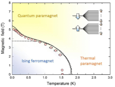
The compound is widely regarded as a prototypical system realizing the transverse-field Ising model Sachdev (1999). The groundstate in zero field is ferromagnetically ordered, while applying a relatively small transverse field induces a zero-temperature quantum phase transition at T into a quantum paramagnet Bitko et al. (1996), as shown in Fig. 1. Meanwhile, the hyperfine coupling strength of a Ho3+ ion is exceptionally large with a coupling constant mK Magariñno et al. (1980); Mennenga et al. (1984). The resulting strong hybridization between the electronic and nuclear magnetic moments hyb leads to two dramatic effects close to the quantum critical point: (i) significant modification of the low-temperature magnetic phase boundary (see Fig. 1) Bitko et al. (1996); (ii) incomplete mode softening of the low energy electronic excitations at the critical point Rønnow et al. (2005). Therefore, this system provides a rare opportunity to explore the quantum phase transition of a magnet coupled to a nuclear spin bath Bitko et al. (1996); Rønnow et al. (2005, 2007); Babkevich et al. (2015).
The impact of strong hybridization has also been highlighted for magnetic-ion diluted insulators, such as LiYF4:Ho3+ using magnetic resonance Giraud et al. (2001, 2003). A similar line of effort has achieved more recently single-molecule magnetic resonance with a rare-earth ion Müllegger et al. (2014). Furthermore, strong hybridization is of great interest in quantum information science Morley et al. (2010, 2013); Shiddiq et al. (2016). As much as these examples focus on the single-ion limit, the other limiting case of many-body systems, such as LiHoF4, provides a very different and complementary perspective. While in the long-range-ordered state the hybridization is suppressed, an applied transverse field introduces quantum fluctuations enhancing the hybridization towards .
However, probing directly the strongly hybridized states in LiHoF4 using spectroscopic methods, at the lowest energy scale, has so far been restricted to the thermal paramagnetic phase in the single-ion limit. The involved energy scale is too low to be resolved by the neutron scattering Rønnow et al. (2005, 2007). Magnetic resonance on 165Ho nuclei would provide a direct way of probing the hybridized nuclear-electronic states. However, the resonance in the ordered phase is expected around the frequency of 4.5 GHz in zero field, which does not fall into the operating frequencies of conventional nuclear magnetic resonance (NMR) nor electron spin resonance (ESR) instrumentation. Some studies have reported a hyperfine structure in ESR Magariñno et al. (1980); Janssen et al. (1985), but all in the paramagnetic regime above the ordering temperature K Bitko et al. (1996). To date, microscopic evidence for the realization of the unique nuclear-electronic Ising model Schechter and Stamp (2005, 2008) is absent.
Here we demonstrate experimentally nuclear-electronic magnetic resonance in LiHoF4 using coplanar microwave resonators and a vector network analyzer (VNA). We successfully trace the temperature and field evolution of the spectrum over the entire phase diagram, and show that it is remarkably well reproduced by a mean-field calculation with parameters set by independent spectroscopic measurements Babkevich et al. (2015); Magariñno et al. (1980); Mennenga et al. (1984); Rønnow et al. (2007).
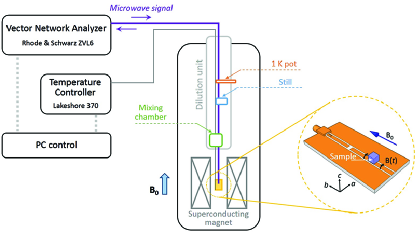
We begin with a description of our experimental setup shown in Fig. 2. A series of microwave coplanar resonators with different fundamental frequencies from 1.7 to 5.6 GHz were prepared. The impedance of the resonator is matched to the rest of the system by optimizing the gap size between the conductors. The oscillating magnetic field, B(t), generated at the sample position is parallel to the surface. A cube shaped sample of mm3 was placed at the center of the active strip, with a sub-millimeter gap in-between to avoid unwanted heating. The measurement geometry was chosen such that the applied magnetic field, B0, is along the crystallographic b axis of the tetragonal Scheelite structure, and B(t) is perpendicular to both B0 and the c axis to satisfy the magnetic resonance condition. We measured the parameter, which is defined as the ratio of the reflected to the input power, using a VNA which is connected through a low-loss cryogenic coaxial cable to the coplanar resonator. The coaxial cable was thermally anchored at each stage of the dilution refrigerator including the 1 K pot, Still, and mixing chamber to ensure thermalisation. The sample thermometer was located only 5 mm away from the sample which was thermally anchored to the mixing chamber. With an input power of -16 dBm applied by the network analyzer, the sample base temperature was 0.15 K to within 0.01 K.
To guide and interpret our experimental investigation, we perform a model calculation using a mean-field approximation. The full Hamiltonian has been well characterized through a number of different experiments Magariñno et al. (1980); Rønnow et al. (2007) and is given by,
| (1) | |||||
where () and () are the electronic and nuclear angular momentum operators at site , the dipolar coupling constant mK, is the dimensionless coupling parameter for the dipole-dipole interaction Jensen and Mackintosh (1991), and the negligible exchange constant mK. The nuclear Zeeman and quadrupole interactions are assumed to be negligible Giraud et al. (2001). The crystal field interaction with the surrounding ions splits the electronic states resulting in a groundstate which is a non-Kramers doublet with a strong Ising-like anisotropy and the first excited state 11 K above. In the ordered state, dipolar coupling lifts the groundstate degeneracy resulting in pseudo-spins up and down which we label as and states. Each state is further split into 8 nuclear-electronic states by the hyperfine interaction (Fig. 1(a), inset).
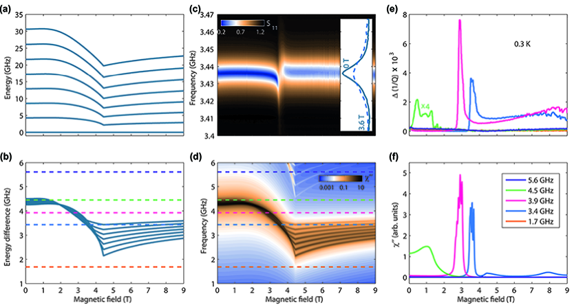
The total Hamiltonian can be diagonalized in the basis of nuclear-electronic states. The evolution of the lowest states with the applied transverse field is shown in Fig. 3(a). The energy level difference between consecutive states, , changes dramatically with the field as illustrated in Fig. 3(b). In the first approximation is proportional to , where is the magnitude of the total angular momentum, hence decreases with the field and reaches a minimum at . The diagram shown in Fig. 3(b) allows us to predict at which field the magnetic resonance occurs for a given frequency.
Experimentally we observed magnetic transitions between the adjacent nuclear-electronic levels through resonant absorption of continuous microwaves by the sample on a coplanar resonator. Figure 3(c) presents a typical frequency-field map at 0.3 K of the parameter using a resonator with the unloaded frequency of 3.4 GHz. The map shows a clear anomaly around 3.6 T indicative of magnetic resonance. This field value indeed agrees with the one predicted by mean-field calculations, which can be seen in Fig. 3(b) by taking an intersect of blue dashed line for 3.4 GHz with the solids lines for the energy level difference.
For an in-depth comparison between experiments and calculations, we proceed to directly calculate the imaginary part of the frequency-dependent susceptibility which is responsible for magnetic resonance absorption Cowan (2005); Abragam and Goldman (1982). The calculations were performed within the linear-response framework Jensen and Mackintosh (1991) using the mean-field wavefunctions and ,
| (2) |
where is the energy of the hybridized nuclear-electronic eigenstates in the presence of the mean-field, is the thermal population factor and is the partition function. The subscript refers to the oscillating field direction. The lifetime in the linear-response calculation of the states is assumed to be independent of field and temperature, and was fixed to 40 ns, corresponding to a damping of GHz, which provided the best match to our data. The lifetime broadening may result from direct or indirect contributions from the electronic dipolar and exchange or nuclear dipolar couplings Abragam and Goldman (1982), which we leave for future study. We note that the contribution to susceptibility from electronic moments, , is 500 times larger than the contribution from nuclear moments . Therefore, despite the predominantly nuclear-spin nature of the levels, the response we measure comes mainly from the electrons. This gives a tremendous enhancement of the signal from the nuclear states amplified by electronic moments. Figure 3(d) presents the calculated frequency-field map of intensity at 0.3 K, which shows a drastic change upon approaching from below. Resonant absorption is expected from our calculations to be in the 2 to 4.5 GHz bandwidth.
The absorptive part of the susceptibility is experimentally estimated as Cowan (2005), where the quality factor is defined as the loaded frequency divided by the full-width-half-maximum in the absorption profile in frequency as shown in the inset of Fig. 3(c). In Fig. 3(e) we show the experimental magnetic resonance spectra at 0.3 K for several different frequencies by plotting , where is a uniform background, which can be compared to the calculated spectra at 0.3 K in Fig. 3(f). Both calculations and measurements at frequencies of 3.4 and 3.9 GHz show resonant peaks around 3.6 and 3.0 T, respectively. Conversely, no resonance features are visible for the frequency of 1.7 GHz in both calculations and experiments. The predicted transitions between second-nearest neighbouring levels at 5.6 GHz is too weak to be observed experimentally. The calculated spectrum for 4.5 GHz appears as a broad hump at fields below 2 T, which can be expected from Fig. 3(d) where the frequency line cuts along the strongest intensity. For a better comparison the value was slightly reduced by 3%, which is nearly within the uncertainty from the reported one Magariñno et al. (1980). In principle, the uncertainty in the crystal field parameters can influence our calculations Babkevich et al. (2015). Nevertheless, excellent agreement with the experiments is remarkable considering that the model is essentially parameter-free. Some minor discrepancies such as the fine structure in the 4.5 GHz experimental spectrum are likely due to fixed lifetime of all levels in our model. However, since the modes around 4.5 GHz lie very close in the relevant field range, that structure would depend critically on the tiny variations of parameters. We therefore consider it more prudent to use a constant damping. The high-field tails in 3.4 and 3.9 GHz spectra are possibly due to the neglected effects of fluctuations.
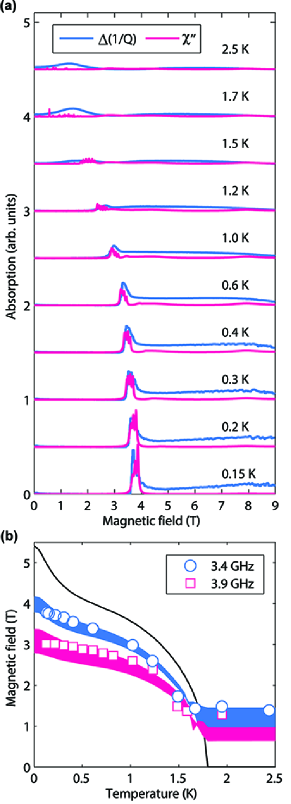
Furthermore, we investigate the temperature evolution of the spectrum for 3.4 GHz from 0.15 to 2.5 K as shown in Fig. 4(a). At base temperature a resonance peak appears around 3.7 T, which on warming decreases in amplitude and shifts to lower fields. The former is due to redistribution of the thermal population of states at higher temperatures. The latter reflects the decreasing size of the ordered electronic moment with increasing temperature, sensed by the nuclei through the hyperfine interactions. In Fig. 4(b) we track the resonance field as a function of temperature. Our measurements are shown to be very sensitive to small variations of the hyperfine coupling as depicted by the bands.
As shown in Fig. 3 and 4, all the salient features of the experimental results are well reproduced by the model calculations, thereby validating the transverse-field nuclear-electronic Ising model Schechter and Stamp (2005, 2008). The excellent description of the experimental results by our model implies that the probed states have a strongly hybridized character of both nuclear and electronic degrees of freedom. While this has been only hinted by previous bulk measurements Bitko et al. (1996) and neutron spectroscopy Rønnow et al. (2005), here we show directly the transitions between the strongly hybridized nuclear-electronic states. Likewise, the presented magnetic resonance should be distinguished from conventional NMR and ESR where the electronic and nuclear moments are approximated to product states Abragam and Goldman (1982); Abragam and Bleaney (2012); Cowan (2005).
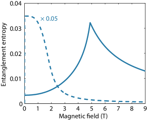
To highlight qualitative difference in the hybridized states between those in the many-body system and in the single-ion limit, we calculate the groundstate entanglement entropy Nielsen and Chuang (2010); Bennett et al. (1996) between the electronic and nuclear moments as a measure of the hybridization. We employ the Schmidt decomposition of the mean-field wavefunction, , where and , where the entanglement entropy is given by the von Neumann entropy . The calculated entropy in the absence of dipolar interactions decreases smoothly with a transverse field (Fig. 5) in agreement with those reported by Ref. Schechter and Stamp (2008). However, by turning on dipolar coupling the model produces a cusp-like peak at , that is, the hybridization in the ordered state of LiHoF4 increases with the applied field until it reaches a peak at the critical point. The field essentially mixes the higher excited states into the groundstate, thereby enhancing the hybridization. Increasingly larger field, , magnetizes the electronic and nuclear moments along the field direction such that the groundstate approaches a product state.
To summarize, we have demonstrated Ho nuclear-electronic magnetic resonance of LiHoF4 in a transverse magnetic field over the entire field-temperature phase diagram. The spectral evolution is remarkably well reproduced by mean-field calculations, validating the transverse-field nuclear-electronic Ising model. Taking advantage of the well-characterized model nature of LiHoF4, we have successfully probed the strongly hybridized states and their evolution in the long-range-ordered state. Our experimental scheme will find direct applications not only in the LiF4 (=rare earth) family Kraemer et al. (2012); Babkevich et al. (2015, 2016), but also other containing compounds including spin glass Schechter and Stamp (2005, 2008); Silevitch et al. (2007); Ancona-Torres et al. (2008); Piatek et al. (2014) and spin ice Harris et al. (1997); Sala et al. (2012).
We are grateful to M. Graf, S. S. Kim and P. Jorba Cabre for their contribution in building experimental setup at initial stage, B. Dalla Piazza for sharing his insight into the mean-field and linear-response theory. We also thank J. Jensen, A. Feofanov and D. Yoon for helpful discussions. M.J. is grateful to support by European Commission through Marie Skłodowska-Curie Action COFUND (EPFL Fellows). This work was supported by the Swiss National Science Foundation, the MPBH network and European Research Council grant CONQUEST. I.K., P.B., and M.J. contributed equally to this work.
References
- Sachdev (1999) S. Sachdev, Quantum Phase Transitions (Cambridge University Press, Cambridge, England, 1999).
- Bitko et al. (1996) D. Bitko, T. F. Rosenbaum, and G. Aeppli, Phys. Rev. Lett. 77, 940 (1996).
- Magariñno et al. (1980) J. Magariñno, J. Tuchendler, P. Beauvillain, and I. Laursen, Phys. Rev. B 21, 18 (1980).
- Mennenga et al. (1984) G. Mennenga, L. de Jongh, and W. Huiskamp, J. Magn. Magn. Mater. 44, 59 (1984).
- (5) One may consider the hybridization strong once the hyperfine coupling strength is comparable to the Zeeman energy scale, for instance, see Ref. Morley et al. (2013) .
- Rønnow et al. (2005) H. M. Rønnow, R. Parthasarathy, J. Jensen, G. Aeppli, T. Rosenbaum, and D. McMorrow, Science 308, 389 (2005).
- Rønnow et al. (2007) H. M. Rønnow, J. Jensen, R. Parthasarathy, G. Aeppli, T. F. Rosenbaum, D. F. McMorrow, and C. Kraemer, Phys. Rev. B 75, 054426 (2007).
- Babkevich et al. (2015) P. Babkevich, A. Finco, M. Jeong, B. Dalla Piazza, I. Kovacevic, G. Klughertz, K. W. Krämer, C. Kraemer, D. T. Adroja, E. Goremychkin, T. Unruh, T. Strässle, A. Di Lieto, J. Jensen, and H. M. Rønnow, Phys. Rev. B 92, 144422 (2015).
- Giraud et al. (2001) R. Giraud, W. Wernsdorfer, A. M. Tkachuk, D. Mailly, and B. Barbara, Phys. Rev. Lett. 87, 057203 (2001).
- Giraud et al. (2003) R. Giraud, A. M. Tkachuk, and B. Barbara, Phys. Rev. Lett. 91, 257204 (2003).
- Müllegger et al. (2014) S. Müllegger, S. Tebi, A. K. Das, W. Schöfberger, F. Faschinger, and R. Koch, Phys. Rev. Lett. 113, 133001 (2014).
- Morley et al. (2010) G. W. Morley, M. Warner, A. M. Stoneham, P. T. Greenland, J. van Tol, C. W. Kay, and G. Aeppli, Nature Mat. 9, 725 (2010).
- Morley et al. (2013) G. W. Morley, P. Lueders, M. H. Mohammady, S. J. Balian, G. Aeppli, C. W. Kay, W. M. Witzel, G. Jeschke, and T. S. Monteiro, Nature Mat. 12, 103 (2013).
- Shiddiq et al. (2016) M. Shiddiq, D. Komijani, Y. Duan, A. Gaita-Ariño, E. Coronado, and S. Hill, Nature 531, 348 (2016).
- Janssen et al. (1985) P. Janssen, I. De Wolf, and I. Laursen, J. Phys. Chem. Solids 46, 1387 (1985).
- Schechter and Stamp (2005) M. Schechter and P. C. E. Stamp, Phys. Rev. Lett. 95, 267208 (2005).
- Schechter and Stamp (2008) M. Schechter and P. C. E. Stamp, Phys. Rev. B 78, 054438 (2008).
- Jensen and Mackintosh (1991) J. Jensen and A. R. Mackintosh, Rare Earth Magnetism (Clarendon, Oxford, 1991).
- Cowan (2005) B. Cowan, Nuclear Magnetic Resonance and Relaxation (Cambridge University Press, Cambridge, 2005).
- Abragam and Goldman (1982) A. Abragam and M. Goldman, Nuclear Magnetism (Clarendon, Oxford, 1982).
- Abragam and Bleaney (2012) A. Abragam and B. Bleaney, Electron Paramagnetic Resonance of Transition Ions (Oxford University Press, Oxford, 2012).
- Nielsen and Chuang (2010) M. A. Nielsen and I. L. Chuang, Quantum computation and quantum information (Cambridge university press, 2010).
- Bennett et al. (1996) C. H. Bennett, H. J. Bernstein, S. Popescu, and B. Schumacher, Phys. Rev. A 53, 2046 (1996).
- Kraemer et al. (2012) C. Kraemer, N. Nikseresht, J. O. Piatek, N. Tsyrulin, B. Dalla Piazza, K. Kiefer, B. Klemke, T. F. Rosenbaum, G. Aeppli, C. Gannarelli, et al., Science 336, 1416 (2012).
- Babkevich et al. (2016) P. Babkevich, M. Jeong, Y. Matsumoto, I. Kovacevic, A. Finco, R. Toft-Petersen, C. Ritter, M. Månsson, S. Nakatsuji, and H. M. Rønnow, Phys. Rev. Lett. 116, 197202 (2016).
- Silevitch et al. (2007) D. Silevitch, D. Bitko, J. Brooke, S. Ghosh, G. Aeppli, and T. Rosenbaum, Nature 448, 567 (2007).
- Ancona-Torres et al. (2008) C. Ancona-Torres, D. M. Silevitch, G. Aeppli, and T. F. Rosenbaum, Phys. Rev. Lett. 101, 057201 (2008).
- Piatek et al. (2014) J. O. Piatek, I. Kovacevic, P. Babkevich, B. Dalla Piazza, S. Neithardt, J. Gavilano, K. W. Krämer, and H. M. Rønnow, Phys. Rev. B 90, 174427 (2014).
- Harris et al. (1997) M. J. Harris, S. T. Bramwell, D. F. McMorrow, T. Zeiske, and K. W. Godfrey, Phys. Rev. Lett. 79, 2554 (1997).
- Sala et al. (2012) G. Sala, C. Castelnovo, R. Moessner, S. L. Sondhi, K. Kitagawa, M. Takigawa, R. Higashinaka, and Y. Maeno, Phys. Rev. Lett. 108, 217203 (2012).