font=rm,small SISSA]Scuola Internazionale Superiore di Studi Avanzati (SISSA), via Bonomea 265, 34136, Trieste, Italy
Empirical corrections to the Amber RNA force field with Target Metadynamics
Abstract
The computational study of conformational transitions in nucleic acids still faces many challenges.
For example, in the case of single stranded RNA tetranucleotides, agreement between simulations
and experiments is not satisfactory due to inaccuracies in the force fields commonly used in molecular
dynamics simulations. We here use experimental data collected from high-resolution X-ray structures to attempt an improvement of the latest version of the AMBER force field. A modified metadynamics algorithm is used to calculate correcting potentials designed to enforce experimental distributions of backbone torsion angles. Replica-exchange simulations of
tetranucleotides including these correcting potentials show significantly better agreement with
independent solution experiments for the oligonucleotides containing pyrimidine bases.
Although the proposed corrections do not seem to be portable to generic RNA systems,
the simulations revealed the importance of the and backbone angles on the modulation of the RNA conformational ensemble. The correction protocol presented here suggests a systematic procedure for force-field refinement.
Introduction
Molecular dynamics is a powerful tool that can be used as a virtual microscope to investigate the structure and dynamics of biomolecular systems.1 However, the predictive power of molecular dynamics is typically limited by the accuracy of the employed energy functions, known as force fields. Whereas important advances have been made for proteins,2, 3 their accuracy for nucleic acids is still lagging behind.4, 5 Force fields for RNA have been used since several years in many applications to successfully model the dynamics around the experimental structures.6 Traditionally, the functional form and parameters of these energy functions have been assessed by checking the stability of the native structure. This has lead for instance to the discovery of important flaws in the parametrization of the backbone7 and of the glycosidic torsion.8 However, to properly validate a force field it is necessary to ensure that the entire ensemble is consistent with the available experimental data. This can be done only using enhanced sampling techniques or dedicated hardware. Recent tests9, 5 have shown that state-of-the-art force fields for RNA are still not accurate enough to produce ensembles compatible with NMR data in solution in the case of single stranded oligonucleotides. Similar issues have been reported for DNA and RNA dinucleosides.10, 11
Previous studies have shown that the distribution of structures sampled from the protein data bank (PDB) may approximate the Boltzmann distribution to a reasonable extent12, 13, 14, 2 and could even highlight features in the conformational landscape that are not reproduced by state-of-the-art force fields.15, 16 This has been exploited in the parametrization of protein force fields. For example, a significant improvement of the force fields of the CHARMM family has been obtained by including empirical corrections commonly known as CMAPs based on distributions from the PDB.17, 18
In this work, we apply these ideas to the RNA field and show how it is possible to derive force-field corrections using an ensemble of X-ray structures. At variance with the CMAP approach, we here correct the force field using a self-consistent procedure where metadynamics is used to enforce a given target distribution.19, 20 Correcting potentials are obtained for multiple dihedral angles using the metadynamics algorithm in a concurrent fashion. Since the target distributions are multimodal, we also use a recently developed enhanced sampling technique, replica exchange with collective-variable tempering (RECT),21 to accelerate the convergence of the algorithm. The correcting potentials are obtained by matching the torsion distributions for a set of dinucleoside monophosphates. The resulting corrections are then tested on tetranucleotides where standard force field parameters are known to fail in reproducing NMR data.
Methods
In this Section we briefly describe the target metadynamics approach and discuss the details of the performed simulations.
Targeting Distributions with Metadynamics
Metadynamics (MetaD) has been traditionally used to enforce an uniform distribution for a properly chosen set of collective variables (CV) that are expected to describe the slow dynamics of a system.22 However, it has been recently shown that the algorithm can be modified so as to target a preassigned distribution which is not uniform.19, 20 In this way a distribution taken from experiments, such as pulsed electron paramagnetic resonance, or from an X-ray ensemble, can be enforced to improve the agreement of simulations with empirical data. We refer to the method as target metadynamics (T-MetaD), following the name introduced in ref 19. For completeness, we here briefly derive the equations. It is also important to notice that the same goal could be achieved using a recently proposed variational approach. 23, 24
In our implementation of T-MetaD a history dependent potential acting on the collective variable at time is introduced and evolved according to the following equation of motion
| (1) |
Here , is the Boltzmann constant, the temperature, is the initial deposition rate of the kernel function which is here defined as a Gaussian with width , (s) is the free energy landscape associated to the target distribution, indicates the maximum value of the function , and is a constant damping factor. The target distribution is thus proportional to . We define where is the characteristic time of bias deposition. The term adjusts the height of the bias potential, making Gaussians higher at the target free-energy maximum and lower at its minimum. This forces the system to spend more time on regions where the targeted free-energy is lower. We notice that a similar argument has been used in the past to derive the stationary distribution of both well-tempered metadynamics, where Gaussian height depends on already deposited potential,25 and of adaptive-Gaussian metadynamics, where Gaussian shape and volume is changed during the simulation.26 The subtraction of sets an intrinsic upper limit for the height of each Gaussian, thus avoiding the addition of large forces on the system. We notice that other authors used terms such as the minimum of or the partition function to set an intrinsic lower limit for the prefactor . 19, 20 At the same time, the term acts as a global tempering factor27 and makes the Gaussian height decrease with the simulation time so as to make the bias potential converge instead of fluctuating. As observed in ref 19, the tempering approach used in well-tempered MetaD in this case would lead to a final distribution that is a mixture of the target one with the one from the original force field. For this reason, we prefer to use here a global tempering approach.27
In the long time limit (quasi-stationary condition) the bias potential will on average grow as25, 27
| (2) |
where is the probability distribution of the biased ensemble. Defining the function we can see this equation is a convolution of a Gaussian and a positive definite function.
| (3) |
As shown in ref 25, 27 this average should be independent of in stationary conditions, so that the function should be also independent of , though still dependent on time
| (4) |
By recognizing that and do not depend on , one can transform the last equation to
| (5) |
which implies that
| (6) |
Thus, the system will sample a stationary distribution of that is identical to the enforced one.
Whereas the equations are here only described for a single CV, this method can be straightforwardly applied to multiple CVs in a concurrent manner. In this case, the total bias potential is the sum of the one-dimensional bias potentials applied to each degree of freedom. Indeed, similarly to the concurrent metadynamics used in RECT,21 all the distributions are self-consistently enforced.20 This is particularly important when biasing backbone torsion angles in nucleic acids since they are highly correlated.28, 29 In this situation it is also convenient to use a biasing method that converges to a stationary potential through a tempering approach, to include in the self-consistent procedure of MetaD an additional effective potential associated to the correlation between the dihedral angles that is as close as possible to convergence.
Simulation Protocols
RNA dinucleoside monophosphates
Fragments of dinucleoside monophosphate with the sequence CC, AA, CA, and AC were extracted from the PDB database of RNA X-ray structures at medium and high resolution (resolution < 3 Å). The selected structures were protonated using pdb2gmx tool from GROMACS 4.6.7.30 Free-energy profiles along the backbone dihedral angles were calculated with the driver utility of PLUMED 2.1.31
Molecular dynamics simulations of the chosen RNA dinucleoside monophosphate sequences were performed using the Amberff99bsc0χOL3 force field (named here Amber14).32, 7, 8 The systems were solvated in an octahedron box of TIP3P water molecules 33 with a distance between the solute and the box wall of 1 nm. The system charge was neutralized by adding 1 Na+ counterion. The LINCS34 algorithm was used to constrain all bonds containing hydrogens and equations of motion were integrated with a timestep of 2 fs. All the systems were coupled to a thermostat through the stochastic velocity rescaling algorithm.35 For all non-bonded interactions the direct space cutoff was set to 0.8 nm and the electrostatic long-range interactions were treated using the default particle-mesh Ewald36 settings. An initial equilibration in the NPT ensemble was done for 2 ns, using the Parrinello-Rahman barostat.37 Production simulations were ran in the NVT ensemble. All the simulations were run using GROMACS 4.6.730 patched with a modified version of the PLUMED 2.1 plugin.31
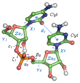
T-MetaD simulations were run to enforce the probability distributions of the angles , , and (see Fig. 1), which were calculated from the X-ray fragments. The target free-energy profiles were calculated with PLUMED 2.1. Distributions were estimated as combination of Gaussian kernels, with a bandwidth of 0.15 rad, and written on a grid with 200 bins spanning the range. The bias potential used for the T-MetaD was grown using a characteristic time ps and a dampfactor . Gaussians with a width of 0.15 rad were deposited every 500 steps.
We underline that simulations performed using T-MetaD could be non ergodic for two reasons. First, there could be significant barriers acting on CVs that are not targeted and thus not biased at this stage (e.g. dihedral angles). Second, if the enforced distribution of a CV is bimodal it will be necessary to help the system in exploring both modes with the correct relative probability. It is thus necessary to combine the T-MetaD approach with an independent enhanced-sampling scheme. Here we used RECT, a replica exchange method where a group of CVs is biased concurrently using a different bias factor for each replica and one reference replica is used to accumulate statistics.21 When T-MetaD and RECT are combined, in each replica a T-MetaD is run with the same settings, including the reference replica. The T-MetaD/RECT simulation was run with 4 replicas for 1 s each. For each residue the dihedrals of the nucleic acid backbone (, , , , ), together with one of the Cartesian coordinates of the ring puckering38 () and the glycosidic torsion angle () were chosen as accelerated CVs (see Fig. 1). To help the free rotation of the nucleotide heterocyclic base around the glycosidic bond, the distance between the center of mass of nucleobases was also biased. For the dihedral angles the Gaussian width was set to 0.25 rad and for the distance it was set to 0.05 nm. The Gaussians were deposited every 500 steps. The initial Gaussian height was adjusted to the biasfactor of each replica, according to the relation , in order to maintain the same ps across the entire replica ladder. The biasfactor ladder was chosen in the range from 1 to 2, following a geometric distribution. In replicas with the target free energy was scaled by a factor . Exchanges were attempted every 200 steps. Statistic was collected from the unbiased replica. A sample input file is provided as supplementary material (see Fig S1).
Finally, a new RECT simulation was run for each dinucleoside with the bias potentials obtained from the T-MetaD applied statically on each replica. These calculations represent the results obtained with a force field that includes the corrections from the PDB distributions and are thus labeled as Amberpdb. Statistics from these simulations were collected to evaluate the effects of the corrections. The simulation time was 1 s per replica.
RNA Tetranucleotides
To test the force field corrections derived on dinucleoside monophosphates, temperature replica-exchange molecular dynamics (T-REMD) simulations 39 were performed on different tetranucleotide systems with sequence CCCC, GACC and AAAA. The correcting potentials calculated for the AA and CC dinucleosides were applied to all the backbone angles of AAAA and CCCC tetranucleotides, respectively. For the GACC tetranucleotide we combined the correcting potentials from the T-MetaD simulations of AA, AC and CC, assuming a similarity between purines A and G.
The T-REMD data related to the Amber14 force field and the protocol for the new simulations performed using the Amberpdb force field were taken from ref 16. The systems were solvated with TIP3P waters and neutral ionic conditions. We used 24 replicas with a geometric distribution of temperatures from 300 to 400 K. Exchanges were attempted every 200 steps. The simulation length was 2.2 s per replica.
Analysis
The result of the molecular dynamics simulations was compared to NMR experimental data of dinucleosides 10, 40, 41, 42 and tetranucleotides.43, 44, 9 vicinal coupling constants were calculated using Karplus expressions.45, 46 We took into account the analysis made in refs 47, 48, 10 to select the most precise sets of parameters. Calculations were performed using the software tool baRNAba 49. Details are given in the supplementary information, subsection 1.1.
Results
As a first step we used our approach to enforce the dihedral distribution from the X-ray fragments on monophosphate dinucleosides AA, AC, CA, and CC. Then, we show that the corrections are partly transferable and could improve agreement with solution experiments for tetranucleotides.
Calculation of correcting potentials for dinucleoside monophosphates
The Amber14 force field is considered to be one of the most accurate ones for RNA, though it is failing to reproduce solution experiments for short flexible oligomers. Recent benchmarks of different Amber force field modifications based on reparametrization of the torsion angles and non-bonded terms have shown that these changes did not lead to a satisfactory agreement with solution experiments for tetranucleotides.5, 9 On the other hand, ensembles of tetranucleotides taken from the PDB have a very good agreement with NMR data.16 We thus decided to add correcting potentials to the dihedral angle terms of Amber14, based on information recovered from high-resolution X-ray structures of RNA deposited in the PDB. We analyzed enhanced sampling simulations of dinucleosides (described in this paper) and tetranucleotides (described in a previous publication16), to select a minimal amount of degrees of freedom to modify. This analysis indicated the backbone angles , , and could benefit from a correction (a full description is presented in supplementary information, section 2). We used T-MetaD to enforce on those dihedrals the probability distributions obtained from fragments of X-ray structures. RNA dinucleoside monophosphates were chosen as model systems to obtain the correcting potentials. As the corrections are sequence dependent, for each nucleobase combination we generated an ensemble of experimental conformations from the PDB database that had the same sequence as the dinucleoside monophosphates.
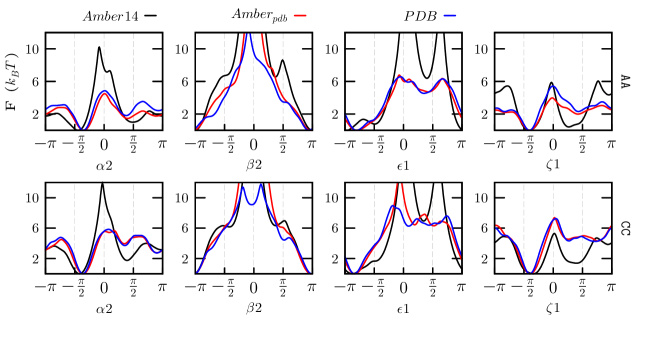
In Fig. 2 we show the free energy profiles of AA and CC dinucleosides projected on the , , and angles. Amber14, Amberpdb, as well as the target PDB ensembles are represented. The profiles of AC and CA are shown in Fig S7. The similarity between the PDB and Amberpdb profiles makes it clear that the corrections efficiently enforce the distributions taken from the X-ray ensemble. Although some differences are visible around the free-energy barriers, they are expected not to be relevant for room temperature properties at equilibrium. Nevertheless, the transition times and the behavior of the Amberpdb potential at high temperatures could be affected by these barriers. In general, barriers in the experimental ensemble are several lower than those from the Amber14 force field. In the corrected ensemble the multimodal character of the force field probability distributions for the angles , and is reduced, to favor the conformations corresponding to the canonical A-form. The observed agreement between the PDB and Amberpdb one-dimensional probability distributions for the selected angles is not necessarily translated into equivalence of the respective ensembles. This is seen for example in the two-dimensional distributions shown in Figs S8-11.
Correcting potentials might in principle also affect the distribution of non-biased degrees of freedom if the latter ones are correlated with the former ones. The distribution of non-biased degrees of freedom, such as the angles , and puckering coordinate , is shown in Fig. S12. Overall, no difference is observed between the Amber14 and Amberpdb free-energy profiles, with the exception of the ratio between the C3’-endo and C2’-endo conformations in CC. This is a consequence of the significant correlation between the backbone angle and the puckering.
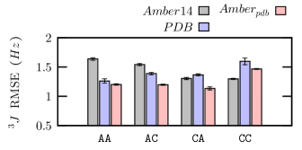
To asses the validity of the corrections, we compared all the ensembles against NMR experimental data10 (Fig 3). Individual vicinal coupling values from the experiments and the simulations are reported in Table S2. In the case of AA, AC and CA dinucleosides the agreement of Amberpdb with the experimental data is better than that of Amber14 and of the X-ray ensemble. This can be explained noticing that Amberpdb combines the good agreement with NMR experiments of Amber14 for angles in the nucleoside (dihedrals and ) with that of the PDB distribution for angles in the backbone (dihedrals and ), as shown in Fig S13. A notable exception is the CC dinucleoside, where the correlation of backbone angles with puckering mentioned above leads to slightly larger deviation in Amberpdb with respect to Amber14. It should be noticed that the NMR observables analyzed here cannot be used to directly determine the conformation around the phosphodiester backbone (/), so the comparison with the NMR vicinal coupling dataset does not take into account the distribution of these angles.
We noticed that, whereas the NMR data was measured at 293 K (AA, CA and AC) and 320 K (CC), simulations were performed at 300 K. However, the agreement between the data for CC obtained at 320K and similar NMR data obtained for a smaller number of couplings at 280K 42 shows that deviations induced by temperature changes are expected to be much smaller than the typical deviations between molecular dynamics and experiment observed here. It is also important to mention that these RMSE values do not take into account systematic errors in the Karplus formulas employed in this study.
It is also interesting to measure the effect of the proposed backbone corrections on the stacking interactions. Stacking free energies computed according to the definition used in a recent paper9 show that the correcting potential have barely no effect on stacking (Fig S14). These numbers can also be compared with experimental values,42, 41, 50 and indicate that Amber force field is likely overestimating stacking interactions as suggested by several authors.51, 52 This comparison is however affected by the definition of stacked conformation, which introduces a large arbitrariness in the estimation of stacking free energies from MD.
Validation of Amberpdb potential on RNA tetranucleotides
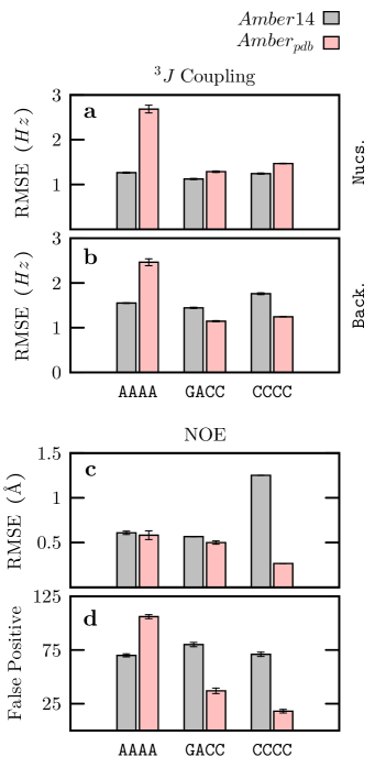
The correcting potentials discussed above are designed so as to enforce the PDB distribution on dinucleosides monophosphates. We here used these corrections to perform simulations on larger oligonucleotides. In particular, we performed extensive simulations of tetranucleotides, which are considered as good benchmarks for force-field testing, as their small size makes the generation of converged ensembles accessible to modern enhanced sampling techniques. We performed three T-REMD simulations with the Amberpdb potential for the tetranucleotide sequences AAAA, GACC and CCCC. These systems have been used before in very long (hundred of s) simulations53, 54, 55, 5, 56 and NMR experimental data is available.43, 44, 9 The Amber14 T-REMD data were taken from ref 16.
The coupling RMSE, the NOE-distance RMSE, and the number of distance false positives, i.e. the MD predicted NOEs not observed in the experiment, are presented in Fig 4. For these systems the number of false positives is one of the most important parameters to assess the quality of the MD ensembles.9 In the case of tetranucleotides containing pyrimidines (GACC and CCCC), the correcting potential improves significantly the agreement with the experimental data, mostly for the NOEs (see Fig S15). This is confirmed by the root-mean-square deviation (RMSD) distribution shown in Figure S16 where it can be appreciated that for these two sequences the corrections lead to an overall improvement of the ensemble by disfavoring the intercalated and inverted structures with a large RMSD from native. A completely different scenario is found for the Amberpdb ensemble of AAAA, where the corrections surprisingly diminish the agreement with experiments. This can be also appreciated in a shift of the Amberpdb RMSD distribution peaks to higher RMSD values due to an increase in the population of compact structures (Fig S16). It should be noticed that the effect of the correcting potentials in purines and pyrimidines depends strongly on the sequence length. Whereas the AAAA tetranucleotide is negatively affected by the corrections, the AA dinucleoside is the one that benefits the most from them.
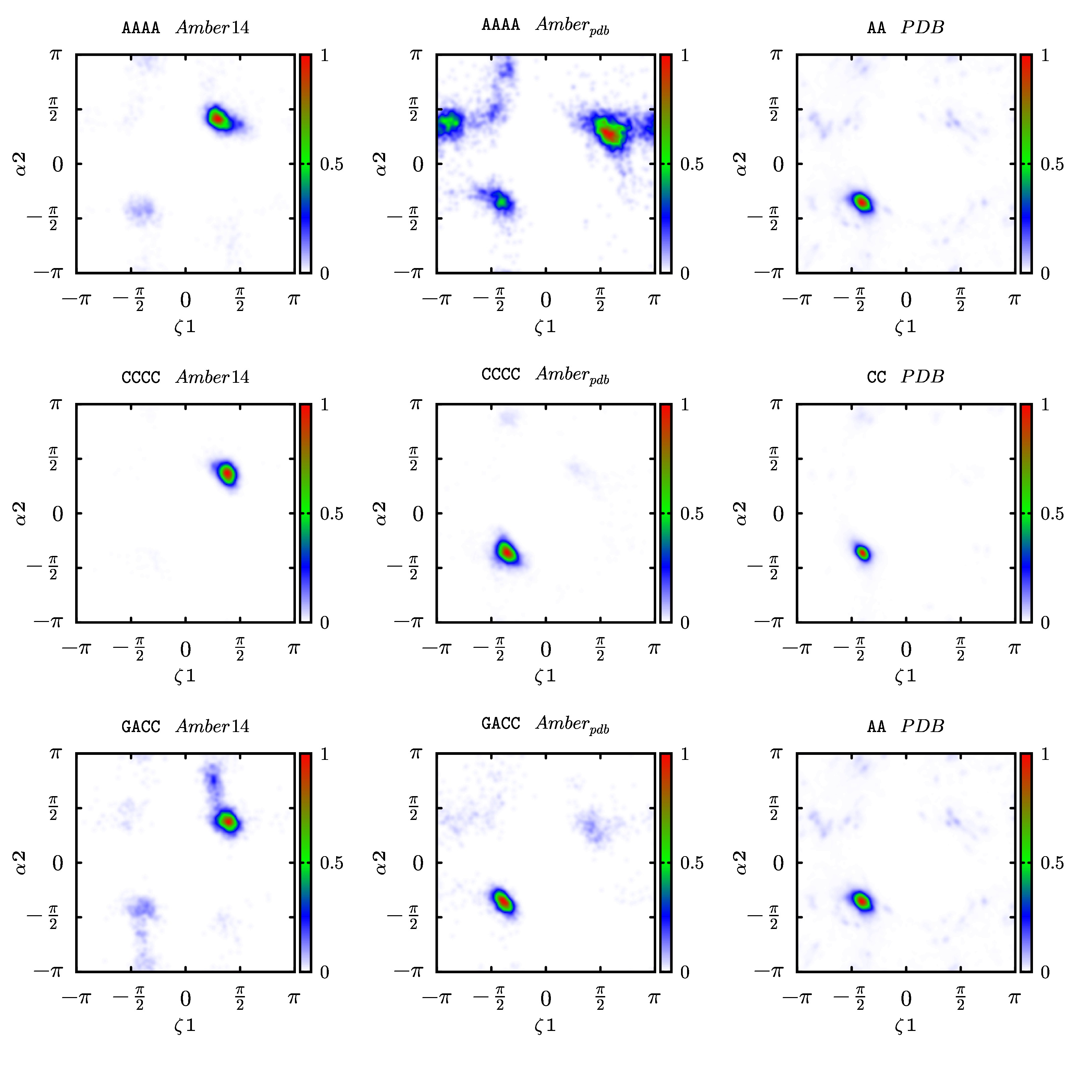
As discussed in the section 2 of the SI, the conformation along the phosphodiester backbone is very different between compact and extended tetranucleotide structures. The probability distribution maps of the / backbone dihedral angles from the tetranucleotides T-REMD simulations and the dinucleosides X-ray ensembles used to generate the corrections are depicted in Fig 5. Only phosphodiester backbone torsion angles are shown, because they are the ones mostly affected by the correction. The other backbone angles maps are shown in the SI (Figs S17-25). In the PDB ensembles the distributions are always unimodal, independently of the sequence, with a peak at the (g-)/(g-) conformation, whereas in the Amber14 ensemble the (g+)/(g+) and (g-)/(g-) conformations are both significantly populated. The effects of the corrections, as seen before, are highly sequence dependent. In case of GACC and CCCC, the (g-)/(g-) rotamer is stabilized in the Amberpdb distributions, with the population of (g+)/(g+) significantly decreased with respect to Amber14. On the contrary, for AAAA the (g+)/(g+) conformation is not unfavored by the correcting potentials, despite not being significantly present in the PDB ensemble. This could be due to the fact that the one dimensional target free-energy profile for dihedrals and for the AA (Fig 2) exhibits barriers which are approximately 4 smaller with respect to the ones from the Amber14 force field. The effect of the decreased barrier height can be appreciated in the / probability distribution of AAAA, where the amount of torsional space explored is increased by the corrections.
Consequences on future force field refinements
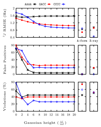
The good agreement of the Amberpdb ensembles with the NMR observables, in the case of CCCC and GACC tetranucleotides, suggests that the RNA conformational space sampled by state-of-the-art force field could be modified to better match experimental solution data by penalizing rotamers of the and angles. As a further test, we reweighted the T-REMD Amber14 ensembles with an additional two-dimensional penalizing Gaussian potential centered on the (g+)/(g+) conformation. Results are shown in Fig 6 for different Gaussian heights. Overall, the agreement with the NMR experimental data improves considerably with respect to the original force field as the Gaussian height increases. The relative population of the / conformations has an important impact on the number of false positive NOE contacts which indicates the presence of intercalated structures. This improvement is achieved without changing the non bonded interactions as it has also been proposed.51 It is however important to observe that these results are obtained by performing a reweighting, and that corrections should be validated by performing separate simulations with this bias potential.
Discussion
In this paper we apply targeted metadynamics to sample preassigned distributions taken from experimental data.19, 20 At variance with the original applications, we here combine T-MetaD with enhanced sampling showing that these protocols can also be used when the investigated ensembles have non-trivial energy landscapes separated by significant barriers .
We apply the method to RNA oligonucleotides, for which the Amber14 force field was proven to be in significant disagreement with solution NMR data.43, 54, 44, 53, 57, 9, 5, 56 Since tetranucleotide fragments extracted from high resolution structures in the PDB were shown to match NMR experiments better than Amber14 force field,16 we here used X-ray structures to build reference distributions of backbone dihedral angles that are then used to devise correcting potentials. More precisely, we use T-MetaD to enforce the empirical distribution of the dihedral angles in the phosphate backbone (, , and ) on four dinucleoside monophosphates.
We calculated the correcting potentials concurrently for all the four angles in order to change the distribution of these consecutive dihedrals along the backbone chain taking into account their correlation. The method successfully enforced the distributions taken from the PDB on all the angles. The new ensemble generated by the corrected force field (Amberpdb) was independently validated against solution NMR data that was not used in the fitting of the corrections. For three of the four dinucleosides studied, Amberpdb showed a better agreement with the NMR data compared with Amber14 and with the X-ray ensemble.
We then tested the portability of the correcting potentials by simulating three tetranucleotides, GACC, CCCC and AAAA. In the case of GACC and CCCC the agreement with NMR data is significantly improved by the corrections. Surprisingly, for AAAA the corrections have the opposite effect and increase the probability of visiting compact structures making the simulated ensemble less compatible with solution experiments. It should be noticed here that this is a non obvious result since the PDB database is expected to have an intrinsic bias towards A-form structures and should thus in principle increase the agreement with solution experiments in this specific case. This indicates that porting the corrections from dinucleosides to tetranucleotides is not straightforward because the coupling between the multiple corrected dihedrals could affect the resulting ensemble in an non-trivial way. Additionally, corrections applied to dihedral angles alone might be not sufficient to compensate errors arising from inexact parametrization of van der Waals or electrostatic interactions.51 Overall, the tests we performed indicate that the corrections derived here should not be considered as portable corrections for the simulation of generic RNA sequences.
Nevertheless, by comparing the backbone angle distributions on the different RNA simulations and the X-ray ensembles, we were able to find possible hints pointing at where refinement of dihedral potentials could lead to an advancement in RNA force fields. In this respect, the results for GACC an CCCC show the significant improvement observed in the Amberpdb simulations for those systems could be reproduced by simply penalizing the (g+)/(g+) conformation, which is overpopulated in Amber14. By a straightforward reweighting procedure, we showed that simple Gaussian potentials that disfavor this conformation significantly improved the experimental agreement with solution experiments for all the three tetranucleotides. Recent modifications of the Lennard-Jones parameters for phosphate oxygens58 and different water models56 were shown to affect the conformational ensemble of RNA tetranucleotides.5, 56 It might be interesting to combine these modified parameters for non-bonded interactions with the here introduced procedure for dihedral angle refinement.
The nature of the correction methodology discussed in this paper is very different from the classical approach to force field parametrization, as it aims to correct the free energy of the system, instead of fitting the potential energy landscape of the dihedral angles while constraining the other degrees of freedom. It is important to notice that the dihedral angle distributions taken from the fragments of the PDB structures do not necessarily represent the conformational ensembles of dinucleosides or tetranucleotides in solution. Indeed, some of the interaction patterns that are present in large structures crystallized in the PDB do not exist in short oligonucleotides. For this reason, in this work the distributions were validated against independent solutions NMR experiments. This allowed the dihedral angles from the PDB distributions that performed better than the force field to be identified. We also recall that in our procedure the force-field torsion energy function is not refitted, but a bias potential is added to the total energy of the system in order to match the free-energy profile of the torsion angles with target ones. Thus, a major advantage of this approach is that it takes explicitly into account the entropic contributions, the cross correlations between torsional angles, and inaccuracies in the non-bonded interactions, among other effects.
Conclusion
In conclusion, in this work we applied the target metadynamics protocol to modify dihedral distributions in dinucleosides. The procedure successfully enforces reference distributions taken from the PDB without affecting the distribution of the dihedral angles that were not biased. However, the attempt to port these corrections to tetranucleotides lead to ambiguous results when applied to different sequences. This could be partly due to the fact that distribution form the PDB are not necessarily a good reference for refinement.
Nevertheless, the simulations revealed the importance of the / angles rotamers on the modulation of the conformational ensemble, and that by only penalizing the (g+)/(g+) rotamer the quality of the ensemble is significantly improved to levels not reported before.
Thomas Cheatham III, Fabrizio Marinelli, and Jiří Šponer are acknowledged for carefully reading the manuscript and providing several useful suggestions. The research leading to these results has received funding from the European Research Council under the European Union’s Seventh Framework Programme (FP/2007-2013) / ERC Grant Agreement n. 306662, S-RNA-S.
References
- Dror et al. 2012 Dror, R. O.; Dirks, R. M.; Grossman, J.; Xu, H.; Shaw, D. E. Annu. Rev. Biophys. 2012, 41, 429–452
- Lindorff-Larsen et al. 2010 Lindorff-Larsen, K.; Piana, S.; Palmo, K.; Maragakis, P.; Klepeis, J.; Dror, R.; Shaw, D. Proteins: Struct., Funct., Bioinf. 2010, 78, 1950–1958
- Lindorff-Larsen et al. 2012 Lindorff-Larsen, K.; Maragakis, P.; Piana, S.; Eastwood, M. P.; Dror, R. O.; Shaw, D. E. PloS one 2012, 7, e32131
- Šponer et al. 2014 Šponer, J.; Banáš, P.; Jurecka, P.; Zgarbova, M.; Kührová, P.; Havrila, M.; Krepl, M.; Stadlbauer, P.; Otyepka, M. J. Phys. Chem. Lett. 2014, 5, 1771–1782
- Bergonzo et al. 2015 Bergonzo, C.; Henriksen, N. M.; Roe, D. R.; Cheatham, T. E. RNA 2015, 21, 1578–1590
- Cheatham and Case 2013 Cheatham, T. E.; Case, D. A. Biopolymers 2013, 99, 969–977
- Pérez et al. 2007 Pérez, A.; Marchán, I.; Svozil, D.; Šponer, J.; Cheatham III, T. E.; Laughton, C. A.; Orozco, M. Biophys. J. 2007, 92, 3817–3829
- Zgarbová et al. 2011 Zgarbová, M.; Otyepka, M.; Šponer, J.; Mládek, A.; Banáš, P.; Cheatham III, T. E.; Jurecka, P. J. Chem. Theory Comput. 2011, 7, 2886–2902
- Condon et al. 2015 Condon, D. E.; Kennedy, S. D.; Mort, B. C.; Kierzek, R.; Yildirim, I.; Turner, D. H. J. Chem. Theory Comput. 2015, 11, 2729–2742
- Vokáčová et al. 2009 Vokáčová, Z.; Budesinsky, M.; Rosenberg, I.; Schneider, B.; Šponer, J.; Sychrovský, V. J. Phys. Chem. B 2009, 113, 1182–1191
- Nganou et al. 2016 Nganou, C.; Kennedy, S. D.; McCamant, D. W. J. Phys. Chem. B 2016, 120, 1250–1258
- Butterfoss and Hermans 2003 Butterfoss, G. L.; Hermans, J. Protein Sci. 2003, 12, 2719–2731
- MacKerell et al. 2004 MacKerell, A. D.; Feig, M.; Brooks, C. L. J. Comput. Chem. 2004, 25, 1400–1415
- Morozov et al. 2004 Morozov, A. V.; Kortemme, T.; Tsemekhman, K.; Baker, D. Proc. Natl. Acad. Sci. U.S.A. 2004, 101, 6946–6951
- Brereton and Karplus 2015 Brereton, A. E.; Karplus, P. A. Sci. Adv. 2015, 1, e1501188
- 16 Bottaro, S.; Gil-Ley, A.; Bussi, G. Nucleic Acids Res. 2016, doi:10.1093/nar/gkw239
- MacKerell et al. 2004 MacKerell, A. D.; Feig, M.; Brooks, C. L. J. Am. Chem. Soc. 2004, 126, 698–699
- Buck et al. 2006 Buck, M.; Bouguet-Bonnet, S.; Pastor, R. W.; MacKerell, A. D. Biophys. J. 2006, 90, L36–L38
- White et al. 2015 White, A.; Dama, J.; Voth, G. A. J. Chem. Theory Comput. 2015, 11, 2451–2460
- Marinelli and Faraldo-Gómez 2015 Marinelli, F.; Faraldo-Gómez, J. D. Biophys. J. 2015, 108, 2779–2782
- Gil-Ley and Bussi 2015 Gil-Ley, A.; Bussi, G. J. Chem. Theory Comput. 2015, 11, 1077–1085
- Laio and Parrinello 2002 Laio, A.; Parrinello, M. Proc. Natl. Acad. Sci. U. S. A. 2002, 99, 12562–12566
- Valsson and Parrinello 2014 Valsson, O.; Parrinello, M. Phys. Rev. Lett. 2014, 113, 090601
- Shaffer et al. 2016 Shaffer, P.; Valsson, O.; Parrinello, M. Proc. Natl. Acad. Sci. U.S.A. 2016, 113, 1150–1155
- Barducci et al. 2008 Barducci, A.; Bussi, G.; Parrinello, M. Phys. Rev. Lett. 2008, 100, 020603
- Branduardi et al. 2012 Branduardi, D.; Bussi, G.; Parrinello, M. J. Chem. Theory Comput. 2012, 8, 2247–2254
- Dama et al. 2014 Dama, J. F.; Parrinello, M.; Voth, G. A. Phys. Rev. Lett. 2014, 112, 240602
- Saenger 1984 Saenger, W. Principles of Nucleic Acid Structure; Springer-Verlag, New York, 1984
- Richardson et al. 2008 Richardson, J. S.; Schneider, B.; Murray, L. W.; Kapral, G. J.; Immormino, R. M.; Headd, J. J.; Richardson, D. C.; Ham, D.; Hershkovits, E.; Williams, L. D.; Keating, K. S.; Pyle, A. M.; Micallef, D.; Westbrook, J.; Berman, H. M. RNA 2008, 14, 465–481
- Hess et al. 2008 Hess, B.; Kutzner, C.; Van Der Spoel, D.; Lindahl, E. J. Chem. Theory Comput. 2008, 4, 435–447
- Tribello et al. 2014 Tribello, G. A.; Bonomi, M.; Branduardi, D.; Camilloni, C.; Bussi, G. Comput. Phys. Commun. 2014, 185, 604–613
- Cornell et al. 1995 Cornell, W. D.; Cieplak, P.; Bayly, C. I.; Gould, I. R.; Merz, K. M.; Ferguson, D. M.; Spellmeyer, D. C.; Fox, T.; Caldwell, J. W.; Kollman, P. A. J. Am. Chem. Soc. 1995, 117, 5179–5197
- Jorgensen 1981 Jorgensen, W. L. J. Am. Chem. Soc. 1981, 103, 335–340
- Hess et al. 1997 Hess, B.; Bekker, H.; Berendsen, H. J.; Fraaije, J. G. J. Comput. Chem. 1997, 18, 1463–1472
- Bussi et al. 2007 Bussi, G.; Donadio, D.; Parrinello, M. J. Chem. Phys. 2007, 126, 014101
- Darden et al. 1993 Darden, T.; York, D.; Pedersen, L. J. Chem. Phys. 1993, 98, 10089–10092
- Parrinello and Rahman 1981 Parrinello, M.; Rahman, A. J. Appl. Phys. 1981, 52, 7182–7190
- Huang et al. 2014 Huang, M.; Giese, T. J.; Lee, T.-S.; York, D. M. J. Chem. Theory Comput. 2014, 10, 1538–1545
- Sugita and Okamoto 1999 Sugita, Y.; Okamoto, Y. Chem. Phys. Lett. 1999, 314, 141–151
- Olsthoorn et al. 1982 Olsthoorn, C. S.; Doornbos, J.; Leeuw, H. P.; Altona, C. Eur. J. Biochem. 1982, 125, 367–382
- Ezra et al. 1977 Ezra, F. S.; Lee, C.-H.; Kondo, N. S.; Danyluk, S. S.; Sarma, R. H. Biochemistry 1977, 16, 1977–1987
- Lee et al. 1976 Lee, C.-H.; Ezra, F. S.; Kondo, N. S.; Sarma, R. H.; Danyluk, S. S. Biochemistry 1976, 15, 3627–3639
- Yildirim et al. 2011 Yildirim, I.; Stern, H. A.; Tubbs, J. D.; Kennedy, S. D.; Turner, D. H. J. Phys. Chem. B 2011, 115, 9261–9270
- Tubbs et al. 2013 Tubbs, J. D.; Condon, D. E.; Kennedy, S. D.; Hauser, M.; Bevilacqua, P. C.; Turner, D. H. Biochemistry 2013, 52, 996–1010
- Karplus 1959 Karplus, M. J. Chem. Phys. 1959, 30, 11–15
- Karplus 1963 Karplus, M. J. Am. Chem. Soc. 1963, 85, 2870–2871
- Sychrovskỳ et al. 2006 Sychrovskỳ, V.; Vokácová, Z.; Šponer, J.; Špacková, N.; Schneider, B. J. Phys. Chem. B 2006, 110, 22894–22902
- Vokáčová et al. 2009 Vokáčová, Z.; Bickelhaupt, F. M.; Šponer, J.; Sychrovský, V. J. Phys. Chem. A 2009, 113, 8379–8386
- Bottaro et al. 2014 Bottaro, S.; Di Palma, F.; Bussi, G. Nucleic Acids Res. 2014, 42, 13306–14
- Frechet et al. 1979 Frechet, D.; Ehrlich, R.; Remy, P.; Gabarro-Arpa, J. Nucleic Acids Res. 1979, 7, 1981–2001
- Chen and García 2013 Chen, A. A.; García, A. E. Proc. Natl. Acad. Sci. U. S. A. 2013, 110, 16820–16825
- Brown et al. 2015 Brown, R. F.; Andrews, C. T.; Elcock, A. H. J. Chem. Theory Comput. 2015, 11, 2315–2328
- Bergonzo et al. 2013 Bergonzo, C.; Henriksen, N. M.; Roe, D. R.; Swails, J. M.; Roitberg, A. E.; Cheatham III, T. E. J. Chem. Theory Comput. 2013, 10, 492–499
- Henriksen et al. 2013 Henriksen, N. M.; Roe, D. R.; Cheatham III, T. E. J. Phys. Chem. B 2013, 117, 4014–4027
- Roe et al. 2014 Roe, D. R.; Bergonzo, C.; Cheatham III, T. E. J. Phys. Chem. B 2014, 18, 3543–52
- Bergonzo and III 2015 Bergonzo, C.; III, T. E. C. J. Chem. Theory Comput. 2015, 11, 3969–3972
- Condon et al. 2014 Condon, D. E.; Yildirim, I.; Kennedy, S. D.; Mort, B. C.; Kierzek, R.; Turner, D. H. J. Phys. Chem. B 2014, 118, 1216–1228
- Steinbrecher et al. 2012 Steinbrecher, T.; Latzer, J.; Case, D. J. Chem. Theory Comput. 2012, 8, 4405–4412