Origin of Pressure-induced Superconducting Phase in studied by Synchrotron X-ray Diffraction and Spectroscopy
Abstract
Pressure dependence of the electronic and crystal structures of , which has pressure-induced two superconducting domes of SC I and SC II, was investigated by x-ray emission spectroscopy and diffraction. X-ray diffraction data show that compressibility along the -axis changes around 12 GPa, where a new superconducting phase of SC II appears. This suggests a possible tetragonal to collapsed tetragonal phase transition. X-ray emission spectroscopy data also shows the change in the electronic structure around 12 GPa. These results can be explained by the scenario that the two SC domes under pressure originate from the change of Fermi surface topology. Present results here show that the nesting condition plays a key role in stabilizing the superconducting state helping to address outstanding fundamental question as to why the SC II appears under pressure.
Introduction
Since the discovery of high-temperature superconductivity in F-doped LaFeAsO in 2008,Kamihara08 many iron-based superconductors with different crystal structures have been synthesized and are still hot topics in condensed matter physics. Most iron-superconductor families have FeAs or FeSe planes as the common layers, which correlate to the superconductivity. The crystal structure of FeSe is the simplest of these iron-based superconductors with .Hsu08 Moreover, it was recently found that a single FeSe layer on showed high of 65–100 K.He13 ; Ge15
Intercalation to FeSe layers by alkaline atoms also raised to 30–46 K in bulk iron-based superconductors of ().Guo10 ; Liu11 ; Ying12 ; Dagotto13 Therefore, in these systems electron-doping to the FeSe layer may play an important role in superconductivity. The electron-doping causes a Fe-deficiency of the FeSe layer to keep the charge valance, and this system is called 122* phase. These new iron-defected systems (122* family) have attracted many interests because of the following several unique features, which are very different from other iron-based superconductors.Dagotto13 (i) This system shows intrinsic phase separation.Ricci15 It consists of 122-type superconductor and 245-type AFM insulator with vacancy order which disappears around 10GPa.FChen11 ; Ding13 ; Li12 ; Bendele14 ; Saini14 ; Guo12 (ii) They have an unprecedented high Néel temperature of 559 K and large magnetic moment of 3.3. Bao11 ; Ye11 This magnetic moment is the largest among pnictide and chalcogenide iron-superconductors. (iii) Unlike the usual iron-based superconductor, there are no hole pockets at Fermi surface which enhances the Fermi surface nesting.Zhang11 (iv) of gradually drops with pressure, and superconductivity (SC I) disappears around 10 GPa. However, interestingly, further pressure induces a new superconductivity (SC II) suddenly around 11 GPa. The SC II phase shows higher than the SC I phase.Sun12 ; Gao14
Recently, single phase non-superconducting was synthesized, and the pressure-temperature phase diagram was revealed.Gao14 By comparing the and phase diagrams, the phase separation in the SC II region was suggested, and the superconducting phase attributed to the 122 phase. Therefore, this means that superconducting phase with and non-superconducting phase with co-exist in the SC II phase.
A theoretical study of the SC I and SC II phases in the 122* system suggested that superconducting symmetry is -wave without -point hole pocket at SC I and -pairing at SC II.Das13 In these systems, however, since no experimental study of not only the electronic structure, but also the crystal structure under pressure has been reported so far, the issue of the appearance of SC II dome under pressure remains unclear.
In this paper we report a systematic study of with x-ray diffraction (XRD) and x-ray emission spectroscopy (XES) under pressure. The purpose of this work is to reveal both the crystal and electronic structures of under pressure to clarify the existence of the two superconducting domes. XES technique has made it possible to probe local magnetic moment under pressure by detecting Fe emission spectra for iron-based superconductor. Tsutsumi59 ; Vanko06 ; JMChen11 ; Gretarsson11 ; Gretarsson13 We also performed the bulk sensitive x-ray absorption (XAS) measurements with partial fluorescence (PFY) mode at the Fe absorption edge. Hamalainen91 We have used the PFY-XAS method where a decay process with shorter life time is selected, resulting spectra are narrower, and making fine electronic structure near the absorption edge better visible.Hamalainen91 ; Dallera02 ; Yamaoka14 Our results show the change in the -axis compressibility around boundary pressure of the SC I and SC II phases, suggesting a crystal structure change at this pressure, probably a tetragonal (T) to collapsed tetragonal (cT) transition. The Fe XES also shows a pressure-induced change in the electronic structure at the transition pressure.
Results
- phase diagram
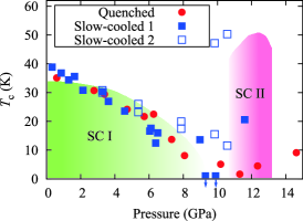
We prepared two kinds of single crystals: a sample quenched at 550 ∘C (quenched sample) and one cooled slowly (slow-cooled sample). A - phase diagram of the quenched and slow-cooled samples is shown in Fig. 1. was determined from the onset temperature of the electrical resistivity measurements. Both samples show the decreases with pressure monotonically in the SC I phase. This behavior agrees well with the reports published.Sun12 ; Gao14 However, the maximum of SC II phase depends on the samples. of the quenched and slow-cooled samples are 5 K and 20 K at the SC II phase, respectively, Fujita15 while of SC II was 50 K in the reports published.Sun12 ; Gao14 These results suggest that the of SC II depends strongly on the sample preparation. Actually, island- and mesh-shape morphology were observed in the back-scattered electron (BSE) image in the slow-cooled and the quenched samples, respectively.Tanaka15 These morphologies were caused by the difference of iron concentration.Tanaka15
X-ray diffraction
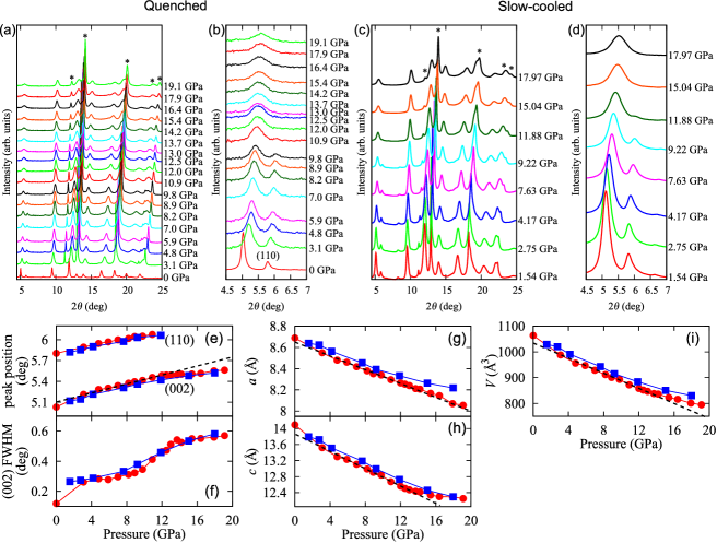
We measured x-ray diffraction patterns under pressure up to 19.1 GPa for the quenched sample and 18.0 GPa for the slow-cooled sample at room temperature as shown in Fig. 2. Both samples consist of a symmetry of the 245 phase and a symmetry of the 122 phase at ambient pressure. Fe vacancy order-disorder transition was reported in the non-superconducting 245 phase at SC II, and crystal symmetry after the transition becomes which is the same as the superconducting phase.Guo12 ; Bendele13 Figures 2(a) and 2(c) show the XRD pattern of the quenched and slow-cooled samples, and the enlarge views are shown in Figs. 2(b) and 2(d). Intensity of the superstructure peak (110) attributed to the Fe vacancy order disappears around 12 GPa, indicating a clear structural phase transition from to symmetry at 245 phase. The same feature has been observed previously.Guo12 ; Bendele13 Seemingly, the above structural transition pressure of 12 GPa coincides with the appearance of the SC II phase as seen in Fig. 1.
Although a Rietveld refinement was not performed because of the restriction of the observed range, we performed peak fits by using the several peaks with the Voigt functions in order to derive the lattice constants. Figure 2(e) indicates (002) and (110) peak position vs pressure. Trend of the pressure evolution of (002) peak position changes around 12 GPa. This system consists of the 122 and 245 phases and thus only the average lattice constant of two phases could be analyzed. Here, we assumed symmetry at all pressures because symmetry can express symmetry. Figures 2(e) and 2(f) show pressure evolution of the lattice constants. Pressure evolution of the -axis shows a monotonic decrease, while that of the -axis changes the slope around 12 GPa. Thus the compressibility along the -axis becomes lower above 12 GPa. This means that the bond along the -axis at the SC II phase is stronger than that at the SC I phase. This suggests a crystal structure change at 12 GPa, probably T cT structural phase transition analogous to .Yu14
Pressure induced change in the emission spectra
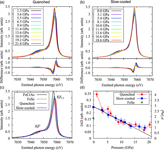
Figures 3(a) and 3(b) show pressure evolution of emission spectra of the quenched and slow-cooled samples, respectively. A spectrum consists of a main peak of and a satellite peak of , which correspond to low-spin and high-spin states, respectively.Tsutsumi59 In Fig. 3, pressure evolution of spectrum shows a shift from the high-spin to the low-spin state with pressure.
Figure 3(c) shows a comparison among the spectra of the quenched sample, the slow-cooled sample, FeCrAs (0), and FeSe (2). As seen in Fig. 3(c), comparison of spectra between and FeCrAs concludes that is in the higher-spin state because of larger intensity. The local moment of Fe can be extracted by the the integrated absolute difference (IAD) analysis of the Fe emission spectra to a reference spectrum.Vanko06 ; Gretarsson11 It is known that the IAD values are proportional to the local magnetic moments.Gretarsson11
Figure 3(d) shows the local magnetic moment estimated by the IAD analysis of the spectra in Figs. 3(a) and 3(b). The local magnetic moment decreases from at ambient pressure to at the SC II phase with pressure. Two samples show roughly the same trend under pressure. Especially the pressure evolution of the local magnetic moment of slow-cooled sample changes the slope at 12 GPa. This coincides with the change in the compressibility along the -axis shown in Fig. 2(h).
Pressure induced change in the PFY-XAS spectra
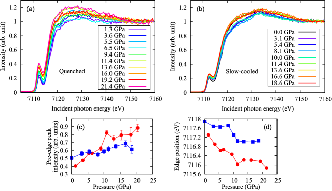
Figures 4(a) and 4(b) show a pressure evolution of the PFY-XAS spectra setting the emitted photon energy to peak of the quenched and slow-cooled samples, respectively. The intensity is normalized to that at 7160 eV. The PFY-XAS spectra show large pre-edge peaks. The pre-edge and the main edge peaks correspond to quadrupole and dipole transitions, respectively. The strong pre-edge peak intensity includes the information of the hybridization between the Fe and Se orbitals.Groot09 The edge position of the PFY-XAS spectra shifts toward lower energy with pressure in both samples, indicating the decrease of the Fe valence. The system includes and Simonelli12 and thus the above result indicates a change in the Fe valence from . The decrease of the Fe valence with pressure may be due to the electron supply from K to FeSe layer caused by the shrink along the -axis. Figures 4(c) shows that the pre-edge peak intensity of the PFY-XAS spectra increases with pressure. Another point we would like to emphasize is that the intensity around 7125 eV changes at 12GPa in the slow-cooled sample, although it is not clear in the quenched one (Fig. 4(a) and (b)). This pressure also coincides with the pressure where the compressibility of the -axis changes.
Discussion
The XRD and XES studies under pressure have been performed for the 122* system, which have pressure-induced two superconducting domes. The XRD results show that the compressibility along the -axis changes at 12 GPa and the superlattice diffraction disappears at the same pressure. Pressure dependence of the lattice constant along the -axis and the volume becomes gentle at the SC II phase. The same -axis evolution has been observed in where Ca, Sr, Ba and Eu.Yu14 This was interpreted as the T cT structural phase transition. Therefore, it is reasonable to expect that system, which has the same crystal structure, also shows the T cT transition.
The change in the crystal structure affects the magnetic property. Actually, the XES results indicate that the trend of the pressure evolution of the magnetic moment and the electronic state shown in Fig. 3(d) changes also at 12 GPa, which seems to correlate to the T cT transition. The average local magnetic moment changes from at ambient pressure to at the SC II phase with pressure. The change in the magnetic moment at 12 GPa is not large in system, probably because the collapse along the -axis at 12 GPa is small.
The PFY-XAS spectra show that the Fe valence decreases with pressure, which may correspond to the increase of the carrier density at the SC II phase due to the supply of the electrons from K to the FeSe layer caused by the shrink along the -axis. The pre-edge peak intensity in the PFY-XAS spectra increases with pressure, indicating the increase of the hybridization between Fe and Se and also the density of states (DOS) near the Fermi surface. The pressure-induced change in the pre-edge peak intensity also correlates to the shift from high-spin to low-spin states.
In the 122 system the superconductivity emerged suddenly at the cT phase when the T cT structural phase transition occurred.Ying15 The phase diagram of the 122 system is similar to that of the 122* system.Nakajima15 The DFT calculations showed the change in the electronic structure between the T phase and cT phase.Guterding15 In , the T cT transition changed the superconductivity symmetry from -wave to -wave. This is a Lifshitz transition which is known to change the Fermi surface drastically from the electronic state with only hole pocket to that with electron and hole pockets. Other calculations of also showed the -wave in the SC I and -wave in the SC II phase.Das13 Therefore, together with these theoretical calculations we conclude that there is the T cT transition in under pressure and thus the high- at the SC II phase could be explained by the strong Fermi surface nesting.
Methods
Sample preparation and characterizations
We prepared two kinds of single crystals.Ozaki13 ; Tanaka15 Single crystals were grown by a simple one-step synthesis. Fe (99.9%), (99%) powders and Se (99.999%) grains were put into an alumina crucible and sealed into an evacuated quartz tube. The mixture was slowly heated up to 900 ∘C and held for 3 hours. The melting mixture was, then, cooled down to room temperature slowly (slow-cooled sample) and quenched at 550 ∘C (quenched sample). Back-scattered electrons (BSE) images were obtained to observe micro-structure. Island- and mesh-shape structure were shown in the slow-cooled and quenched samples, and the chemical composition determined by using energy dispersive x-ray spectrometry (EDX) were and , respectively.Tanaka15 The area ratios between the superconducting region and non-superconducting region is 10-13% in the slow-cooled sample and 30-35% in the quenched sample. of the present samples under pressure were measured at Osaka University.Fujita15
XRD, XES, and PFY-XAS measurements under pressure
We performed XRD, XES, and PFY-XAS experiments for the slow-cooled and quenched samples. For XRD, XES, and PFY-XAS measurement, these samples with NaCl as the pressure medium were loaded into a sample chamber of the gasket in the glove box of pure Ar atmosphere because these samples are chemically unstable in the air. Pressure was monitored by ruby fluorescence method.Mao76
Pressure dependence of the XRD patterns were measured at SPring-8 BL12B2 using a 3-pin plate diamond anvil cell (DAC, Almax Industries) with a CCD detection system at room temperature. We took an arrangement of both incoming and outgoing x-ray beams passed through the diamonds with incident photon energy of 20 keV. NaCl was loaded as the pressure medium and well-mixed with the sample because of reduction of preferred orientation of the sample. 2D image of CCD was integrated by using FIT2D program.Hammersley96
The PFY-XAS and XES measurements were performed at the Taiwan beam line BL12XU at SPring-8. The undulator beam was monochromatized by a cryogenically-cooled double crystal Si(111) monochromator. A Johann-type spectrometer equipped with a spherically bent Si(531) analyzer crystal (radius of 1 m) and a Si solid state detector (Amptech) were used to analyze the Fe emission of the de-excitation at the Fe absorption edge. At the emitted photon energy of 7.6 keV the overall energy resolution was estimated to be 0.9 eV. The intensities of the measured spectra were normalized using the incident beam that was monitored just before the sample.
For the high-pressure XES experiments the x-ray beam was focused to 20-30 (horizontal) 30-40 (vertical) at the sample position using a toroidal and a Kirkpatrick-Baez mirror. High-pressure conditions were achieved at room temperature using a diamond anvil cell coupled with a gas-membrane. A Be-gasket with 3 mm in diameter and approximately 100 thick was pre-indented to approximately 35-40 thickness around the center. The diameter of the sample chamber in the gasket was approximately 100 and the diamond anvil culet size was 300 . We used the Be gasket in-plane geometry with a scattering angle of 90∘, where both incoming and outgoing x-ray beams passed through the Be gasket. Be was used due to its higher transmittance to x-rays in comparison to other high- materials.
IAD analyses
The IAD analysis is performed in the following way: (i) match the center of mass between the sample and reference spectra, (ii) take the difference between them, and (iii) integrate the absolute value of the difference. The intensity is normalized by the area of the spectrum.
References
- (1) Kamihara, Y., Watanabe, T., Hirano, M. & Hosono, H. Iron-based layered superconductor (-) with . J. Am. Chem. Soc. 130, 3296 (2008). URL http://dx.doi.org/10.1021/ja800073m.
- (2) Hsu, F.-C. et al. Superconductivity in the PbO-type structure -FeSe. Proc. Nat. Acad. Sci. (USA) 105, 14262 (2008). URL http://dx.doi.org/10.1073/pnas.0807325105.
- (3) He, S. et al. Phase diagram and electronic indication of high-temperature superconductivity at 65 K in single-layer FeSe films. Nat. Mater. 12, 605 (2013). URL http://dx.doi.org/10.1038/nmat3648.
- (4) Ge, J.-F. et al. Superconductivity above 100 K in single-layer FeSe films on doped . Nat. Mater. 14, 285 (2015). URL http://dx.doi.org/10.1038/nmat4153.
- (5) Guo, J. et al. Superconductivity in the iron selenide . Phys. Rev. B 82, 180520 (2010). URL http://link.aps.org/doi/10.1103/PhysRevB.82.180520.
- (6) Liu, R. H. et al. Coexistence of superconductivity and antiferromagnetism in single crystals (, / and /): Evidence from magnetization and resistivity. EPL 94, 27008 (2011). URL http://stacks.iop.org/0295-5075/94/i=2/a=27008.
- (7) Ying, T. P. et al. Observation of superconductivity at 30-46 in (, , , , , , and ). Sci. Rep. 2, 426 (2012). URL http://dx.doi.org/10.1038/srep00426.
- (8) Dagotto, E. The unexpected properties of alkali metal iron selenide superconductors. Rev. Mod. Phys. 85, 849 (2013). URL http://link.aps.org/doi/10.1103/RevModPhys.85.849.
- (9) Ricci, A. et al. Direct observation of nanoscale interface phase in the superconducting chalcogenide with intrinsic phase separation. Phys. Rev. B 91, 020503 (2015). URL http://link.aps.org/doi/10.1103/PhysRevB.91.020503.
- (10) Chen, F. et al. Electronic identification of the parental phases and mesoscopic phase separation of superconductors. Phys. Rev. X 1, 021020 (2011). URL http://link.aps.org/doi/10.1103/PhysRevX.1.021020.
- (11) Ding, X. et al. Influence of microstructure on superconductivity in and evidence for a new parent phase . Nat. Commun. 4, 1897 (2013). URL http://dx.doi.org/10.1038/ncomms2913.
- (12) Li, W. et al. Phase separation and magnetic order in K-doped iron selenide superconductor. Nat. Phys. 8, 126 (2012). URL http://www.nature.com/nphys/journal/v8/n2/abs/nphys2155.html.
- (13) Bendele, M. et al. Spectromicroscopy of electronic phase separation in superconductor. Sci. Rep. 4, 5592 (2014). URL http://dx.doi.org/10.1038/srep05592.
- (14) Saini, N. L. et al. X-ray absorption and photoemission spectroscopy of electronic phase separation in . Phys. Rev. B 90, 184510 (2014). URL http://link.aps.org/doi/10.1103/PhysRevB.90.184510.
- (15) Jing, J. et al. Pressure-driven quantum criticality in iron-selenide superconductors. Phys. Rev. Lett. 108, 197001 (2012). URL http://dx.doi.org/10.1103/PhysRevLett.108.197001.
- (16) Bao, W. et al. A novel large moment antiferromagnetic order in superconductor. Chin. Phys. Lett. 28, 086104 (2011). URL http://stacks.iop.org/0256-307X/28/i=8/a=086104.
- (17) Ye, F. et al. Common crystalline and magnetic structure of superconducting () single crystals measured using neutron diffraction. Phys. Rev. Lett. 107, 137003 (2011). URL http://link.aps.org/doi/10.1103/PhysRevLett.107.137003.
- (18) Zhang, Y. et al. Nodeless superconducting gap in (, ) revealed by angle-resolved photoemission spectroscopy. Nat. Mater. 10, 273 (2011). URL http://dx.doi.org/10.1038/nmat2981.
- (19) Sun, L. et al. Re-emerging superconductivity at 48 kelvin in iron chalcogenides. Nature (London) 483, 67 (2012). URL http://dx.doi.org/10.1038/nature10813.
- (20) Gao, P. et al. Role of the 245 phase in alkaline iron selenide superconductors revealed by high-pressure studies. Phys. Rev. B 89, 094514 (2014). URL http://dx.doi.org/10.1103/PhysRevB.89.094514.
- (21) Das, T. & Balatsk, A. V. Origin of pressure induced second superconducting dome in (). New J. Phys. 15, 093045 (2013). URL http://dx.doi.org/10.1088/1367-2630/15/9/093045.
- (22) Tsutsumi, K. The x-ray non-diagram lines of some compounds of the iron group. J. Phys. Soc. Jpn. 14, 1696 (1959). URL http://journals.jps.jp/doi/abs/10.1143/JPSJ.14.1696.
- (23) Vankó, G. et al. Probing the spin momentum with x-ray emission spectroscopy: the case of molecular-spin transitions. Phys. Chem. B 110, 11647 (2006). URL http://dx.doi.org/10.1021/jp0615961.
- (24) Chen, J. M. et al. Pressure dependence of the electronic structure and spin state in superconductors probed by x-ray absorption and x-ray emission spectroscopy. Phys. Rev. B 84, 125117 (2011). URL http://link.aps.org/doi/10.1103/PhysRevB.84.125117.
- (25) Gretarsson, H. et al. Revealing the dual nature of magnetism in iron pnictides and iron chalcogenides using x-ray emission spectroscopy. Phys. Rev. B 84, 100509 (2011). URL http://dx.doi.org/10.1103/PhysRevB.84.100509.
- (26) Gretarsson, H. et al. Spin-state transition in the fe pnictides. Phys. Rev. Lett. 110, 047003 (2013). URL http://link.aps.org/doi/10.1103/PhysRevLett.110.047003.
- (27) Hämäläinen, K., Siddons, D. P., Hastings, J. B. & E.Berman, L. Elimination of the inner-shell lifetime broadening in x-ray-absorption spectroscopy. Phys. Rev. Lett. 67, 2850 (1991). URL http://link.aps.org/doi/10.1103/PhysRevLett.67.2850.
- (28) Dallera, C. et al. New spectroscopy solves an old puzzle: the kondo scale in heavy fermions. Phys. Rev. Lett. 88, 196403 (2002). URL http://link.aps.org/doi/10.1103/PhysRevLett.88.196403.
- (29) Yamaoka, H. et al. Role of valence fluctuations in the superconductivity of 122 compounds. Phys. Rev. Lett. 113, 086403 (2014). URL http://link.aps.org/doi/10.1103/PhysRevLett.113.086403.
- (30) Fujita, H. et al. Pressure dependence of superconductive transition temperature on . J. Phys.: Conf. Ser. 592, 012070 (2015). URL http://dx.doi.org/10.1088/1742-6596/592/1/012070.
- (31) Tanaka, M. et al. Origin of the Higher- Phase in the system. J. Phys. Soc. Jpn. 85, 044710 (2016). URL http://dx.doi.org/10.7566/JPSJ.85.044710.
- (32) Bendele, M. et al. Interplay of electronic and lattice degrees of freedom in superconductors under pressure. Phys. Rev. B 88, 180506 (2013). URL http://dx.doi.org/10.1103/PhysRevB.88.180506.
- (33) Yu, Z. et al. Conventional empirical law reverses in the phase transitions of 122-type iron-based superconductors. Sci. Rep. 4, 7172 (2014). URL http://dx.doi.org/10.1038/srep07172.
- (34) de Groot, F., Vanko, G. & Glatzel, P. The 1s x-ray absorption pre-edge structures in transition metal oxides. Journal of Physics: Condensed Matter 21, 104207 (2009). URL http://stacks.iop.org/0953-8984/21/i=10/a=104207.
- (35) Simonelli, L. et al. Coexistence of different electronic phases in the superconductor: A bulk-sensitive hard x-ray spectroscopy study. Phys. Rev. B 85, 224510 (2012). URL http://link.aps.org/doi/10.1103/PhysRevB.85.224510.
- (36) Ying, J.-J. et al. Tripling the critical temperature of by carrier switch. arXiv:1501.00330 (2015). URL http://arxiv.org/abs/1501.00330.
- (37) Nakajima, Y. et al. High-temperature superconductivity stabilized by electron-hole interband coupling in collapsed tetragonal phase of under high pressure. Phys. Rev. B 91, 060508 (2015). URL http://link.aps.org/doi/10.1103/PhysRevB.91.060508.
- (38) Guterding, D., Backes, S., Jeschke, H. O. & Valentí, R. Origin of the superconducting state in the collapsed tetragonal phase of . Phys. Rev. B 91, 140503 (2015). URL http://dx.doi.org/10.1103/PhysRevB.91.140503.
- (39) Ozaki, T. et al. Evolution of superconductivity in isovalent Te-substituted crystals. Supercond. Sci. Technol. 26, 055002 (2013). URL http://stacks.iop.org/0953-2048/26/i=5/a=055002.
- (40) Mao, H.-K. & Bell, P. M. High-pressure physics: The 1-megabar mark on the ruby static pressure scale. Science 191, 851 (1976). URL http://dx.doi.org/10.1126/science.191.4229.851.
- (41) Hammersley, A. P., Svensson, S. O., Hanfland, M., Fitch, A. N. & Hausermann, D. Two-dimensional detector software: From real detector to idealised image or two-theta scan. High Pressure Research 14, 235 (1996). URL http://dx.doi.org/10.1080/08957959608201408.
Acknowledgements
The experiments were performed at Taiwan beam line BL12XU and BL12B2 at SPring-8 under SPring-8 Proposals No. 2013B4127, No. 2013B4156, & 2014A4257 (corresponding NSRRC Proposal No. 2013-3-007). We are grateful to Yumiko Zekko, Satomi Kawase and Yu Ohta in Kwansei Gakuin University and Yuki Sumi in Doshisha University for their help in the experiment. We deeply thank Young-June Kim at University of Toronto for the preparation of the FeCrAs sample. We also deeply appreciate Jin-Ming Chen and Jenn-Min Lee for the use of the diamond anvil cell system in the XRD experiment and Akihiko Machida for use of the glove box in JAERI. This work was partly supported by JSPS KAKENHI Grant Number 15K05194 and 26400322. This work at UT Austin was supported as part of EFree, an Energy Frontier Research Center funded by the U. S. Department of Energy Office of Science, Office of Basic Energy Sciences under Award DE-SC0001057.