Optical–nanofiber–based interface for single molecules
Abstract
Optical interfaces for quantum emitters are a prerequisite for implementing quantum networks. Here, we couple single molecules to the guided modes of an optical nanofiber. The molecules are embedded within a crystal that provides photostability and, due to the inhomogeneous broadening, a means to spectrally address single molecules. Single molecules are excited and detected solely via the nanofiber interface without the requirement of additional optical access. In this way, we realize a fully fiber–integrated system that is scalable and may become a versatile constituent for quantum hybrid systems.
pacs:
42.50.-p, 42.81.Qb, 33.80.-b, 78.67.BfI Introduction
In recent years, single molecules in solids Siyushev et al. (2014); Basché et al. (1995, 1992); Celebrano et al. (2011); Gerhardt et al. (2007, 2010); Kozankiewicz and Orrit (2014) and other solid state quantum emitters such as color centers in diamond Jelezko and Wrachtrup (2004); Maurer et al. (2012); Neumann et al. (2008); Pingault et al. (2014) and quantum dots Hansom et al. (2014a); Delteil et al. (2016); Yalla et al. (2014); Arcari et al. (2014); Javadi et al. (2015) have gained increasing interest as building blocks for quantum networks Nemoto et al. (2014); Kimble (2008), quantum metrology Doherty et al. (2014); Goldstein et al. (2011); Acosta et al. (2013) and nanosensors Faez et al. (2014a); Mazzamuto et al. (2014); Kucsko et al. (2013). For all these applications a strong light–matter interaction is essential. This can be achieved by coupling to a large ensemble of quantum emitters Lukin and Imamoğlu (2001); Reim et al. (2010), by employing a cavity Volz et al. (2014); Thompson et al. (2013); Ritter et al. (2012); Schietinger et al. (2008) or by decreasing the mode area of the interacting light field Faez et al. (2014b); Hwang and Hinds (2011); Yalla et al. (2012); Vetsch et al. (2010); Kirs̆anskė et al. (2017); Lombardi et al. and hence achieving a significant overlap between the absorption cross–section of the emitter and the respective light field. A versatile platform to achieve such a small mode area of the light field are optical nanofibers Vetsch et al. (2010); Garcia-Fernandez et al. (2011). An optical nanofiber is the waist of a tapered optical fiber (TOF) and has a diameter smaller than the wavelength of the light it is guiding. Therefore, an appreciable fraction of the light propagates outside the fiber in the form of an evanescent wave. Due to the strong transverse confinement of the light field, which prevails over the entire length of the nanofiber, the interaction with emitters close to the surface can be significant Le Kien et al. (2005a); Reitz et al. (2013); Nayak et al. (2007); Yalla et al. (2012); Liebermeister et al. (2014).
Single molecules in crystalline solids are efficient quantum emitters that exhibit strong zero phonon lines (ZPL) which can be lifetime-limited and as narrow as tens of MHz at cryogenic temperatures Kummer et al. (1994); Moerner (2004). Due to inhomogeneous broadening caused by the host crystal such molecules can be spectrally discerned and individually addressed using a narrowband laser Moerner and Orrit (1999). For a low concentration of molecules, this makes it possible to circumvent the additional spatial selection that has been used for numerous single molecule experiments in the past Michaelis et al. (1999); Tamarat et al. (2000), even if a large fraction of the crystal is illuminated. This was also exploited in recent experiments Faez et al. (2014b); Türschmann et al. (2017), where single dibenzoterrylene molecules have been coupled to light propagating through a nanocapillary.
Single molecules come in a large variety and they are small quantum emitters which is useful when coupling them to nano- and microcavities Wang et al. (2017); Chizhik et al. (2009); Norris et al. (1997) and also offers the possibility to study collective phenomena of quantum emitters. Additionally, single molecules such as polycyclic aromatic hydrocarbons can be spectrally very stable and do not suffer from photobleaching when embedded in the right host matrix. In addition to a near-unity quantum yield, these are very important features when working with solid state emitters. Here, we show for the first time that single organic molecules can be interfaced with an optical nanofiber. This presents a new platform based on solid state emitters that can be used for quantum optics and that is naturally integrated into optical fiber networks.
II Experimental Setup
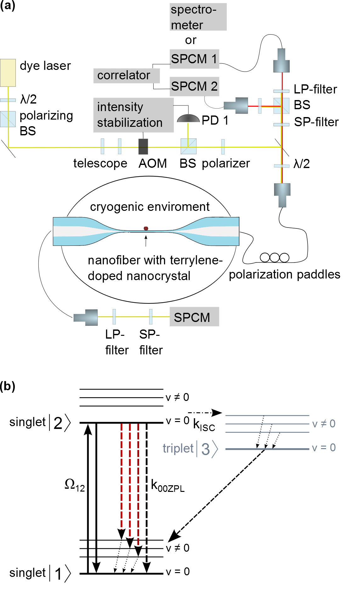
In our experimental setup, the TOF resides inside a cryostat [Fig. 1(a)] and we interface terrylene molecules in a para–terphenyl (p–terphenyl) crystal with the evanescent light field surrounding its nanofiber. The latter has a total length of and a diameter of . The TOF is produced in a heat and pull process using a custom–made pulling rig Warken et al. (2008). In the long tapered section of the fiber, the weakly guided LP01 mode of the standard single mode optical fiber is adiabatically transformed into the strongly guided HE11 mode of the nanofiber waist and back yielding transmission losses of less than 2% from 520–. For our purpose, a broadband transmission is crucial as the excitation and detection wavelengths can differ by more than 100 nm. This requires a careful choice of tapering angles and waist diameter Stiebeiner et al. (2010). Terrylene in p–terphenyl can exhibit four different electronic transition frequencies from the ground to the first excited state termed X1–X4, corresponding to four possible orientations of the molecules in the crystal. Molecules in the X4 orientation are resonant with light at and have been shown to be very photostable Bordat and Brown (2002). Hence, all measurements presented here use molecules in this site. A simplified level diagram is depicted in Fig. 1(b). The laser excites the molecule on the zero phonon line 00ZPL that connects the ground and the excited electronic state without any vibrational contribution of the molecule. After excitation, the molecule will decay into any of the vibrational states in the electronic ground state with a probability determined by the Franck Condon and Debye–Waller factors. From these states it will nonradiatively decay into the vibrational ground state within picoseconds. Hence, the absorption cross–section Loudon (2000) of a single molecule in a solid is
| (1) |
where the product represents the projection of the polarization vector of the excitation light on the unit vector of the molecular dipole and denotes the excitation wavelength. is the lifetime–limited linewidth and the homogeneously broadened linewidth of the 00ZPL, respectively. If the molecular dipole is aligned with the polarization of the excitation light and the homogeneous broadening is negligible, the absorption cross–section will approach that of a simple two–level atom and is comparable to the effective mode area Warken et al. (2007) of our optical nanofiber of about (see Appendix B). This ensures a strong effect of a single molecule on the light field. The probability of a terrylene molecule to decay to the triplet state after excitation rather than to the singlet ground state is very low and has experimentally been found to be at cryogenic temperatures Banasiewicz et al. (2005); Basché et al. (1995).
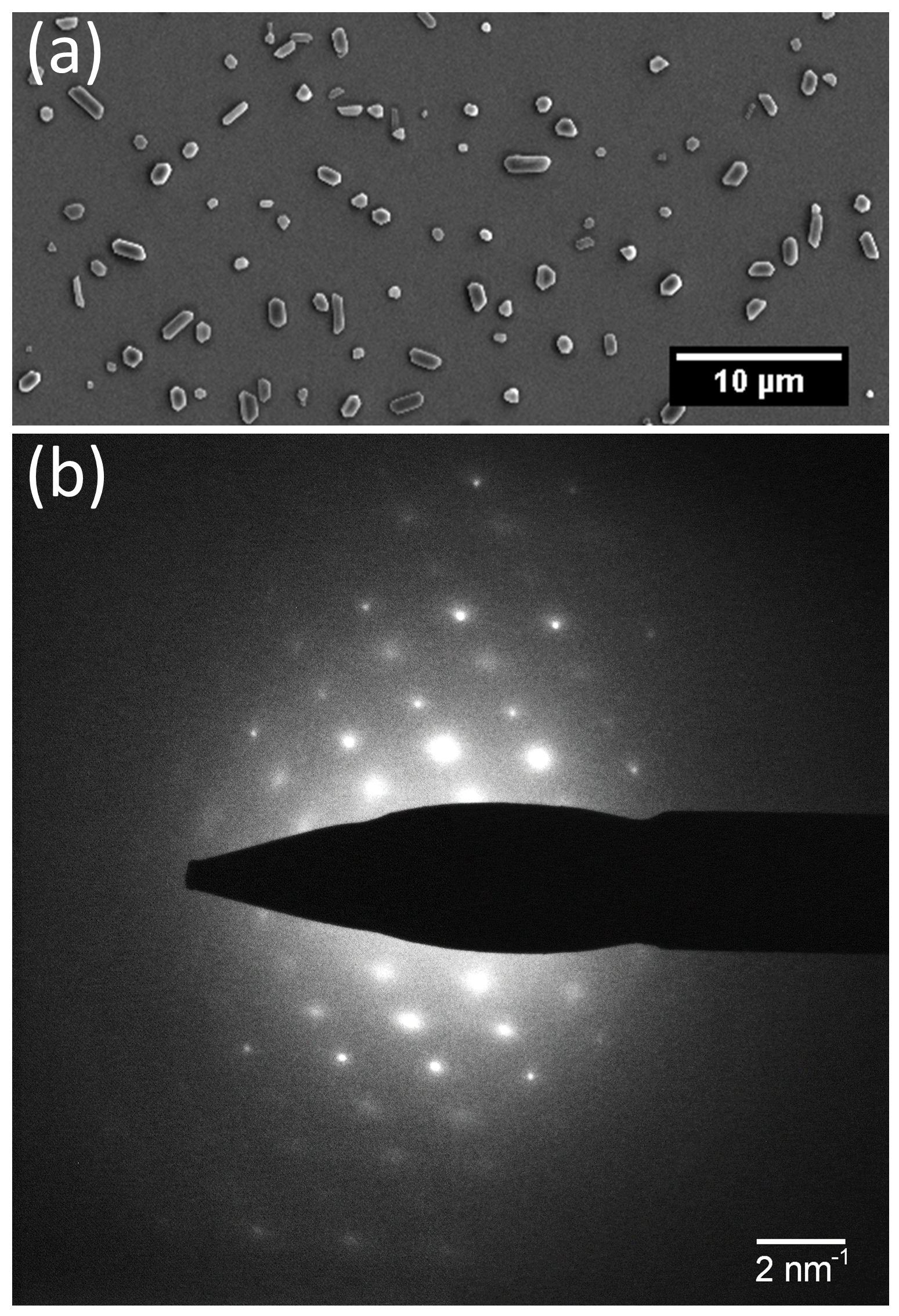
To maintain sufficient light guiding capabilities of the nanofiber–crystal system for spectroscopy and to ensure that the crystal stays tightly adhered to the vertically mounted nanofiber, the crystals have to be on the order of a few hundred nanometers in size. Such nanocrystals are grown by a reprecipitation method Kasai et al. (1992) from an oversaturated solution of a molar mixture of terrylene/p–terphenyl in toluene. The solution is heated until both compounds are dissolved and then isopropanol is added as a reprecipitation agent. This procedure results in terrylene doped p–terphenyl crystals of platelet morphology as seen in Fig. 2(a), which shows a scanning electron microscope (SEM) image of such crystals deposited on a silicon substrate. The majority of crystals have base dimensions in the range of 200 to 2000 nm and a base width to height ratio of 2.5:1 to 5:1 as determined by atomic force microscopy (AFM) measurements. The single-crystalline nature of these crystals has been verified by performing selected area electron diffraction (SAED) measurements using a transmission electron microscope (TEM), see Fig. 2(b). SAED also indicates that the substrate–supported crystal platelet base face is of (001) orientation, i.e. the crystal’s c-axis is perpendicular to the substrate. It is known that the dipole moment of the transition to the lowest electronically excited state in terrylene is linear and lies along the long axis of the molecule Bordat and Brown (2002). When inserted into a p–terphenyl crystal, the molecule’s long axis and thus the dipole moment lie nearly parallel to the crystal’s c-axis and therefore in our configuration nearly perpendicular to the substrate.
A terrylene–doped p–terphenyl nanocrystal is deposited on the nanofiber by a drop–touch method: a drop of the suspension of doped p–terphenyl nanocrystals is briefly brought into contact with the nanofiber via a pipette. During this process, the transmission of the excitation laser and the fluorescence of the nanofiber is monitored with a power meter and a spectrometer, respectively. When a doped crystal has adhered to the nanofiber surface during the contact with the suspension droplet, a typical fluorescence signal and some loss in transmission is observed. As crystals of a variety of sizes are produced during our growth process, we have to post–select the size of the deposited crystal. However, since the largest crystals sediment faster, we usually find suitably small ones in the suspension supernatant. If it is nevertheless found that the transmission deteriorates too much during the deposition process, the crystal can be washed off with acetone and another one deposited.
The TOF is mounted on a steel holder with two NdFeB magnets. This ensures that the fiber is firmly held and that it stays intact during the cooling process down to cryogenic temperatures. This fiber setup is mounted in the cold–pot of a custom–made cryostat that can cool the sample to . To ensure efficient thermalisation of the nanofiber and the crystal with the walls of the cold–pot, helium buffer gas at a pressure of a few mbar is introduced into the cold–pot before cool–down, after it has been evacuated.
To excite the molecules, light of a dye laser (Spectra-Physics Matisse-DS) is coupled into the optical fiber that connects to the TOF and that enters the cryostat via a teflon feed–through [Fig. 1(a)]. To compensate for intensity fluctuations and drifts, the laser beam is sent through an acousto–optic modulator (AOM) and partly onto a photodiode (PD) and is actively intensity stabilised. A fraction of the Stokes–shifted laser-induced fluorescence (LIF) of the molecule is collected by the nanofiber, fiber–guided out of the cryostat, and can be monitored by a spectrometer (Shamrock SR-303i, Andor Technology) or single photon counting modules (SPCMs) at either end of the optical fiber.

Contributions from the excitation light and fluorescence on the 00ZPL are filtered out by a long–pass (LP) filter. Together with a short–pass (SP) filter to block Raman scattering from the fiber, this leaves a transmission window in the range of 630–650 nm. Due to the inhomogeneous line shifts induced by the host crystal matrix, single molecules can be spectrally selected with the narrowband dye laser if the terrylene concentration in the host crystal is small enough.
III Experimental Results
Figure 3 shows the fluorescence excitation spectrum of a molecular ensemble in the X4 orientation. The excitation line of what has been verified to be a single molecule is highlighted in red.
To characterize the optical interface created by individual molecules and the optical nanofiber, we investigate several molecules that are located in the same nanocrystal. The transition frequencies of solid state quantum emitters are known to be very sensitive to the environment, which can be a favourable effect if controlled well Dolde et al. (2011) or lead to unwanted spectral diffusion. In order to evaluate the stability of the transition frequency in our system, we record the same spectral line of the spectra of individual molecules over time. Figure 4 shows the evolution of a spectral line over the timescale of minutes. The excitation power corresponds to a saturation parameter of and, over all spectra, the linewidth was measured to be MHz. A slow drift of MHz/s is observable.

This could be a result of a drift in the laser frequency, the wavemeter, or the molecular transition frequency. The faster frequency scatter on top of this drift is found to be MHz rms. This value is well below the lifetime-limited linewidth of the molecular transition (see Appendix A). Even for a lifetime-limited molecular spectrum and without any active stabilization this implies that the molecule would be resonant with a fixed-frequency laser for about two minutes. The measured stability of the molecule is thus found to be superior to other solid state emitters, which may require active stabilization of the resonance frequency on a faster timescale Robledo et al. (2010); Hansom et al. (2014b). By cooling the doped crystal to 1.7 K, we can also achieve stable lifetime-limited spectra of our single molecules recorded on either end of the TOF, as is experimentally shown in Appendix A.
To verify the interaction with single quantum emitters, second order fluorescence intensity correlation measurements are performed using a Hanbury-Brown-Twiss setup [Fig. 1(a)]. The backscattered fluorescence is split by a 50/50 beamsplitter (BS) and directed to two SPCMs. Their counts are then recorded by means of a field programmable gate array and correlated. For a single quantum emitter, photon antibunching is observed, which manifests itself as an antibunching dip in the intensity correlation measurements at zero time delay between two detection events. Experimental imperfections such as incoherent background scatter will reduce the contrast of the dip but a dip deeper than 50% is prove of a single quantum emitter. Figure 5 shows second order fluorescence intensity correlation measurements of a molecule on the nanofiber where the antibunching dip is clearly visible.
As the excitation intensity is increased, the onset of Rabi oscillations is apparent. Since the decay into the triplet state is negligible for short times, the measurements are fitted to the correlation function that is obtained by solving the optical Bloch equations for a two level system with an overall amplitude , a spontaneous decay rate and a Rabi frequency , where is the dipole moment of the molecule’s transition and E the electric field at the molecule’s location Loudon (2000).
| (2) | ||||
| with | ||||
An incoherent Poissonian background added to a signal with average intensity reduces the contrast of the intensity correlation measurements. This is taken into account when analyzing our data with the modified intensity correlation function Verberk and Orrit (2003)
| (3) |
We fit eqn.(3) to the data for different excitation powers while using as a global fit parameter for all measurements on a given molecule. This yields the respective Rabi frequencies and the non–power broadenend homogeneous linewidth of the molecule. From these fits we also obtain the saturation intensity of the molecule as .

Figure 6 shows this expected linear increase of the squared Rabi frequency as a function of excitation power for three different molecules in the same crystal. The error bars obtained from fitting the intensity correlation measurements are smaller than the depicted datapoints in Fig. 6. From the fits, the saturation power corresponding to is obtained. We obtain saturation powers for molecule A of nW, molecule B of nW and molecule C of nW. The biggest contribution to the error in the saturation powers arises from the uncertainty in determining the power in the fiber. We convert to the maximum intensity at the surface of the nanofiber by considering the fundamental quasilinearly polarised HE11 mode that is supported by our optical nanofiber with a diameter of 320 nm Le Kien et al. (2005a). Without the exact knowledge of the orientation of the molecule’s transition dipole moment and its distance from the nanofiber surface, this gives an upper limit for the saturation intensity. The measured saturation intensities are Wcm-2 for molecule A, Wcm-2 for molecule B and Wcm-2 for molecule C. These results compare well with results obtained by other groups who studied terrylene in bulk p–terphenyl Kummer et al. (1994); Tamarat et al. (1995). To our knowledge a saturation intensity of Wcm-2 as for molecule C is the lowest measured so far for terrylene in p–terphenyl. As opposed to measurements on terrylene in bulk p–terphenyl using a confocal microscope, the excitation light in our case enters through the side of the thin host crystal platelets. As the transition dipole moment of the molecules lies nearly perpendicular to the base of these platelets, this suggests an improved overlap between the polarization of the nanofiber–guided excitation light and the transition dipole moment of the molecules.

An independent measurement of the saturation power is obtained by recording the resonant fluorescence rate as a function of excitation power as plotted in Fig. 7, which includes a fit to . The error bars on the fluorescence rate that are obtained by taking the standard error of the amplitude from fits over several molecular spectra are smaller than the depicted datapoints. This measurement yields a saturation power of nW for molecule B, where the error stems from the fit and from the uncertainty in the excitation power inside the fiber. This translates into a saturation intensity of Wcm-2 for molecule B in good agreement with Wcm-2 as obtained with the HBT setup.

We performed a further measurement on a fourth molecule (molecule D) that yielded a lower fluorescence rate and indeed was measured to have a higher saturation power of nW and therefore a saturation intensity of Wcm-2. This suggests that this molecule is located further away from the nanofiber surface such that its fluorescence does not couple back to the nanofiber–guided modes as efficiently. Alternatively, the alignment between polarization of the excitation light and the dipole moment of molecule D may be less favorable. Although all molecules are embedded in a single crystal and therefore have the same orientation, the inherent birefringence of the host crystal can cause a less favorable alignment: The phase shift between the corresponding polarization components is , where is the vacuum wavelength, the propagation distance and is the effective birefringence that can reach 0.32 in our case.

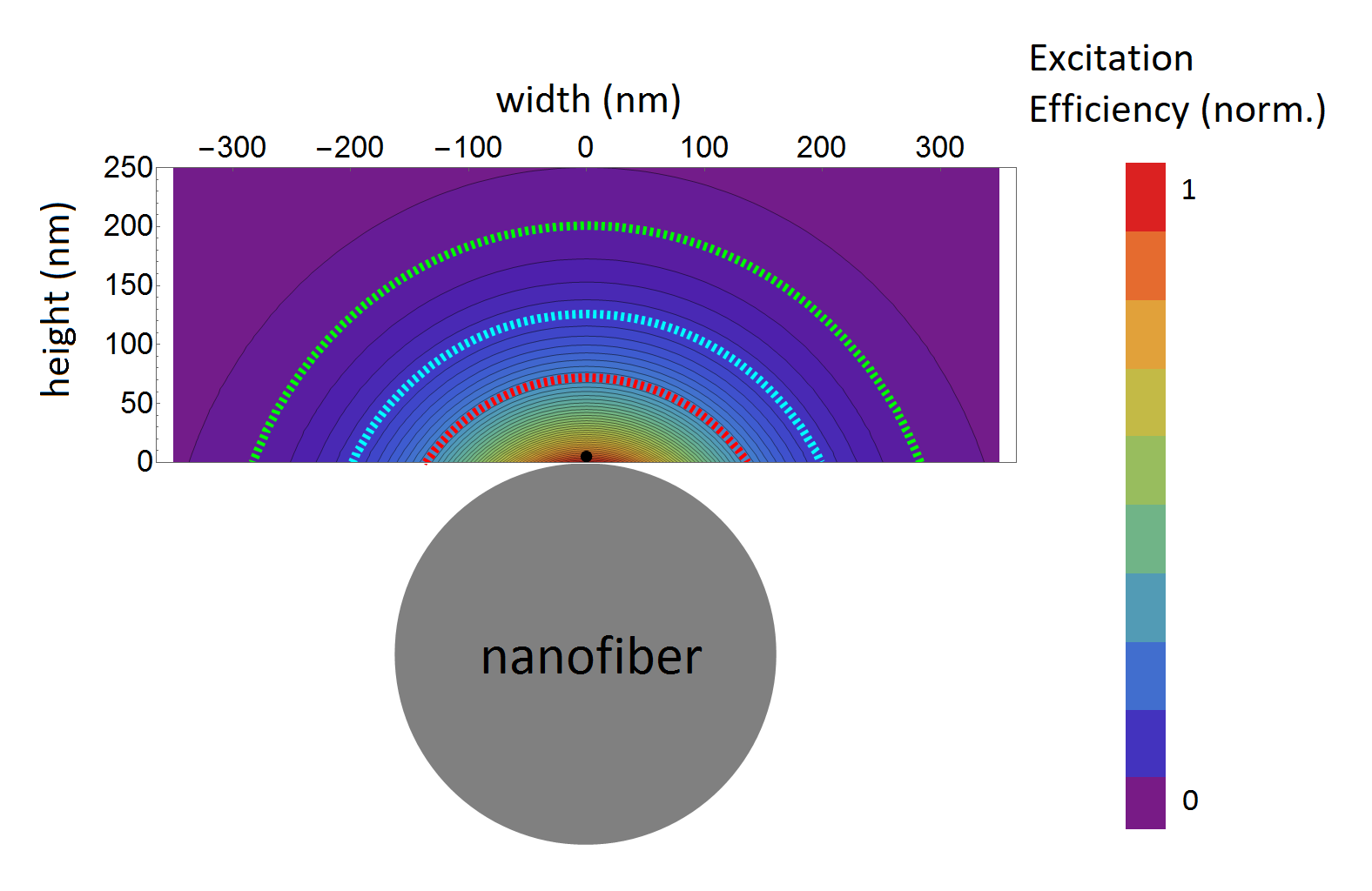
The efficiency of exciting the different molecules is given by , where is the effective mode area at the position of the molecule. More details can be found in Appendix B. The molecules are also detected via the nanofiber interface and hence the overall efficiency for fluorescence excitation and detection via the nanofiber interface is then given as , where is the coupling efficiency of dipole radiation to the nanofiber modes. This coupling efficiency depends on the radiated wavelength, distance and orientation of the dipole with respect to the nanofiber surface. Here, is the scattering rate into guided modes and is the total scattering rate of the dipole. For our case of a nanofiber with radius and Stokes shifted fluorescence in the range of 630–650 nm, we calculated this coupling efficiency for a radially, azimuthally and axially oriented dipole following Le Kien et al. (2005a), see Fig. 8.
Since the power needed to saturate molecule C is the lowest yet measured for terrylene in p–terphenyl, we assume that this molecule is very close to the surface of the nanofiber. An upper limit for the other molecules from the nanofiber surface can then be estimated by comparing their saturation intensities. Because the host crystal is birefringent, we only give an upper limit on the radial distance between the different molecules. Figure 9 shows the calculated efficiency of fluorescence excitation via the nanofiber interface for different positions of the dipole with respect to the nanofiber surface. The positions are chosen to lie within the volume of a platelet crystal with its base on the nanofiber surface. The molecular dipole is oriented perpendicular to the crystal’s base and excited by quasilinearly polarised light via the nanofiber–based interface. Assuming unpolarised light instead of quasilinearly polarised light affects the relative distances between the different molecules by less than 3%. We did not incorporate the refractive indices of the crystal into this model because they would make the local efficiencies very dependent on the crystal’s geometry and we are only interested in assigning the maximum radial distance of the measured molecules. The maximum radial distances of the investigated molecules are depicted in their respective colours by dashed contours [Fig. 9]. These results show that they radially all lie within less than of each other and less than from the nanofiber surface. This translates into coupling efficiencies for a radial dipole to the nanofiber mode between 5-30% [Fig. 8]. This means that our set-up can be a superior choice for coupling single photons to single mode optical fibers compared to using conventional confocal microscopes Fujiwara et al. (2011); Schröder et al. (2011); Karlsson et al. (2016) and thus opens the way for fully fiber–coupled single photon sources. It is also an important step towards strong coupling of single molecules to optical waveguide structures.
IV Conclusion
Summarizing, we have shown how single molecules can be optically interfaced via the evanescent field surrounding an optical nanofiber. This is an important addition to the toolbox of quantum emitters such as atoms Vetsch et al. (2010); Goban et al. (2012); Hafezi et al. (2012), quantum dots Yalla et al. (2012); Fujiwara et al. (2011) and NV centers in nanodiamonds Liebermeister et al. (2014); Fujiwara et al. (2015) that have been fiber–integrated by coupling to optical nanofibers. Each of these systems has its own intrinsic advantages for their usage in quantum networks. Single molecules in solids are efficient quantum emitters that come in a large variety of emission wavelengths. This makes them suitable to be interfaced with other quantum emitters Siyushev et al. (2014). They have an advantageous level structure for the implementation of triggered single photon sources Moerner (2004); Lounis and Moerner (2000) and have proven their versatility in quantum optics Hwang et al. (2009); Pototschnig et al. (2011); Maser et al. (2016). Further, single waveguide–coupled molecules allow the investigation of photon–mediated interactions between two quantum emitters even when they are separated by much more than the excitation wavelength Rist et al. (2008); Loo et al. (2013); Le Kien et al. (2005b). These interactions can be further enhanced by using a nanofiber between two fiber Bragg gratings and thereby realizing a high–Q cavity Wuttke et al. (2012). Single molecules that are coupled to the evanescent field of optical nanofibers therefore not only offer a rich experimental platform for investigating entanglement and correlations between quantum emitters, they also provide a means for implementing components of quantum networks such as fiber–coupled single photon sources Ahtee et al. (2009); Hwang and Hinds (2011); Polisseni et al. (2016) or photon sorters Witthaut et al. (2012); Ralph et al. (2015).
Acknowledgements.
The scanning electron microscope imaging has been carried out using facilities at the University Service Centre for Transmission Electron Microscopy (USTEM), Vienna University of Technology, Austria. We gratefully acknowledge financial support by the Austrian Science Fund (FWF Lise Meitner project No. M 2114-N27) and the Federal Ministry of Science, Research and Economy of Austria.Appendix A Lifetime-limited single photons from a fully fiber-integrated single molecule
To show that we can readily produce fiber-coupled lifetime-limited photons from single molecules in nanocrystals with our set-up, we have measured molecular spectra and corresponding fluorescence correlation measurements at a temperature of 1.7 K. These measurements were performed on a different
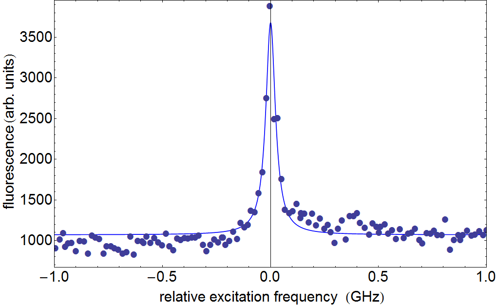
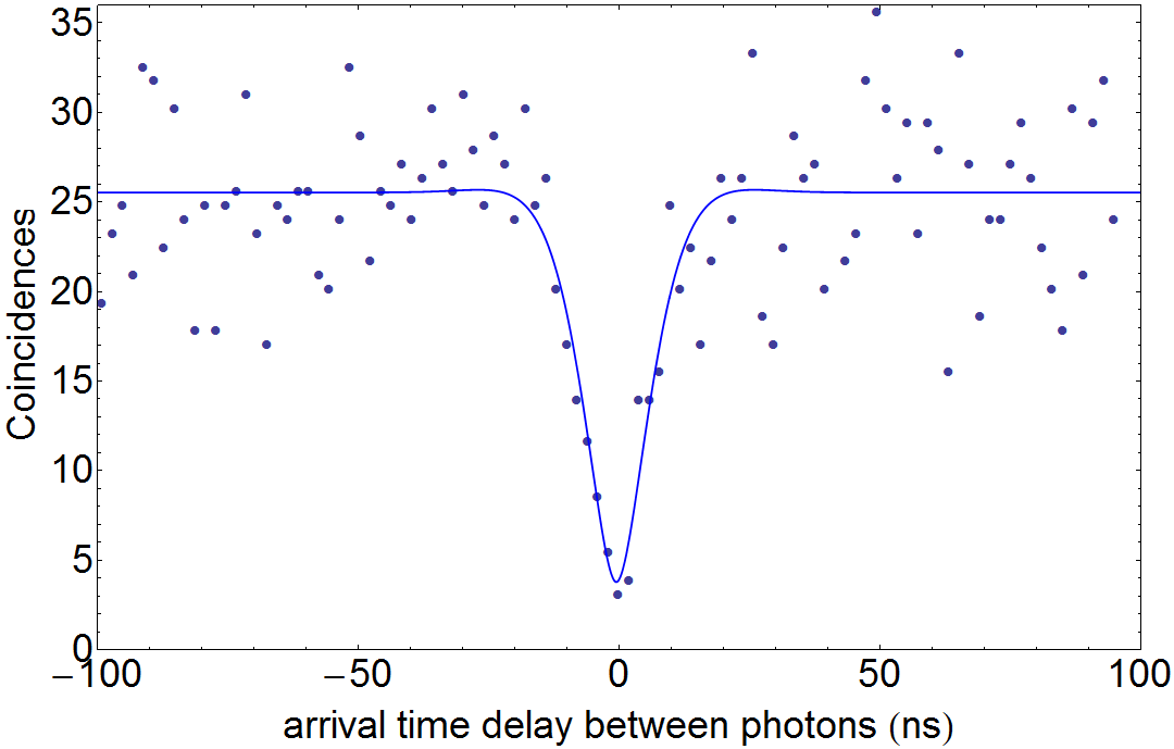
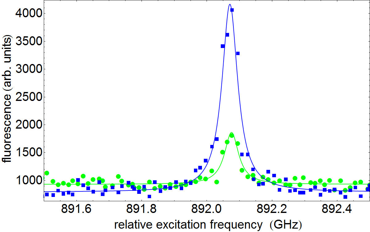
sample mounted in a helium flow cryostat (Janis) that can cool samples continuously below 2 K. Figure 10 shows a molecular spectrum at 1.7 K that has a linewidth of MHz and Figure 11 shows the corresponding second order fluorescence correlation measurement yielding a linewidth of MHz in agreement with that obtained from the spectrum. We can measure such spectra on both ends of the tapered optical fiber as shown in Figure 12. This underpins the fully fiber-coupled nature of our single molecules.
Appendix B Excitation efficiency of a molecule on the optical nanofiber surface
The efficiency of exciting a molecule via the nanofiber interface is given by , where is the effective mode area of the light field at the position of the molecule. Here, we want to estimate the excitation efficiency for a single molecule that is situated on the surface of the optical nanofiber at position (see Figure 9 in main text) and has a radially oriented transition dipole moment with respect to the nanofiber. It is excited by a mode that is quasilinearly polarised and for which the transverse polarisation component is aligned with the direction of the molecule’s dipole moment. The effective mode area on the surface is then given as , where is the surface intensity at and is the overlap between the unit vector of the molecular dipole moment and the polarisation vector at the position of the molecule. On the surface this then yields an effective mode area of 0.4 , with an excitation wavelength of 580 nm and for a nanofiber with a diameter of 320 nm. We deduce a value for the absorption cross-section of the molecule from our saturation intensity measurements. The saturation intensity of the molecule closest to the nanofiber has been Wcm-2. The intensity needed to saturate a molecular transition depends on its molecular dipole moment as Jelezko and Wrachtrup (2004)
| (4) |
Here, to obtain a correct value for the magnitude of the dipole moment , the metastable triplet state, which is split into two levels without an external field, has to be taken into account. In this case, . are the transition rates from level to level and the superscripts ′ and ′′ represent the two non degenerate levels of the triplet level . represents the total dephasing rate of the excited state. Using equation 4 and the triplet state parameters for terrylene in p-terphenyl from Hegerfeldt and Seidel (2003), we obtain Debye for the transition dipole moment of terrylene in p-terphenyl from the ground state to the first electronically excited state. The molecular dipole moment yields a minimum oscillator strength for this transition as Bransden and Joachain (2003):
| (5) |
This gives an effective absorption cross-section of and hence an excitation probability of a single molecule on the nanofiber surface of 17%.
References
- Siyushev et al. (2014) P. Siyushev, G. Stein, J. Wrachtrup, and I. Gerhardt, “Molecular photons interfaced with alkali atoms,” Nature 509, 66–70 (2014).
- Basché et al. (1995) Th Basché, S. Kummer, and C. Bräuchle, “Direct spectroscopic observation of quantum jumps of a single molecule,” Nature 373, 132–134 (1995).
- Basché et al. (1992) Th. Basché, W. E. Moerner, M. Orrit, and H. Talon, “Photon antibunching in the fluorescence of a single dye molecule trapped in a solid,” Phys. Rev. Lett. 69, 1516–1519 (1992).
- Celebrano et al. (2011) M. Celebrano, P. Kukura, A. Renn, and V. Sandoghdar, “Single-molecule imaging by optical absorption,” Nat. Photon. 5, 95 (2011).
- Gerhardt et al. (2007) I. Gerhardt, G. Wrigge, P. Bushev, G. Zumofen, M. Agio, R. Pfab, and V. Sandoghdar, “Strong Extinction of a Laser Beam by a Single Molecule,” Phys. Rev. Lett. 98, 033601 (2007).
- Gerhardt et al. (2010) I. Gerhardt, G. Wrigge, J. Hwang, G. Zumofen, and V. Sandoghdar, “Coherent nonlinear single-molecule microscopy,” Phys. Rev. A 82, 063823 (2010).
- Kozankiewicz and Orrit (2014) B. Kozankiewicz and M. Orrit, “Single-molecule photophysics, from cryogenic to ambient conditions,” Chem. Soc. Rev. 43, 1029–1043 (2014).
- Jelezko and Wrachtrup (2004) F. Jelezko and J. Wrachtrup, “Read-out of single spins by optical spectroscopy,” J. Phys.: Condens. Matter 16, R1089 (2004).
- Maurer et al. (2012) P. C. Maurer, G. Kucsko, C. Latta, L. Jiang, N. Y. Yao, S. D. Bennett, F. Pastawski, D. Hunger, N. Chisholm, M. Markham, D. J. Twitchen, J. I. Cirac, and M. D. Lukin, “Room-Temperature Quantum Bit Memory Exceeding One Second,” Science 336, 1283–1286 (2012).
- Neumann et al. (2008) P. Neumann, N. Mizuochi, F. Rempp, P. Hemmer, H. Watanabe, S. Yamasaki, V. Jacques, T. Gaebel, F. Jelezko, and J. Wrachtrup, “Multipartite Entanglement Among Single Spins in Diamond,” Science 320, 1326–1329 (2008).
- Pingault et al. (2014) B. Pingault, J. N. Becker, C. H. H. Schulte, C. Arend, C. Hepp, T. Godde, A. I. Tartakovskii, M. Markham, C. Becher, and M. Atatüre, “All-Optical Formation of Coherent Dark States of Silicon-Vacancy Spins in Diamond,” Phys. Rev. Lett. 113, 263601 (2014).
- Hansom et al. (2014a) J. Hansom, C. H. H. Schulte, C. Le Gall, C. Matthiesen, E. Clarke, M. Hugues, J. M. Taylor, and M. Atatüre, “Environment-assisted quantum control of a solid-state spin via coherent dark states,” Nat. Phys. 10, 725–730 (2014a).
- Delteil et al. (2016) A. Delteil, Z. Sun, W. Gao, E. Togan, S. Faelt, and A. Imamoğlu, “Generation of heralded entanglement between distant hole spins,” Nat. Phys. 12, 218–223 (2016).
- Yalla et al. (2014) R. Yalla, M. Sadgrove, K. P. Nayak, and K. Hakuta, “Cavity quantum electrodynamics on a nanofiber using a composite photonic crystal cavity,” Phys. Rev. Lett. 113, 143601 (2014).
- Arcari et al. (2014) M. Arcari, I. Söllner, A. Javadi, S. Lindskov Hansen, S. Mahmoodian, J. Liu, H. Thyrrestrup, E. H. Lee, J. D. Song, S. Stobbe, and P. Lodahl, “Near-unity coupling efficiency of a quantum emitter to a photonic crystal waveguide,” Phys. Rev. Lett. 113, 093603 (2014).
- Javadi et al. (2015) A. Javadi, I. Söllner, M. Arcari, S. Lindskov Hansen, L. Midolo, S. Mahmoodian, G. Kirs̆anskė, T. Pregnolato, E. H. Lee, J. D. Song, S. Stobbe, and P. Lodahl, “Single-photon non-linear optics with a quantum dot in a waveguide,” Nat. Commun. 6, 8655 (2015).
- Nemoto et al. (2014) K. Nemoto, M. Trupke, S. J. Devitt, A. M. Stephens, B. Scharfenberger, K. Buczak, T. Nöbauer, M. S. Everitt, J. Schmiedmayer, and W. J. Munro, “Photonic Architecture for Scalable Quantum Information Processing in Diamond,” Phys. Rev. X 4, 031022 (2014).
- Kimble (2008) H. J. Kimble, “The quantum internet,” Nature 453, 1023–1030 (2008).
- Doherty et al. (2014) M. W. Doherty, V. V. Struzhkin, D. A. Simpson, L. P. McGuinness, Y. Meng, A. Stacey, T. J. Karle, R. J. Hemley, N. B. Manson, Lloyd C. L. Hollenberg, and S. Prawer, “Electronic Properties and Metrology Applications of the Diamond NV- Center under Pressure,” Phys. Rev. Lett. 112, 047601 (2014).
- Goldstein et al. (2011) G. Goldstein, P. Cappellaro, J. R. Maze, J. S. Hodges, L. Jiang, A. S. Sørensen, and M. D. Lukin, “Environment-Assisted Precision Measurement,” Phys. Rev. Lett. 106, 140502 (2011).
- Acosta et al. (2013) V. M. Acosta, K. Jensen, C. Santori, D. Budker, and R. G. Beausoleil, “Electromagnetically Induced Transparency in a Diamond Spin Ensemble Enables All-Optical Electromagnetic Field Sensing,” Phys. Rev. Lett. 110, 213605 (2013).
- Faez et al. (2014a) S. Faez, S. J. van der Molen, and M. Orrit, “Optical tracing of multiple charges in single-electron devices,” Phys. Rev. B 90, 205405 (2014a).
- Mazzamuto et al. (2014) G. Mazzamuto, A. Tabani, S. Pazzagli, S. Rizvi, A. Reserbat-Plantey, K. Schädler, G. Navickaite, L. Gaudreau, F. S. Cataliotti, F. Koppens, and C. Toninelli, “Single-molecule study for a graphene-based nano-position sensor,” New J. Phys. 16, 113007 (2014).
- Kucsko et al. (2013) G. Kucsko, P. C. Maurer, N. Y. Yao, M. Kubo, H. J. Noh, P. K. Lo, H. Park, and M. D. Lukin, “Nanometre-scale thermometry in a living cell,” Nature 500, 54–58 (2013).
- Lukin and Imamoğlu (2001) M. D. Lukin and A. Imamoğlu, “Controlling photons using electromagnetically induced transparency,” Nature 413, 273–276 (2001).
- Reim et al. (2010) K. F. Reim, J. Nunn, V. O. Lorenz, B. J. Sussman, K. C. Lee, N. K. Langford, D. Jaksch, and I. A. Walmsley, “Towards high-speed optical quantum memories,” Nat. Photon. 4, 218–221 (2010).
- Volz et al. (2014) J. Volz, M. Scheucher, C. Junge, and A. Rauschenbeutel, “Nonlinear π phase shift for single fibre-guided photons interacting with a single resonator-enhanced atom,” Nat. Photon. 8, 965–970 (2014).
- Thompson et al. (2013) J. D. Thompson, T. G. Tiecke, N. P. de Leon, J. Feist, A. V. Akimov, M. Gullans, A. S. Zibrov, V. Vuletic, V., and M. D. Lukin, “Coupling a Single Trapped Atom to a Nanoscale Optical Cavity,” Science 340, 1202–1205 (2013).
- Ritter et al. (2012) S. Ritter, C. Nölleke, C. Hahn, A. Reiserer, A. Neuzner, M. Uphoff, M. Mücke, E. Figueroa, J. Bochmann, and G. Rempe, “An elementary quantum network of single atoms in optical cavities,” Nature 484, 195–200 (2012).
- Schietinger et al. (2008) S. Schietinger, T. Schröder, and O. Benson, “One-by-One Coupling of Single Defect Centers in Nanodiamonds to High-Q Modes of an Optical Microresonator,” Nano Lett. 8, 3911–3915 (2008).
- Faez et al. (2014b) S. Faez, P. Türschmann, H. R. Haakh, S. Götzinger, and V. Sandoghdar, “Coherent Interaction of Light and Single Molecules in a Dielectric Nanoguide,” Phys. Rev. Lett. 113, 213601 (2014b).
- Hwang and Hinds (2011) J. Hwang and E. A. Hinds, “Dye molecules as single-photon sources and large optical nonlinearities on a chip,” New J. Phys. 13, 085009 (2011).
- Yalla et al. (2012) R. Yalla, Fam Le Kien, M. Morinaga, and K. Hakuta, “Efficient Channeling of Fluorescence Photons from Single Quantum Dots into Guided Modes of Optical Nanofiber,” Phys. Rev. Lett. 109, 063602 (2012).
- Vetsch et al. (2010) E. Vetsch, D. Reitz, G. Sagué, R. Schmidt, S. T. Dawkins, and A. Rauschenbeutel, “Optical Interface Created by Laser-Cooled Atoms Trapped in the Evanescent Field Surrounding an Optical Nanofiber,” Phys. Rev. Lett. 104, 203603 (2010).
- Kirs̆anskė et al. (2017) G. Kirs̆anskė, H. Thyrrestrup, R. S. Daveau, C. L. Dree en, T. Pregnolato, L. Midolo, P. Tighineanu, A. Javadi, S. Stobbe, R. Schott, A. Ludwig, A. D. Wieck, S. I. Park, J. D. Song, A. V. Kuhlmann, I. Söllner, C. Löbl, R. J. Warburton, and P. Lodahl, “Indistinguishable and efficient single photons from a quantum dot in a planar nanobeam waveguide,” arXiv1701.08131 (2017).
- (36) P. E. Lombardi, A. P. Ovvyan, S. Pazzagli, G. Mazzamuto, G. Kewes, O. Neitzke, N. Gruhler, O. Benson, W. H. P. Pernice, F. S. Cataliotti, and C. Toninelli, “Photostable molecules on chip: Integrated sources of nonclassical light,” ACS Photonics, doi:10.1021/acsphotonics.7b00521 .
- Garcia-Fernandez et al. (2011) R. Garcia-Fernandez, W. Alt, F. Bruse, C. Dan, K. Karapetyan, O. Rehband, A. Stiebeiner, U. Wiedemann, D. Meschede, and A. Rauschenbeutel, “Optical nanofibers and spectroscopy,” Appl. Phys. B 105, 3–15 (2011).
- Le Kien et al. (2005a) Fam Le Kien, S. Dutta Gupta, V. I. Balykin, and K. Hakuta, “Spontaneous emission of a cesium atom near a nanofiber: Efficient coupling of light to guided modes,” Phys. Rev. A 72, 032509 (2005a).
- Reitz et al. (2013) D. Reitz, C. Sayrin, R. Mitsch, P. Schneeweiss, and A. Rauschenbeutel, “Coherence Properties of Nanofiber-Trapped Cesium Atoms,” Phys. Rev. Lett. 110, 243603 (2013).
- Nayak et al. (2007) K. P. Nayak, P. N. Melentiev, M. Morinaga, Fam Le Kien, V. I. Balykin, and K. Hakuta, “Optical nanofiber as an efficient tool for manipulating and probing atomicFluorescence,” Opt. Express 15, 5431–5438 (2007).
- Liebermeister et al. (2014) L. Liebermeister, F. Petersen, A. v Münchow, D. Burchardt, J. Hermelbracht, T. Tashima, A. W. Schell, O. Benson, T. Meinhardt, A. Krueger, A. Stiebeiner, A. Rauschenbeutel, H. Weinfurter, and M. Weber, “Tapered fiber coupling of single photons emitted by a deterministically positioned single nitrogen vacancy center,” Appl. Phys. Lett. 104, 031101 (2014).
- Kummer et al. (1994) S. Kummer, Th Basché, and C. Bräuchle, “Terrylene in p-terphenyl: a novel single crystalline system for single molecule spectroscopy at low temperatures,” Chem. Phys. Lett. 229, 309 – 316 (1994).
- Moerner (2004) W. E. Moerner, “Single-photon sources based on single molecules in solids,” New J. Phys. 6, 88 (2004).
- Moerner and Orrit (1999) W. E. Moerner and Michel Orrit, “Illuminating Single Molecules in Condensed Matter,” Science 283, 1670–1676 (1999).
- Michaelis et al. (1999) J. Michaelis, C. Hettich, A. Zayats, B. Eiermann, J. Mlynek, and V. Sandoghdar, “A single molecule as a probe of optical intensity distribution,” Opt. Lett. 24, 581 (1999).
- Tamarat et al. (2000) Ph. Tamarat, A. Maali, B. Lounis, and M. Orrit, “Ten Years of Single-Molecule Spectroscopy,” J. Phys. Chem. A 104, 1–16 (2000).
- Türschmann et al. (2017) P. Türschmann, N. Rotenberg, J. Renger, I. Harder, O. Lohse, T. Utikal, S. Götzinger, and V. Sandoghdar, “On-chip linear and nonlinear control of single molecules coupled to a nanoguide,” Nano Lett. 17, 4941–4945 (2017).
- Wang et al. (2017) D. Wang, H. Kelkar, D. Martin-Cano, T. Utikal, S. Götzinger, and V. Sandoghdar, “Coherent coupling of a single molecule to a scanning fabry-perot microcavity,” Physical Review X 7, 021014 (2017).
- Chizhik et al. (2009) A. Chizhik, F. Schleifenbaum, R. Gutbrod, A. Chizhik, D. Khoptyar, and A. J. Meixner, “Tuning the fluorescence emission spectra of a single molecule with a variable optical subwavelength metal microcavity,” Phys. Rev. Lett. 102, 073002 (2009).
- Norris et al. (1997) D. J. Norris, M. Kuwata-Gonokami, and W. E. Moerner, “Excitation of a single molecule on the surface of a spherical microcavity,” Appl. Phys. Lett. 71 (1997).
- Warken et al. (2008) F. Warken, A. Rauschenbeutel, and T. Bartholomaus, “Fiber Pulling Profits from Precise Positioning-Precise motion control improves manufacturing of fiber optical resonators,” Photonics Spectra 42, 73 (2008).
- Stiebeiner et al. (2010) A. Stiebeiner, R. Garcia-Fernandez, and A. Rauschenbeutel, “Design and optimization of broadband tapered optical fibers with a nanofiber waist,” Opt. Express 18, 22677–22685 (2010).
- Bordat and Brown (2002) P. Bordat and R. Brown, “Molecular mechanisms of photo-induced spectral diffusion of single terrylene molecules in p terphenyl,” J. Chem. Phys. 116, 229 (2002).
- Loudon (2000) R. Loudon, The Quantum Theory of Light (OUP Oxford, 2000).
- Warken et al. (2007) F. Warken, E. Vetsch, D. Meschede, M. Sokolowski, and A. Rauschenbeutel, “Ultra-sensitive surface absorption spectroscopy using sub-wavelength diameter optical fibers,” Opt. Express 15, 11952 (2007).
- Banasiewicz et al. (2005) M. Banasiewicz, O. Morawski, D. Wiącek, and B. Kozankiewicz, “Triplet population and depopulation rates of single terrylene molecules in p-terphenyl crystal,” Chem. Phys. Lett. 414, 374–377 (2005).
- Li and Egerton (2004) P. Li and R. F. Egerton, “Radiation damage in coronene, rubrene and p-terphenyl, measured for incident electrons of kinetic energy between 100 and 200 kev,” Ultramicroscopy 101, 161–172 (2004).
- Rice et al. (2012) Andrew P. Rice, Fook S. Tham, and Eric L. Chronister, “A Temperature Dependent X-ray Study of the Order–Disorder Enantiotropic Phase Transition of p-Terphenyl,” J. Chem. Crystallogr. 43, 14–25 (2012).
- Kasai et al. (1992) H. Kasai, H. S. Nalwa, H. Oikawa, S. Okada, H. Matsuda, N. Minami, A. Kakuta, K. Ono, A. Mukoh, and H. Nakanishi, “A Novel Preparation Method of Organic Microcrystals,” Jpn. J. Appl. Phys. 31, L1132–L1134 (1992).
- Dolde et al. (2011) F. Dolde, H. Fedder, M. W. Doherty, T. Nöbauer, F. Rempp, G. Balasubramanian, T. Wolf, F. Reinhard, L. C. L. Hollenberg, F. Jelezko, and J. Wrachtrup, “Electric-field sensing using single diamond spins,” Nat. Phys. 7, 459–463 (2011).
- Robledo et al. (2010) Lucio Robledo, Hannes Bernien, Ilse van Weperen, and Ronald Hanson, “Control and coherence of the optical transition of single nitrogen vacancy centers in diamond,” Physical Review Letters 105, 177403 (2010).
- Hansom et al. (2014b) Jack Hansom, Carsten H. H. Schulte, Clemens Matthiesen, Megan J. Stanley, and Mete Atat re, “Frequency stabilization of the zero-phonon line of a quantum dot via phonon-assisted active feedback,” Appl. Phys. Lett. 105, 172107 (2014b).
- Verberk and Orrit (2003) R. Verberk and M. Orrit, “Photon statistics in the fluorescence of single molecules and nanocrystals: Correlation functions versus distributions of on- and off-times,” J. Chem. Phys. 119, 2214–2222 (2003).
- Tamarat et al. (1995) Ph. Tamarat, B. Lounis, J. Bernard, M. Orrit, S. Kummer, R. Kettner, S. Mais, and Th. Basché, “Pump-Probe Experiments with a Single Molecule: ac-Stark Effect and Nonlinear Optical Response,” Phys. Rev. Lett. 75, 1514–1517 (1995).
- Fujiwara et al. (2011) M. Fujiwara, K. Toubaru, T. Noda, H. Zhao, and S. Takeuchi, “Highly Efficient Coupling of Photons from Nanoemitters into Single-Mode Optical Fibers,” Nano Lett. 11, 4362–4365 (2011).
- Schröder et al. (2011) T. Schröder, F. Gädeke, M. J. Banholzer, and O. Benson, “Ultrabright and efficient single-photon generation bassed on nitrogen-vacancy centres in nanodiamonds on a solid immersion lens,” New J. Phys. 13, 055017 (2011).
- Karlsson et al. (2016) J. Karlsson, L. Rippe, and S. Kröll, “A confocal optical microscope for detection of single impurities in a bulk crystal at cryogenic temperatures,” Rev. Sci. Instrum. 87, 033701 (2016).
- Goban et al. (2012) A. Goban, K. S. Choi, D. J. Alton, D. Ding, C. Lacroûte, M. Pototschnig, T. Thiele, N. P. Stern, and H. J. Kimble, “Demonstration of a State-Insensitive, Compensated Nanofiber Trap,” Phys. Rev. Lett. 109, 033603 (2012).
- Hafezi et al. (2012) M. Hafezi, Z. Kim, S. L. Rolston, L. A. Orozco, B. L. Lev, and J. M. Taylor, “Atomic interface between microwave and optical photons,” Phys. Rev. A 85, 020302 (2012).
- Fujiwara et al. (2015) M. Fujiwara, H. Zhao, T. Noda, K. Ikeda, H. Sumiya, and S. Takeuchi, “Ultrathin fiber-taper coupling with nitrogen vacancy centers in nanodiamonds at cryogenic temperatures,” Opt. Lett. 40, 5702 (2015).
- Lounis and Moerner (2000) B. Lounis and W. E. Moerner, “Single photons on demand from a single molecule at room temperature,” Nature 407, 491–493 (2000).
- Hwang et al. (2009) J. Hwang, M. Pototschnig, R. Lettow, G. Zumofen, A. Renn, S. Götzinger, and V. Sandoghdar, “A single-molecule optical transistor,” Nature 460, 76–80 (2009).
- Pototschnig et al. (2011) M. Pototschnig, Y. Chassagneux, J. Hwang, G. Zumofen, A. Renn, and V. Sandoghdar, “Controlling the Phase of a Light Beam with a Single Molecule,” Phys. Rev. Lett. 107, 063001 (2011).
- Maser et al. (2016) A. Maser, B. Gmeiner, T. Utikal, S. Götzinger, and V. Sandoghdar, “Few-photon coherent nonlinear optics with a single molecule,” Nat. Photon. 10, 450–453 (2016).
- Rist et al. (2008) S. Rist, J. Eschner, M. Hennrich, and G. Morigi, “Photon-mediated interaction between two distant atoms,” Phys. Rev. A 78, 013808 (2008).
- Loo et al. (2013) A. F. van Loo, A. Fedorov, K. Lalumière, B. C. Sanders, A. Blais, and A. Wallraff, “Photon-Mediated Interactions Between Distant Artificial Atoms,” Science 342, 1494–1496 (2013).
- Le Kien et al. (2005b) Fam Le Kien, S. Dutta Gupta, K. P. Nayak, and K. Hakuta, “Nanofiber-mediated radiative transfer between two distant atoms,” Phys. Rev. A 72, 063815 (2005b).
- Wuttke et al. (2012) C. Wuttke, M. Becker, S. Brückner, M. Rothhardt, and A. Rauschenbeutel, “Nanofiber Fabry–Perot microresonator for nonlinear optics and cavity quantum electrodynamics,” Opt. Lett. 37, 1949–1951 (2012).
- Ahtee et al. (2009) V. Ahtee, R. Lettow, R. Pfab, A. Renn, E. Ikonen, S. Gotzinger, and V. Sandoghdar, “Molecules as sources for indistinguishable single photons,” J. Mod. Opt. 56, 161–166 (2009).
- Polisseni et al. (2016) C. Polisseni, K. D. Major, S. Boissier, S. Grandi, A. S. Clark, and E. A. Hinds, “Stable, single-photon emitter in a thin organic crystal for application to quantum-photonic devices,” Opt. Express 24, 5615 (2016).
- Witthaut et al. (2012) D. Witthaut, M. D. Lukin, and A. S. Sørensen, “Photon sorters and QND detectors using single photon emitters,” EPL (Europhysics Letters) 97, 50007 (2012).
- Ralph et al. (2015) T. C. Ralph, I. Söllner, S. Mahmoodian, A. G. White, and P. Lodahl, “Photon Sorting, Efficient Bell Measurements, and a Deterministic Controlled- Gate Using a Passive Two-Level Nonlinearity,” Phys. Rev. Lett. 114, 173603 (2015).
- Hegerfeldt and Seidel (2003) Gerhard C. Hegerfeldt and Dirk Seidel, “Blinking molecules: Determination of photophysical parameters from the intensity correlation function,” J. Chem. Phys. 118, 7741–7746 (2003).
- Bransden and Joachain (2003) B. H. Bransden and C. J. Joachain, Physics of Atoms and Molecules (Prentice Hall, 2003).