Effect of moiré superlattice reconstruction in the electronic excitation spectrum of graphene-metal heterostructures
Abstract
We have studied the electronic excitation spectrum in periodically rippled graphene on Ru(0001) and flat, commensurate graphene on Ni(111) by means of high-resolution electron energy loss spectroscopy and a combination of density functional theory and tight-binding approaches. We show that the periodic moiré superlattice originated by the lattice mismatch in graphene/Ru(0001) induces the emergence of an extra mode, which is not present in graphene/Ni(111). Contrary to the ordinary intra-band plasmon of doped graphene, the extra mode is robust in charge-neutral graphene/metal contacts, having its origin in electron-hole inter-band transitions between van Hove singularities that emerge in the reconstructed band structure, due to the moiré pattern superlattice.
Keywords graphene/metal interfaces, plasmons, moiré reconstruction, electron energy loss spectroscopy, tight-binding
Large-scale, highly crystalline graphene can be grown on metal substrates by chemical vapour deposition (CVD).[1] Depending on lattice mismatch between graphene (Gr) and the underlying substrate, graphene can grow as commensurate or incommensurate sheets. In the latter case, a moiré superlattice emerges [2]. The moiré-derived superperiodic potential leads to a reconstruction of the electronic band structure, with emergence of mini-bands and Dirac-cone replicas in the moiré superlattice Brillouin zone, leading to a wealth of intriguing phenomena [2, 3, 4, 5, 6]. The spatial modulation in periodically rippled graphene determines the existence of localized electronic states in the high and low areas of the moiré structure at energies close to the Fermi level [7]. The band structure reconstruction imposed by the periodic moiré potential implies a modification of screening properties and, thus, of the plasmonic excitations [8]. Inter-band transitions associated with the superlattice mini-bands contribute to the excitation spectrum of quasi-periodic Gr/hBN superlattices, as recently shown by infrared nano-imaging experiments [9]. Similar effects associated with the moiré pattern have been observed in the phonon spectra of Gr/Ru(0001) and Gr/Ni(111)[10, 11, 12]. Whereas optical experiments typically probe modes of small wave-vectors, of the order of the photon momentum, spectroscopies based on inelastic scattering of impinging electrons allow exploring the excitation spectrum along the whole Brillouin zone.
Herein, we take as model systems periodically rippled Gr/Ru(0001) and flat, commensurate graphene on Gr/Ni(111) to put in evidence the effects of the moiré reconstruction on plasmonic excitations in graphene. For this aim we combine high-resolution electron energy loss spectroscopy (HREELS) with theoretical ab initio density functional theory (DFT) and tight-binding propagation method (TBPM) calculations [13, 14]. Due to the lattice mismatch with the Ru lattice, graphene forms a moiré reconstruction on Ru(0001) [15, 16, 17], well approximated as a (). The geometric corrugation of graphene nanodomes on Ru(0001) has been imaged by scanning tunneling microscopy (STM) [16, 18]. Periodically inhomogeneous electronic properties in valleys and hills of the ripples have been inferred by means of scanning tunneling spectroscopy (STS) [19], which shows the occurrence of (i) localized states [7], (ii) splitting of image states [20] and (iii) local variations of the work function [21]. Peaks observed in STS near the Fermi level arise from localized electronic states in the hills of the moiré pattern [7]. Conversely, the close lattice match between graphene and Ni allows the growth of a commensurate graphene overlayer on Ni(111) [22, 23, 24]. Despite the hybridization of Ni- orbitals with states of graphene [25], the adsorption energy still remains in the range of a typical physisorption (67 meV per carbon atom) [26]. We find that commensurate graphene on Ni(111) exhibits a single plasmon peak, associated to well known standard intra-band collective charge oscillation with dispersion , in terms of the in-plane wave-vector . On the other hand, Gr/Ru(0001) shows (besides the standard intra-band plasmon) an additional gapped and weakly dispersing mode in the spectrum. Our TBPM calculations, based on structures determined by atomistic simulations, suggest that the extra mode is associated to inter-band particle-hole excitations between van Hove singularities generated in the reconstructed band structure of the moiré superlattice.
HREELS experiments have been performed on graphene grown on single crystals of Ru(0001) and Ni(111), by dosing ethylene with the sample kept at 1050 and 790 K, respectively. For the case of moiré-reconstructed Gr/Ru(0001), periodic nanodomes have been imaged by STM, as we reported elsewhere [16]. HREELS experiments were carried out by using an electron energy loss spectrometer, whose energy resolution is 5 meV. The range of primary beam energies used for investigating the dispersion of low-energy plasmons, eV, provided the best compromise among surface sensitivity, high cross-section for mode excitation and resolution. As given by , the parallel momentum transfer depends on , , and according to [27]:
| (1) |
where is the energy loss, is the electron scattering angle and and are the wave-vectors of impinging and of scattered electrons, respectively. All measurements were made at room temperature. Further experimental details are reported in the Supporting Information (SI).
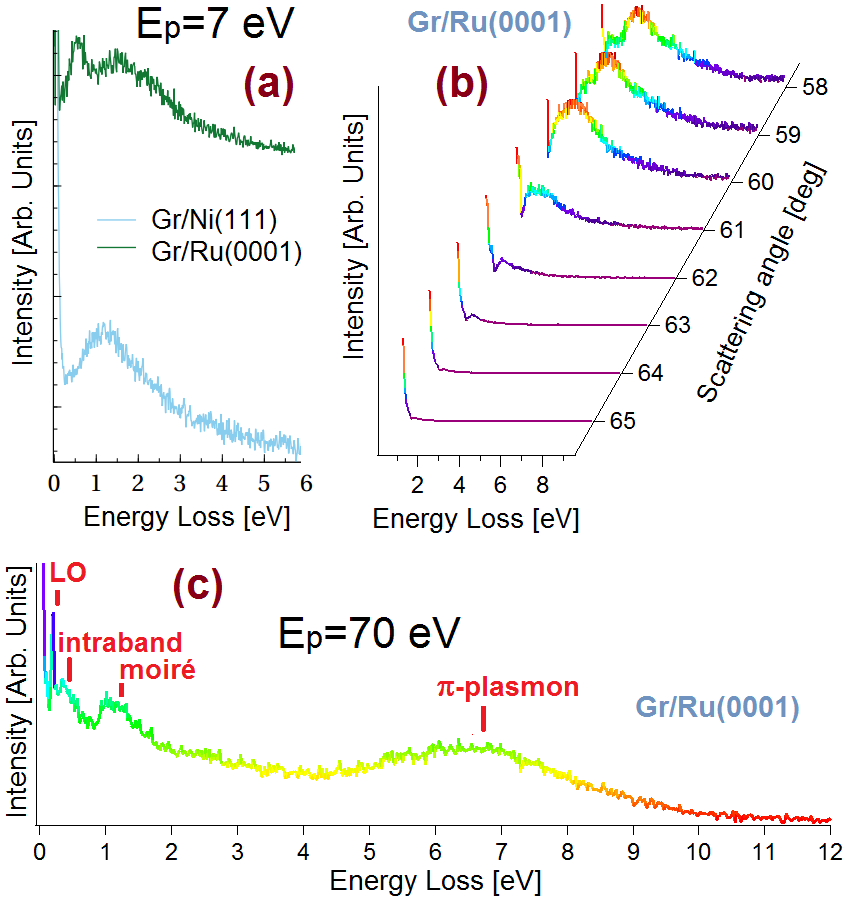
Fig. 1a shows the HREELS spectra acquired for Gr/Ni(111) (blue curve) and Gr/Ru(0001) (green curve) for a primary electron beam energy eV in an off-specular scattering geometry (; ). The loss spectrum of Gr/Ni(111) is characterized by a single peak [at an energy of 1.2 eV for Å-1, as obtained from Eq. (1)], assigned to the standard intra-band plasmon of doped graphene (for a review see e.g. [28, 29, 30]). Such a mode corresponds to the collective excitation of the charge density associated to graphene carriers screened by the Ni substrate [31]. The dispersion relation of this mode is that of plasmons in a two-dimensional electron gas (2DEG) [32] where is the chemical potential, , being the dielectric constant of the substrate and is the Fermi velocity of the carriers. In the long wavelength limit (), the main difference is that the velocity of the plasmon mode in Gr/Ni(111), defined from the slope of the dispersion relation, is smaller as compared to isolated graphene, due to the screening effects of the Ni substrate. At large wave-vectors the dispersion of the mode is dominated by the linear term .
Contrary to the excitation spectrum of graphene on Ni(111) that presents a single peak, our HREELS experiments for periodically rippled Gr/Ru(0001), as shown by the green line in Fig. 1a, show two loss features at 0.6 and 1.5 eV, corresponding to and 0.21 Å-1 in-plane wave-vectors, respectively. This finding is an evident fingerprint of the deep difference in the electronic properties of the two epitaxial-graphene systems. By considering the values of the above-mentioned physical quantities for Gr/Ru(0001), we can assume that the peak at 0.6 eV corresponds to the standard intra-band plasmon of graphene, which is the counterpart in Gr/Ru(0001) of the mode discussed above for Gr/Ni(111). Here we are interested on elucidating the nature of the extra mode observed at eV, which will be the focus of our work. For this we have measured the dispersion relation of plasmonic excitations in Gr/Ru(0001), by collecting loss spectra as a function of the scattering angle at fixed incident angle () and with eV. Fig. 1b shows that in nearly specular conditions (), i.e. for small momenta, the intraband plasmon is the unique peak which can be distinguished in the experimental loss spectrum. At , the extra mode emerges ( Å-1) and becomes the predominant peak of the HREELS spectrum for .
We note that the so-called -plasmon mode at eV, originated from inter-band electron-hole transitions between the van Hove singularities of the graphene -bands [33, 34, 35, 36], cannot be excited in the experimental conditions at which Fig. 1b has been obtained. However, by increasing the impinging energy at 70 eV (Fig. 1c), the -plasmon appears in the HREELS spectrum, while the cross-section for the excitation of low-energy plasmons is reduced at such a high value of the primary electron beam energy.
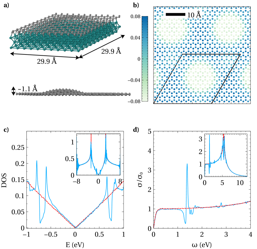
To establish whether the extra mode could be an effect of the moiré superlattice of Gr/Ru(0001), we have constructed a TB model of a deformed graphene layer, considering the positions of the carbon atoms in graphene nanodomes on Ru(0001), as obtained by DFT calculations (Fig. 2a) (see details in Sec. S3 of the SI). We then use this information to modulate the hopping parameters between sites and and on-site potentials as function of interatomic distances of the different carbon atoms [37, 38]. The modulation of the on-site energy is shown in Fig. 2b, which follows the same periodicity as the modulation of the atomic coordinates in the moiré pattern. By repeating the unit cell shown in Fig. 2(a), we extended the superlattices to carbon atoms, which is the typical size of our real space TB Hamiltonian matrix. After constructing the TB Hamiltonian for the Gr/Ru(0001), we calculate numerically the corresponding electronic density of states (DOS) by using the TBPM [13, 14]. Here the DOS is obtained by Fourier transform of the time-dependent correlation function , where is a random initial wave function. The time-evolution in the correlation function is performed by using the Chebychev polynomial algorithm.
The result for the DOS of Gr/Ru(0001) superlattices is shown in Fig. 2c, which clearly indicates the appearance of new singularities in the spectrum as compared to pristine graphene. One could identify some of them according to the super-periodicity of the moiré pattern. For example, the extra singularities around the energy eV (see inset of Fig. 2c) follow the expression , where is the length of the moiré’s pattern in terms of the graphene interatomic distance Å, and is the Fermi velocity in pristine graphene [39, 40]. The emergence of these extra singularities indicates the presence of flat bands in the band structure of the moiré superlattices, and may induce new strong optical excitations between the valence and conduction bands. This is indeed what we obtain in our numerical calculations for the optical conductivity shown in Fig. 2d, which have been obtained by using the Kubo’s formula [41] in the framework of TBPM [13]. A very sharp optical excitation peak (absent in pristine graphene) appears at the energy eV. At the same time, the optical conductivity at an energy smaller than rapidly drops from to , where is the universal optical conductivity of graphene. Such a phenomenon is qualitatively similar to that obtained in the infrared optical spectrum of Gr/h-BN heterostructures, due to the quasi-periodic moiré superlattice reconstruction, as it has been discussed theoretically [38] and observed experimentally [9]. Thus, periodically rippled graphene on Ru(0001) represents a suitable platform to modulate the optical properties of graphene.
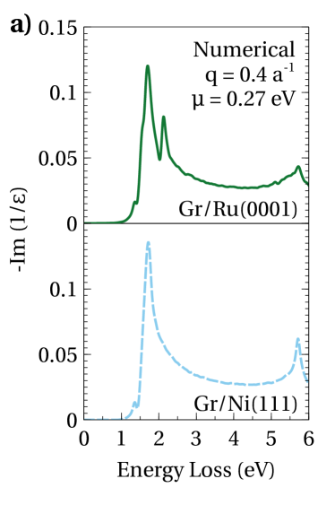
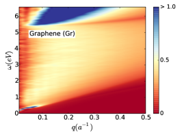
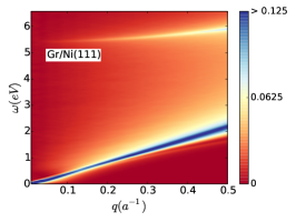
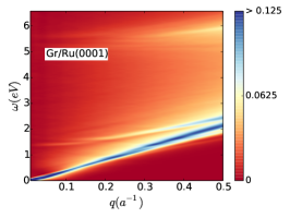
In order to compare to the HREELS experiments of Fig. 1, we calculate the loss function Im, in terms of the dielectric function within the random phase approximation (RPA) where is the polarization function. Our theoretical results for Gr/Ni(111) and for Gr/Ru(0001) are shown in Fig. 3. The calculated loss function of Gr/Ru(0001) (Fig. 3a) well reproduces the emerging extra mode of energy eV, which is instead absent in flat, commensurate Gr/Ni(111). This fact is in good agreement with our HREELS measurements, Fig. 1. This feature is due to a mode associated to transitions between electrons in the saddle points of the mini-bands generated by the moiré superlattice. When devising optoelectronic applications, the behavior of the modes with respect to modifications of the charge doping should be taken into account. We have further studied the effect of doping on the moiré mode. Fig. 3b shows that the extra inter-band mode is not affected by the carrier concentration, being present also in a charge-neutral moiré superlattice on Ru(0001). The robustness of the moiré mode toward changes in electron doping also implies that it would not be influenced by environmental doping, usually observed in air-exposed graphene field-effect transistors [43]. Experimentally, it is not possible to apply gate voltages in an HREELS apparatus and, thus, we are unable to probe plasmons in charge-neutral Gr/Ru(0001). However, HREELS studies on plasmonic modes in moiré-reconstructed, charge-neutral graphene on Pt3Ni(111) reveal that only the moiré mode exists at this interface (Section S4, SI).
We notice that, strictly speaking, the moiré mode cannot be considered as fully coherent plasmons, as it is the case for the low-energy plasmon with dispersion law given by . For doped graphene (free-standing or deposited on any substrate), the intra-band plasmon is undamped above the threshold until it enters the inter-band particle-hole continuum of the spectrum, and then it is damped by decaying into electron-hole pairs. However, the inter-band moiré mode that we have found in Gr/Ru(0001)is a mode which lies inside the continuum of particle-hole excitations: at the inter-band energy . Therefore, the mode is damped even in the limit (see also Sec. S7 of the SI). The phenomenology of this mode is similar to that of the so-called -plasmon in graphene, that has been studied from both experimental [33, 34, 36] and theoretical [35] point of views, and it is observed also in our present experiments (Fig. 1c) and theoretical calculations (Fig. 4) at around eV.
In Fig. 4 we compare the corresponding loss functions in the plane, calculated with our model in the range of wave-vectors () for momentum transfer along the K direction, corresponding to plasmon wavelengths in the range nm. Plasmon dispersion in the M direction (reported in Figure S6 of the SI) is quite similar. Both, free-standing graphene and Gr/Ni(111) (Fig. 4a,b), are characterized by two modes: a low-energy intra-band plasmon (with dispersion relation given by ), and a high-energy inter-band -plasmon [33, 34, 35]. As seen in Fig. 4, the dispersion of the standard low-energy 2D plasmon for all the cases is dominated, at large wave-vectors, by the linear term , which is independent of the dielectric constant of the embedding medium. Periodically rippled Gr/Ru(0001) (Fig. 4c) exhibits, besides the above-mentioned modes, an extra mode dispersing from 1.7 eV at to 2.3 eV at high momenta. As we have discussed above, we associate this mode to the HREELS peak in the loss function at this energy (Fig. 1). It is interesting to notice that a careful inspection of Fig. 4c also indicates the occurrence of moiré-derived splitting of the high-energy inter-band -plasmon. The HREELS spectrum shown in the SI, Fig. S8, indicates that also such a theoretical prediction well reproduces the experimental findings.
In conclusion, we have studied the influence of moiré superlattice on the excitation spectrum of Gr/metal contacts. Whereas Gr/Ni(111) shows commensurate lattices with no relevant effect on the band structure, Gr/Ru(0001) leads to a moiré superlattice with a reconstruction of the energy band spectrum of graphene. In particular, new flat mini-bands are created, which produce the appearance of van Hove singularities in the electronic DOS. Our HREELS experiments reveal extra peaks in the loss spectrum of Gr/Ru(0001), which are well explained by our theoretical model based on TBPM. We associate the extra modes to inter-band excitations between the newly generated van Hove singularities. Our results demonstrate the importance of the band reconstruction in the excitation spectrum of graphene on specific substrates, associated to the moiré-derived superperiodic potential. The evidence of extra modes in graphene quantum dots paves the way toward the control of graphene plasmonic properties through appropriately engineered periodic surface patterns. Moreover, the occurrence of an extra mode in the visible range of the spectrum, robust against environmental doping, opens novel perspectives for graphene optoelectronics.
References
References
- [1] Batzill M 2012 Surf. Sci. Rep. 67 83 – 115
- [2] Pletikosić I, Kralj M, Pervan P, Brako R, Coraux J, N’Diaye A T, Busse C and Michely T 2009 Phys. Rev. Lett. 102(5) 056808
- [3] Xu Y, Semidey-Flecha L, Liu L, Zhou Z and Goodman D W 2011 Farad. Discuss. 152 267–276
- [4] Voloshina E N, Fertitta E, Garhofer A, Mittendorfer F, Fonin M, Thissen A and Dedkov Y S 2013 Sci. Rep. 3 1072
- [5] Wallbank J R, Patel A A, Mucha-Kruczyński M, Geim A K and Fal’ko V I 2013 Phys. Rev. B 87(24) 245408
- [6] San-Jose P, Gutiérrez-Rubio A, Sturla M and Guinea F 2014 Phys. Rev. B 90(7) 075428
- [7] Stradi D, Barja S, Díaz C, Garnica M, Borca B, Hinarejos J J, Sánchez-Portal D, Alcamí M, Arnau A, Vázquez de Parga A L, Miranda R and Martín F 2012 Phys. Rev. B 85(12) 121404
- [8] Tomadin A, Guinea F and Polini M 2014 Phys. Rev. B 90(16) 161406
- [9] Ni G X, Wang H, Wu J S, Fei Z, Goldflam M D, Keilmann F, Özyilmaz B, Castro Neto A H, Xie X M, Fogler M M and Basov D N 2015 Nat. Mater. 14 1217
- [10] Maccariello D, Campi D, Al Taleb A, Benedek G, Farías D, Bernasconi M and Miranda R 2015 Carbon 93 1–10
- [11] Maccariello D, Al Taleb A, Calleja F, Vázquez de Parga A, Perna P, Camarero J, Gnecco E, Farías D and Miranda R 2015 Nano letters 16 2–7
- [12] al Taleb A, Anemone G, Farías D and Miranda R 2016 Carbon 99 416–422
- [13] Yuan S, De Raedt H and Katsnelson M I 2010 Phys. Rev. B 82 115448
- [14] Hams A and De Raedt H 2000 Phys. Rev. E 62 4365
- [15] Wang B, Bocquet M L, Marchini S, Günther S and Wintterlin J 2008 Phys. Chem. Chem. Phys. 10 3530–3534
- [16] Borca B, Barja S, Garnica M, Minniti M, Politano A, Rodriguez-García J M, Hinarejos J J, Farías D, Vázquez de Parga A L and Miranda R 2010 New J. Phys. 12 093018
- [17] Iannuzzi M, Kalichava I, Ma H, Leake S J, Zhou H, Li G, Zhang Y, Bunk O, Gao H, Hutter J, Willmott P R and Greber T 2013 Phys. Rev. B 88(12) 125433
- [18] Marchini S, Günther S and Wintterlin J 2007 Phys. Rev. B 76 075429
- [19] Gyamfi M, Eelbo T, Waśniowska M and Wiesendanger R 2011 Phys. Rev. B 83 153418
- [20] Borca B, Barja S, Garnica M, Sánchez-Portal D, Silkin V M, Chulkov E V, Hermanns C F, Hinarejos J J, Vázquez de Parga A L, Arnau A, Echenique P M and Miranda R 2010 Phys. Rev. Lett. 105(3) 036804
- [21] Feng W, Lei S, Li Q and Zhao A 2011 J. Phys. Chem. C 115 24858–24864
- [22] Shikin A, Farıas D, Adamchuk V and Rieder K H 1999 Surf. Sci. 424 155–167
- [23] Gamo Y, Nagashima A, Wakabayashi M, Terai M and Oshima C 1997 Surf. Sci. 374 61–64
- [24] Dahal A and Batzill M 2014 Nanoscale 6 2548–2562
- [25] Andersen M, Hornekær L and Hammer B 2012 Phys. Rev. B 86(8) 085405
- [26] Mittendorfer F, Garhofer A, Redinger J, Klimeš J, Harl J and Kresse G 2011 Phys. Rev. B 84(20) 201401
- [27] Rocca M 1995 Surf. Sci. Rep. 22 1 – 71
- [28] Grigorenko A N, Polini M and Novoselov K S 2012 Nature Photon. 6 749–758
- [29] Stauber T 2014 J. Phys.: Condens. Matter 26 123201
- [30] Politano A and Chiarello G 2014 Nanoscale 6 10927–10940
- [31] Cupolillo A, Ligato N and Caputi L 2013 Surf. Sci. 608 88 – 91
- [32] Stern F 1967 Phys. Rev. Lett. 18(14) 546–548
- [33] Eberlein T, Bangert U, Nair R R, Jones R, Gass M, Bleloch A L, Novoselov K S, Geim A and Briddon P R 2008 Phys. Rev. B 77(23) 233406
- [34] Politano A, Radović I, Borka D, Mišković Z and Chiarello G 2016 Carbon 96 91 – 97
- [35] Yuan S, Roldán R and Katsnelson M I 2011 Phys. Rev. B 84 035439
- [36] Nelson F J, Idrobo J C, Fite J D, Miskovic Z L, Pennycook S J, Pantelides S T, Lee J U and Diebold A C 2014 Nano Letters 14 3827–3831
- [37] Pereira V M, Castro Neto A H and Peres N M R 2009 Phys. Rev. B 80 045401
- [38] Slotman G J, van Wijk M M, Zhao P L, Fasolino A, Katsnelson M I and Yuan S 2015 Phys. Rev. Lett. 115(18) 186801
- [39] Park C H, Yang L, Son Y W, Cohen M L and Louie S G 2008 Phys. Rev. Lett. 101(12) 126804
- [40] Park C H, Yang L, Son Y W, Cohen M L and Louie S G 2008 Nat. Phys. 4 213–217
- [41] Kubo R 1957 J. Phys. Soc. Jpn. 12 570–586
- [42] Choi W S, Seo S S A, Kim K W, Noh T W, Kim M Y and Shin S 2006 Phys. Rev. B 74 205117
- [43] Yang Y, Brenner K and Murali R 2012 Carbon 50 1727