Mechanism of Threshold Elongation of DNA Macromolecule
Abstract
The mechanism of threshold elongation (overstretching) of DNA macromolecules under the action of external force is studied within the framework of phenomenological approach. When considering the task it is taken into account that double-stranded DNA is a polymorphic macromolecule with a set of metastable states. Accordingly to proposed mechanism, DNA threshold elongation can take place as a cooperative structural transition of macromolecule to metastable form, which is stabilized by the external force. For the description of DNA overstretching, the model included external (stretching) and internal (conformation) displacement components is constructed. As assumed, both components are coupled on the pathway of double helix transformations. It is shown, that under force action DNA deformation proceeds in two stages. First one is the restructuring of the double helix, which leads to formation of conformational bistability in DNA macromolecule. On the second stage, the conformational transition and the deformation induced by it consistently cover the macromolecule as a threshold process. The propagation of overstretched deformation in bistable macromolecule occurs as the motion of transition boundaries (domain walls), which are topologically stable structural excitations. Using the proposed approach, the contributions to DNA overstretching process are determined. This is elastic stretching of the double helix, deformation induced by conformational changes in macromolecule before overstretching transition, and threshold elongation as such. The greatest contribution to overstretching process in DNA (more then 80) is introduced by threshold elongation. As shown, the effect of threshold deformation in DNA double helix takes place due to the critical correspondence of acting force with elastic properties of macromolecule, and achieves by restructuring of the double helix. The comparison of calculated characteristics of overstretching process for heteronomous DNA with experimental data shows good agreement. Analysis of the results obtained, together with the available literature data allow to conclude, that overstretching transition in the double helix is dynamical process and can spread in DNA chain. In the same time, in DNA with AT-rich content due to large dissipation the overstretching process leads to force induced melting transition, and should have nearly static character.
1 Introduction
Mechanical properties of DNA macromolecule are essentially important for understanding the mechanisms of biological processes in cells. To interpret the experimental results on DNA mechanics, the macromolecule has been often modeled as an elastic rod which parameters determined in various indirect experiments [1, 2, 3]. Due to development of single molecule manipulation techniques, the mechanical parameters of DNA double helix (Young’s module, bending and rotating stiffness) have been significantly refined [4, 5, 6, 7, 8, 9]. A new tool has been also used to study such important mechanical processes as DNA unzipping, stretching, unwrapping, packaging, looping and interaction with proteins [7, 8, 9, 10, 11, 12, 13, 14].
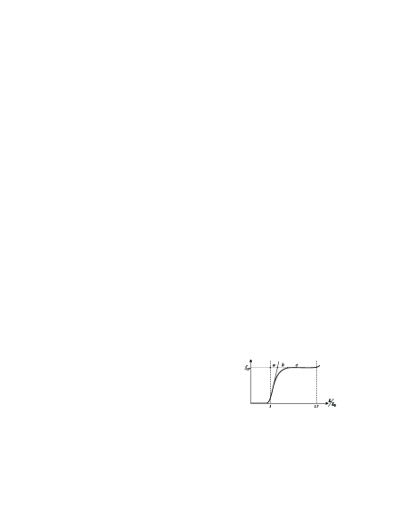
New results of DNA mechanics satisfy the elastic rod model not always. Sometimes, under the action of external force the deformation of double-stranded (ds) DNA occurs cooperatively as a threshold process (see reviews [7, 8, 9, 10]). Most clearly such effects manifest themselves in the experiments on DNA chain stretching. It is shown that at some critical value of applied force ( 65 pN) the rotationally unconstrained macromolecule overstretches, that is, elongates in 1.5-1.7 times [5, 6, 7, 8, 9]. The constrained dsDNA transforms to overstretched state under the action of larger force ( 110 pN) [15, 16]. The typical view of dsDNA overstretching curve drawn by data [6] is shown in Fig. 1.
Similar results are observed for synthetic polynucleotides [17, 18] and oligonucleotides [19, 20, 21]. These macromolecules have a DNA-type structure with two bonded polynucleotide strands. The stretching of single-stranded DNA corresponds to behaviour of extensible elastic rod [7, 9]. For double-stranded RNA macromolecules the effect of overstretching is not observed [22].
A plenty of works focusing on the mechanism of DNA overstretching has been performed. As considered [4, 6, 7, 8, 9], the force-extension curve (Fig. 1) on the interval of small forces (6 pN) describes the process of dsDNA unwrapping, for higher forces - the double helix elastic stretching. Then, on the interval before threshold elongation, the deviation from elastic trajectory of DNA stretching curve is observed, and at critical force value the threshold elongation as such occurs. The nature of the first two contributions to dsDNA elongation has been well studied, but the origin of last two processes remain not clear still.
Basically, there are two scenarios of the process. First one is force-induced internal melting (FIM) of DNA double helix with a gradual slow transition into ss-state [13, 23, 24, 15, 25]. Second scenario is cooperative fast transformation from the usual dsDNA (-form) to the stretched form () with preserved hydrogen bonds in complementary pairs [5, 6, 17, 26, 27, 28]. This is so called - transition. Thermodynamical analysis made in [29, 30] and experiments [19, 20, 28, 21, 31] show that both FIM and - transitions are possible for dsDNA. Their realization depends on temperature, counterions concentration in solution, and DNA nucleotide content. Studying the kinetics of DNA overstretching authors of [20, 28] have come to the conclusion that overstretching process takes place initially as - transition, and then FIM transition can occur.
It is also shown, that threshold elongation of dsDNA is no uniform deformation [15, 16, 27], and the fragments with different states (stretched and overstretched) can alternate in one chain under critical force action.
Recently, one more step in understanding the mechanism of DNA overstretching transition has been done. In paper [21] the appearance of bistability between and forms of dsDNA is observed in the force interval, when overstretching is occurred in dsDNA. The similar event is observed under study of dsDNA stretching through the melting of one DNA strand from another - unpeeling [25](Suppl.).
As it turns out, the course of overstretching process in dsDNA is recognized in general terms. However, this is not the case for the mechanism.
The conformational analysis and molecular dynamics modeling have found few structures of overstretched DNA depending on the value of applied force and the manner of its applying to macromolecule [5, 32, 33, 34, 12]. The most probable structure of -DNA is a narrow fibre with inclined hydrogen bonded base pairs and saved stacking interactions. This structure is obtained in conformational analysis studies [5, 32], and observed in MD simulations of DNA elongation in solvent conditions [33, 34]. The performed calculations demonstrate the opportunities of the double helix to overstretching transitions, but no offer an explanation of their appearance in the double helix.
The mechanism of DNA overstretching is proposed in [28], where authors believe that overstretching deformation in dsDNA happens as two-state reaction process - transition from ground compact to metastable extended state, within the appearance of some domains in macromolecule chain. But authors [28] do not explain, why the monomers in the domains remain in unprofitable metastable states, and do not return to usual compact form.
Authors of [35, 36, 37] have suggested that the coupling of DNA stretching with double helix twisting and bending can be the reason of DNA threshold elongation. It should be noted, that of course, the coupling of deformation components of macromolecule, should give the definite quantitative effect in dsDNA stretching, but cannot be the origin of threshold process.
On the other hand, the threshold effect may be result of the coupling of macromolecule deformation components with the conformational restructuring of the double helix. This possibility is indicated under dsDNA overstretching modeling in [32], where sharp jumps in macromolecule stretching energy are observed. A similar concept has expressed by authors of [28]. Studding the kinetics of overstretching process, they come to conclusion, that dsDNA macromolecule shows two kinds of elasticity in the ranges of acting force lower and higher, then 35 pN. That is, under tension of external force the macromolecule can change its structural organization.
So, the nature of DNA threshold elongation is still being discussed. It is necessary to understand why and how the transition to overstretched form occurs in DNA chain. What the action produces by external force, and how is it connected with the parameters of the double helix? What is the conditions required for threshold deformation of the double helix, and what is the differences between - and FIM transition realization in dsDNA?
To answer these and some other questions in present paper a phenomenological model for DNA deformation study is proposed (2). Under model construction, dsDNA is considered as a polymorphic macromolecule, which has a set of metastable states. Following this thesis, the contributions to deformation of polymorphic macromolecule are determined in 3. The restructuring in polymorphic macromolecule under action of external force, and the possibility of conformational bistability formation are studied in 4. In 5 the dynamics of of conformational transitions from compact to stretched form in dsDNA is under consideration. Using the developed model, the quantitative characteristics of dsDNA overstretching are estimated in 6. The agreement of developed theory with experiments, the dependence of DNA overstretching on nucleotide content, the reasons for of - or FIM transition realization in DNA chain are discussed in (7). At the end, the conclusions and predictions from the performed study are formulated 8.
2 DNA macromolecule under the action of external force
Considering the action of external force on dsDNA, it should be taken into account that DNA double helix is a polymorphic molecule [38, 39]. As in all polymorphic structures, the form of dsDNA and its changes are coupled with the disposition and displacements of macromolecule structural elements in the frame of double helix. This is well illustrated by example of - conformational transition in DNA, where the parameters of the double helix turn (such as length and twist angle) change together with the changes in positions of structural elements inside the double helix [40, 38]. The interrelation between the displacements of DNA structural elements with the changes of the double helix form manifests itself also in the observations of conformationally induced deformations in dsDNA [41, 42, 43]. The importance of coupling between the double helix deformation and its conformational state is evident also from the results of conformational analysis of DNA overstretching in [5, 32, 33, 34]. So, the coupling of macromolecule deformation with its conformation state will be accounted under model construction of DNA overstretching process in the present work.
For understanding the mechanism of dsDNA overstretching deformation the model with two types of displacement components will be used. One component will describe an external deformation of the macromolecule, and another component - internal conformational transformation in the double helix structure. Let external component () will describe a displacement of -th monomer link from its equilibrium position in macromolecule chain under dsDNA stretching. Note, this is one of the components of DNA model as elastic rod, for which the value of elastic constant is well known. Let internal component will describe the change in conformation state of double helix monomer link (). It is well known, that one of the dominant components in specifying the conformation of the double helix is the position of base pair [40, 38]. The displacement of paired DNA bases in monomer unit reflects the changes in the double helix conformation under overstretching transition as well, as testified by the results of [5, 32, 33, 34]. So, the displacement of mass center of DNA base pairs with respect to mass center of monomer unit will be considered as internal component under modeling the overstretching process in dsDNA. In the present paper it will be studied also the dynamics of deformation in DNA macromolecule. So, let both model components depend on time: and .
The expression for energy of DNA macromolecule under the action of external force can be written in two-component approach as follows:
| (1) |
In expression (1) , are derivatives on time; and are the elastic constants, which describe the interactions along macromolecular chain for external and internal components; means monomer mass, is reduced mass of monomer link according to the pathway of conformational transformation. It is significant to note, that for DNA conformational pathways with joint motions of nucleic bases, the masses and have the same value for AT and GC pairs [44, 45, 46].
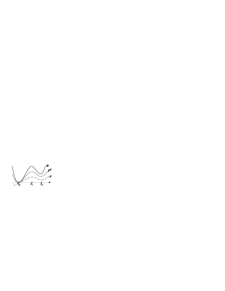
Potential function describes the change of monomer energy on the pathway of conformational transformation from the usual compact to the stretched helix form. Under conditions when compact DNA form is more stable (at the physiological conditions - -form [38]), function must be taken in the shape of double well with non-equivalent stable states: ground stable state - , and metastable one - (Fig. 2). It is assumed, as in [6, 33], that potential barrier in under DNA overstretching transition originates from the conformational barriers in sugar-phosphate backbone of the double helix, and mainly related to the conformation of sugar rings.
The term with coefficient describes the coupling between internal and external components under macromolecule restructuring. The value of this coupling depends on macromolecule conformational state, determined by function , and on value of external force controlled by parameter . The coupling between internal and external components exists and without action of external forces, as seen from many examples in [38, 39]. The presence of this term and its sign in expression (1) reflects the fact, that polymorphic macromolecule can reduce the energy needed for deformation due to the change of its conformation.
Potential functions and due to their physical meaning should increase on the pathway from ground state of the system up to transition barrier, and then decrease near metastable state (Fig. 2). Explicit view of used potential functions can be founded with the help of conformational analysis, but for the goals of present study it is sufficient to determine the shapes of potential functions only.
In expression (1) the last term describes the action of external force on the macromolecule state. It is reasonable to consider, that the force is applied to the external component of macromolecule deformation:
| (2) |
When writing expression (1), it is assumed also that under external force action the macromolecule chain is fully unwrapped up to its contour length before stretching.
Under modeling the process of DNA overstretching, it will be not specified the type of transition, - or FIM, at once. The differences between them will be indicated on the basis of obtained results at the end of the study.
Note, that in this work just the coupling between one external and one internal components of DNA transformation is under consideration. It is clear, that in such deformation as DNA overstretching and other degrees of freedom of the double helix have to take place also. But in the present study the simplest model with two dominated components is considered to understand the basic mechanism of threshold deformation in the double helix. Such two-component approach has been successfully used for the modeling of - transition in dsDNA [44, 45, 46], and under studies of the effects of DNA localized bending and unzipping [47, 48, 49]. The action of external force on DNA macromolecule in last papers is considered indirectly, as the change in external component stiffness. In the present work the external force is seen as a direct participant in the process of dsDNA deformation.
3 Deformation of polymorphic macromolecule
Using model (1,2), let us study the deformation of polymorphic macromolecule, its dependence from the conformational state and from the value of external force. Let us consider firstly the uniform deformation of polymorphic macromolecule. In this case it is true: , , and , . Accordingly to considered problem, the potential energy of macromolecule monomer link is bound to have two minima (), and one maximum () between them. Let one of minima () be a ground state, and for this state the following condition takes place: . For other extremum points () let us assume , and (Fig. 2).
The expression for energy density of the deformed macromolecule can be written in the form:
| (3) |
Here , and is the number of monomer links in macromolecule.
In the presence of acting force equilibrium states of the system can be determined by solution of following equations:
| (4) |
| (5) |
From last equation, the expression for deformation of monomer link of polymorphic macromolecule can be written as:
| (6) |
Here
| (7) |
As might be expected, the deformation of polymorphic macromolecule is caused by force action, elastic properties of the chain, and conformational state of macromolecule. First term in expression (6) describes elastic deformation of macromolecule as in the model of elastic rod (). Second term () appears in expression (6) due to coupling between external and internal components of macromolecule transformation. This term describes the conformationally induced deformation.
Let us consider the action of external force on conformational state of polymorphic macromolecule. After substitution of expression (6) in equation (4) and subsequent integration, the expression for conformational energy of polymorphic macromolecule under tension of external force can be written as:
| (8) |
where is the constant of integration which will be determined later.
It is clear, that the view of conformational energy (8) depends on explicit expressions of used potential functions. However, it is quite enough to study the problem envisaging only their shapes shown in Fig. 2. Notice, as seen from expression (8), the shapes of and should be similar due to description by these functions the different aspects of the same physical process – the energy changes during the transition from one conformational state to another. Thus, let us study the problem within following approximation: . Here parameter accords with the value of energy barrier between the conformations. Such approach does not limit the generality of the study and, as will be showed, it yields sufficiently interesting results. Using proposed approximation, expression (8) can be written as follows:
| (9) |
where
| (10) |
For the double-well shape of energy (9) the value should be positive, thus, it must be considered:
| (11) |
Expression (9) clearly shows, that the action of external force can affect the conformational state of polymorphic macromolecule due to the coupling between external and internal components. Under force action the positions of macromolecule stable states should change, and new extremum points of macromolecule energy (, , and ) should be found. Rewriting equation (4) for macromolecule conformation energy (9), one can obtain the equation for the determination of new extremum states:
| (12) |
Note, as seen from (12), in the absence of external force the positions of stable states of macromolecule are determined by equation:
| (13) |
The extremum is determined by the form of potential function only, from the condition =. As follows from expression (13), the positions of two other extrema can be found from the solution of equation: , and they are and .
When , from equation (12) it follows that new extremum can be obtained from solution of equation:
| (14) |
where
| (15) |
Two other solutions follow again from the equation: , and they are , . When writing expression (15), the inequality (11) is taken into account.
The first derivative of conformational energy (9) on for point is zero, as seen from (14), and the second derivative for remains positive. Thus, this point is energy minimum, a new ground state of the system in the presence of external force. New position of ground state can be found by solving equation (14) with known function , values of acting force, parameters and .
The change of macromolecule ground state position in the presence of external force can be understood directly from equation (14). It is seen, when , then , and . On the other hand, if , then . Accordingly to the shape of function (Fig. 2), it may be so, if macromolecule ground state shifts () under force action.
Another energy minimum of the system is . For this extremum point the condition of minimum is also met, because , due to , and, as it can be showed, the second derivative is positive. It is clear, that between the minima ( and ) the maximum at is realized.
To fully determine the ground state of the system under the action of external force let us assume . Then, in accordance with equation (9) and expression (14), the constant value can be determined as:
| (16) |
Taking into account expression (14), and constant value (16), it is possible to write expression for conformational energy (9) in the form:
| (17) |
where
| (18) |
It is seen, that at , in accordance with condition accepted above for the ground state.
Using the form of function (18), the deformation of macromolecule monomer link due to its conformational transformation (7) can be presented as:
| (19) |
In expression (19)
| (20) |
Under writing expression (19), equation (14) and expression (7) are used.
The value is the primary deformation of macromolecular structure due to the change of macromolecular conformation before the structural transition. One of the manifestations of primary deformation is the shift of ground state of polymorphic macromolecule. As seen from (20) and (14), if , then and .
The second term in (19)
| (21) |
is the deformation accompanied a structural transition as such, when it occurs in macromolecule.
By this means, the deformation of the monomer link of polymorphic macromolecule under the action of external force consists of elastic deformation (), and the deformations induced in macromolecule structure before () and during () the conformational transition.
4 Critical regime: formation of conformational bistability
Thus, polymorphic macromolecule has an additional mechanisms of deformation connected with conformational transformations in its structure. Let us consider the action of external force on conformational state of polymorphic macromolecule and determine the conditions, when the deformation can have a threshold character.
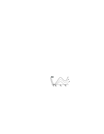
As shown in previous section, the external force can shape the conformation state of polymorphic macromolecule. One of the consequences of this process is the shift of ground state position in macromolecule conformation. Another one can be the changes in energy barrier between ground and metastable state, and in energy difference between conformational states as well. Really, as seen from expression (14), under increasing of external force the value of should increase. So, the difference between and values , and should become smaller, accordingly to (17,18). At some critical value of external force () the macromolecule conformation can transit to form with two equivalent stable states and reduced transition barrier (Fig. 3). Such transformations in macromolecule structure make the transitions between and states very probable.
The conditions of such macromolecule transformation under the action of critical force can be found from the relation:
| (22) |
The realization of equality (22) means, that conformational energy (17) takes the form of double-well function. That is, the macromolecule transforms into bistable conformation (see Fig. 3, curve 3). Such a state of the system, appeared under the external force tension, will be called as critical regime.
Taking into account expressions (17,22), and condition (and hence ), it can be concluded that
| (23) |
As follows from relation (18), in this case it is true:
| (24) |
From relations (22,23,24) it is seen, that expression for macromolecule conformational energy in the critical regime can be approximated by the analytical function. Really, on the interval of () energy (17,18) can be presented in the view:
| (25) |
where .
Thus, in critical regime the expression for conformational energy (17) can be written in the form:
| (26) |
As seen, function (26) has two minima and one maximum (). Substituting the coordinate of energy maximum in expression (26), the value of energy barrier in the critical regime can be determined as:
| (27) |
As a whole, using expressions (6,7,18,20), energy density of polymorphic macromolecule (3) in the conditions of critical force action can be written as:
| (28) |
that is, the sum of critical conformation energy (26) and the work performed by external force to form the bistable conformation of macromolecule. The value of this work accords to the reaction of the system on external action, and per one monomer unit has the following view:
| (29) |
where first term is the energy expended for elastic deformation, and second term is the energy of primary deformation.
The transformation of macromolecule conformational state takes place due to the action of external force. Within the used approach, the force acts on external component of macromolecule deformation, and then, due to the coupling of external and internal components affects the macromolecule conformational state. From the results, obtained in Sec. 3, it is seen, that modification of macromolecule conformational state is going through the changes of parameters and , which values are determined by the magnitudes of and , and by parameter of components coupling . If parameters and are the constants for definite macromolecules, the parameter has to depend on acting force, and to change like a magnitude of external force.
Let us consider the dependencies of and on . It is obviously, that if =0, the definite coupling between macromolecule transformation components exists, let it is . If 0, the force can affect the coupling between the components, as is supposed . Accordingly to (10,11,15), value of can change in definite numerical interval, that is: , where . The change of coupling parameter leads to the changes of and , and to shaping of macromolecule conformation. When , then and . When , then and .
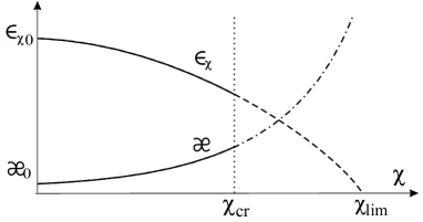
The numerical analysis of expressions (10,15) with possible values of and shows the validity the conclusions drawn (see Fig. 4). From the results presented in the figure, it is seen that parameter grows with peaking under increasing of . Under the critical value of acting force, coupling parameter reaches the magnitude . In its turn, the parameter of primary deformation achieves its critical value , which accords to appearance of conformational bistability in macromolecule structure. In opposite, parameter decreases with increasing of . Such a course of the dependence explains the decreasing in the value of conformational barrier in macromolecule under tension of external force, as shown in Fig. 3. Once the coupling parameter riches its peaking value, the further changes in macromolecule characteristics are terminated, and another stage of threshold deformation begins.
It is useful to determine the area of plausible values of . Really, when parameter accords to it critical value, then from expression (15) should be:
| (30) |
Thus, for example, if observed value of , then following estimation can be done: .
It is of interest to express the value of critical force in terms of the macromolecule characteristics. Using expression (14) and equality (24) one can obtain the value of external force in conditions of critical regime:
| (31) |
The relationship of critical force magnitude with macromolecule parameters will be better seen after rewriting expression (31) for the square of critical force. Taking into account (30), expression (31) can be presented as:
| (32) |
Here is the energy difference between the ground and metastable (stretched) state of macromolecule monomer link itself, in the absence of external force:
| (33) |
Under writing expressions (32,33), it is taken into account, that in the frame of accepted approximation for potential functions it is true: .
So, the square of critical force is proportional to the energy value of monomer metastable state, and to the macromolecule stretch stiffness. As seen, the balance of product of and with acting force can be achieved for account of the changing in parameter . In fact, the growth of to it peaking value is fixed by the magnitude of critical force.
Note, as approaching of acting force to its critical value the contributions of primary and elastic deformation increase. Wherein the elastic deformation increases linearly, the increasing of primary deformation looks like exponential dependence. Both these contributions form the uniform deformation of macromolecule (), but not constitute the threshold process. However, under action of critical force the macromolecule conformation becomes bistable, and the conformational transition from ground to stretched state can occur in macromolecule. Due to appearance of structural transitions, the macromolecule deformation acquires a threshold character, and this can explain the mechanism of DNA chain overstretching.
5 Dynamics of conformationally induced deformation in DNA macromolecule
Let us consider the occurrence of conformational transitions and induced by them deformations in polymorphic macromolecule of DNA type. In addressing the issue, it is necessary keep into account, that such deformation has a local character and occurs there, where the conformational transition occurs. The realization of structural transitions in macromolecule chain occurs through the formation of domains with another structural states [50]. So, the conformationally induced deformation is not uniform, and cannot cover whole macromolecule at once. If energies of different states are the same, the boundaries of domains (domain walls) can move along the chain increasing (or decreasing) the domain area [51, 52, 53].
Of course, the domains with stretched form can appear also in conditions of inequality of the energies of ground and stretched states, at . However, due to the energy difference, these domains will not be stable and can not take part in the process of DNA overstretching. The probability of the reverse transition to ground state will be higher, than the for the direct process. At the same time, the appearance of conformational bistability under promotes the occurrence of stable domains, provides the facility for conformationally induced deformation to propagate along the macromolecule chain, and implements threshold character of deformation.
In this section the dynamics of conformational transition and induced by it deformation in DNA macromolecule are studied for the conditions of critical regime of external force action. Let us go to the continuum approximation, which is usually used for the studying structural transitions dynamics. In this approximation: and , , , and are derivatives on , and is the distance between monomers along the macromolecular chain.
The equations of motion for external and internal components of conformationally induced deformation can be written as:
| (34) | |||||
| (35) |
In equations (34,35) , , and the relation between potential functions ( and ) is used also.
To consider the dynamics of conformational transition along macromolecular chain, let us introduce a wave coordinate: , and look for the solution having asymptotic of ground () or metastable () states of macromolecule.
After wave substitution and one-time integration of equations (34), one can obtain the following:
| (36) |
Here is derivation on , is the constant of integration. Accordingly to accepted initial conditions . Equation (36) determines the dynamics of deformation in macromolecule.
Substituting expression for from equation (36) in equation (35), and integrating it, the equation for conformational component can be obtained:
| (37) |
where:
| (38) |
Under writing expression (38), it is taken into account that the constant of integration accords to expression (16).
Unfortunately, equation (37) with potential (38) is quite complicated for direct analysis. But this equation can be simplified, taking into account that in experiments on DNA threshold deformations the measured processes have the velocities not exceeding m/sec [5], which are much smaller then the velocity of sound in DNA ( m/sec [54]). The velocity should have close order , accordingly to analysis in [44]. Therefore, let us consider the problem in approximation of a small values of transition wave velocity: ,. In this case the potential function (38) takes the form which accords to energy (9) with constant (16), and at to expression (26), so that
| (39) |
The solution of equation (37) with potential (39) for asymptotic of stable states (at , or , and ) has the form of domain wall [51, 52, 53]. After integration of equation (37) with mentioned above boundary conditions, one can obtain the expression for the domain wall (conformational transition wave) in DNA macromolecule:
| (40) |
where constant
| (41) |
has the dimension of inverse length.
In expression (40) the sign has been chosen in accordance to the boundary conditions: plus, if at the boundary state is realized; minus, if . As one can see, solution (40) is a wave in the form of a step - the transition from state to , or reverse process.
Taking into account result (40), let us find the deformation induced in macromolecule by the appearance of conformational transition. The equation for external component (36) in the approximation of small velocities takes the form:
| (42) |
where formula (18) is used as well.
Accordingly, the deformation of monomer link of polymorphic macromolecule can be written as:
| (43) |
where is the uniform deformation of macromolecule chain, and is the deformation of the monomer link caused by conformational transition (Fig. 5a).
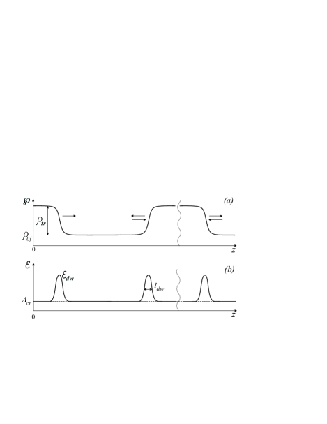
The part of deformation induced by conformational transition is expressed as:
| (44) |
where . Here the solution for (40) and expression (25) for function are used also. To derive expression (44), it is supposed, that at one side of macromolecular chain the state is realized, and at another side - state. Thus, at one side of domain wall (where ), and at another side () (Fig. 5a).
It should be noted, that solutions 40 and 44 obtained for domain walls are the topological solitons. This type of solitins remains stable for unchanged boundary conditions in macromolecule chain [51, 52, 53], and their appearance in the macromolecule happens at the end of the chain only. The equations (37) and (42) have also the two-solitons solutions. These solutions are shown in Fig. 5 as well. Such type of solutions describes the occurrence of the domains of another conformation inside macromolecule. Note, that realization of single-soliton solution is more favorable energetically, because it requires only one domain wall formation at the end of macromolecule chain in this case.
A single-soliton and two-solitons solutions differ from each other not only by the number of domain walls, but also by the boundary conditions for their realization. For the realization of single-soliton solution it is important to have distinct stable states ( or ) at the different ends of transition region. For realization of two-solitons solution it is necessary to have the same stable states (usually ground state ) at the domain boundaries.
6 Properties of overstretching transition: quantitative estimations
The developed approach allows to study the properties of overstretching process in dsDNA on quantitative level. It is of interest to determine amplitude, thickness and energy of domain walls for the regime of dsDNA critical deformation.
The expression for amplitude of macromolecule deformation, induced by conformational transition, can be obtained substituting the value of constant (41) in expression of from (44). So, . Considering that constants and for DNA macromolecule are the same order of values, it can be obtained a simple formula for the estimation of deformation amplitude:
| (45) |
Note, whereas the amplitude of conformational transition is some constant defined by the pathway of transition, the amplitude of macromolecule deformation varies proportionally with square root of .
Using expressions (6,7,20) and (45), the macromolecule deformation per one monomer unit can be written as:
| (46) |
The thickness of domain wall is determined as [51]. So, using expressions (10,15) and (41), it can be obtained the following formula for thickness of domain wall under macromolecule overstretching:
| (47) |
It is seen, that domain wall thickness (47), as well as its amplitude (45), is proportional to root of .
The energy of domain wall can be determined as:
| (48) |
where
| (49) |
To derive expression (49), the solution (40) and expressions (26,27) are used.
To estimate the full energy required to initiate domain wall formation, it is necessary to include in consideration the work of external force to create the bistability in macromolecule chain (29). So, the initiation energy of domain wall formation can be written as:
| (51) |
Here =. In expression (51) the first part of energy is connected with the domain formation as such. Another term accords to the part of work on creation of conformation bistability on the interval of domain wall thickness.
The obtained expressions are used for calculations of the characteristics of dsDNA overstretching process: the contributions from elastic, primary and threshold deformations, the energies and sizes of domain walls, the values of energy barrier and energy difference between ground and stretched states under overstretching transition in macromolecule. The results of calculations are shown in Tabl. 1.
In calculations the following magnitudes of model parameters are used: =3.4 ; =S, where dsDNA stretch modulus S=1100200 pN (accordingly to [36]). That is =4.60.8 kcal/mol . The value of parameter =4 kcal/mol is adopted by the data of conformational analysis of DNA backbone [55, 38]. This choice accords to the assumption of [6, 33] about the main role of conformational transformations in DNA sugar rings during the overstretching transition. Parameter is taken to be equal to 1 . That value by order of magnitude corresponds to the data of conformational analysis of overstretching transformation in dsDNA [32] (stucture 1.6), and [33].
For estimations of other characteristics of dsDNA overstretching it is necessary to know the value of parameter (or ). The estimations of these parameters can be done from observed value of macromolecule deformation in the critical regime. As known [4, 5, 6, 7, 8, 9] for critical regime, when =65 pN, the deformation of dsDNA with average nucleotide content comprises 0.7 of its length. Considering, that the stretch modulus of such dsDNA has average value (S=1100 pN), from expression (46) it can be calculated 1. This estimation is important for the evaluation of coupling parameter . Using formula (30) with known values of and , it can be obtained 3 kcal/mol.
It should be emphasized, that estimated in such way parameters accord to the stretching of DNA chain with average value of stretch modulus. But dsDNA is a heteronomous macromolecule, and its chain consists from the fragments with different nucleotide content (and so with distinct elastic properties). So, it is necessary to consider, that under action of definite critical force the fragments with other (not average) elastic properties are stretched also. Tacking this into account, the calculations of double helix elongation characteristics are performed also for DNA fragments with stretch modules differing from their average value (on 200 pN). These calculations are done with the same values of parameters and for all macromolecule fragments. This is because parameter reflects the conformational transformations in dsDNA backbone and has no change considerably for different nucleotide content. In its turn, coupling parameter , as assumed, is depended on the value of external force, but the force is the same for whole macromolecule. However, the values of parameter for these distinct DNA fragments are calculated by formula (15) with distinct (not average) magnitudes of .
It is of interest also, to estimate the values of energy barrier for macromolecule transition to stretched state, and energy difference between ground and stretched states under overstretching transition for DNA fragments with distinct stretch modules. The value of energy barrier can be calculated as =, using expressions (10,27) and already determined parameters. The energy difference between the states in the frame of developed approach is determined as = accordingly to (17) (note, =0). Needed for these calculations value of is equal to the value of in critical regime and can be found from expression (14). For the fragments with distinct stretch modules value of can be calculated with the same values of and , but with value of calculated by (15) for different stretch modules. As seen, the values of () and () can be calculated as well.
The results of calculations of overstretching characteristics for dsDNA per one monomer unit for distinct stretch modules but with the same value of external force (=65 pN) are shown in Tabl. 1. The data for critical regime realization (for DNA with average stretch modulus) are presented in the second row. All values in the table are calculated with the accuracy to second sing after coma and then are rounded to first sing.
| S | () | () | () | ||
|---|---|---|---|---|---|
| (pN) | |||||
| 900 | 1.5 | 3.0 (0.2; 0.4; 2.4) | 0.8(-0.1) | 3.1 | 2.4 (0.9; 1.5) |
| 1100 | 1.0 | 2.3 (0.2; 0.2; 1.9) | 1.0 (0.0) | 3.0 | 2.4 (0.5; 1.9) |
| 1300 | 0.7 | 2.0 (0.2; 0.1; 1.7) | 1.2 (0.1) | 3.1 | 2.6 (0.4; 2.2) |
7 Discussion: theory and experiment
7.1 Quantitative agreement
As seen, the calculated values of dsDNA overstretching deformation (Tabl. 1) in order of their magnitudes correspond to experimental data [4, 5, 6, 7, 8, 9]. Over the range of DNA stretch modulus variation the contribution of elastic deformation to total value of macromolecule deformation changes moderately, only in second sign after coma. In contrast, the contributions of primary and threshold deformations and the value of parameter change significantly, and are larger for macromolecule fragments with smaller stretch modulus. The deformation gives the largest contribution to the value of macromolecule deformations in each case. The large values of provide the threshold character of macromolecule deformation as a whole. The obtained result accords with the data of overstretching kinetics study in force-clamp experiments [20, 28]. As shown in these works, the amplitudes of double helix elongation increase in times in the interval of critical force action, where DNA macromolecule exhibits threshold deformation.
The results presented in Tabl. 1 also show, that the transition barrier between compact and stretched DNA forms in conditions of critical regime has the order of 1 kcal/mol. The energy difference between transition states is absent for DNA with the average stretch modulus (as expected for critical regime), and is sufficiently small (but not zero) for DNA fragments with distinct stretch modules. It means, that propagation of domain walls in heteronomous macromolecule occurs with minimal losses just for the conditions of critical regime. With increasing the deviation from these conditions, the losses will increase, and the deformation will not be pass as threshold process, and not have large contribution in whole macromolecule deformation. This result explain the relatively small width of the force interval of critical regime realization in dsDNA, observed in experiments [4, 6, 7, 8, 9].
The calculated energies of domain walls initiation (Tabl. 1) are more than twice the energies of transition barriers, that should be facilitated domain walls passage through the barriers. The domain walls energies do not change significantly for macromolecule fragments with distinct stretch modules. In such an effect the simultaneous decreasing of the work and increasing of domain wall energy play its role. Similar effect is seen for the domain walls sizes (), which values are conserved on interval of stretch modulus variation. Here two processes, the decreasing of parameter and increasing of macromolecule stiffness, proceed concurrently (45,47). Thus, the domain walls features are weakly dependent on variations of stretch modulus, under its considered deviations from average value ( 200 pN).
From calculated data it is seen also, that the energy of domain wall formation () introduces the larger contributions to whole initiation energies of overstretching transition. It is of interest to compare the obtained values of with the data of thermodynamical analysis of DNA overstretching transitions in [31]. In this work the free energy difference for overstretched transitions are measured for definite DNA sequences, where are observed - or FIM transitions. By the conditions of experiment the transitions are studied in the critical force interval ([31]). From these data the values of free energy for dsDNA overstretching transitions can be calculated by known formula = -. The calculations give the following values: 1.7 kcal/mol for DNA overstretching in FIM transition, and 1.9 kcal/mol for overstretched DNA resulting in - transition. As seen, these energy values agree with the results listed in Tabl. 1 for the domain wall energy in DNAs with less and more helix stiffness (first and second rows).
The obtained magnitudes of domain walls energies allow also to estimate the cooperativity of - transition in conditions of critical regime. Using formula for cooperativity length [50], which in this case has a view: , and the data on domain energy for the conditions of critical regime (Tabl. 1, second row) one can obtain the length of cooperativity (average domain size) for DNA - transition equal to about 24 base pairs. This estimation fully accords to the value obtained in [28] from the analysis of overstretching transition kinetics in dsDNA.
Of course, performed calculations are the estimations only. But the data of Tabl. 1 show, that proposed theoretical approach can be used for the interpretation of experimental data.
7.2 Dependence on nucleicleotide content. - and FIM overstretching transitions
Up to now in the present study, when considering the process of DNA overstretching, no differences between two scenarios of double helix elongation, - or FIM transitions, have been done. Now, on the basis of obtained results, the realization of two possible processes in heteronomous dsDNA can be analyzed.
From obtained results (Tabl. 1) it is seen, that for double helix fragments with different values of elastic and conformational properties the overstretching deformation should be different. As one can estimate from results of [17, 18], the stretch modulus for AT-rich DNA is smaller than for GC-rich DNA. So, accordingly to the estimations made (Tabl. 1), and expression (46), the overstretching deformation of heteronomous DNA should be larger for macromolecule fragments with smaller stiffness, that is, for AT-rich fragments. Note, this conclusion is in accordance with results of [21], where the larger amplitudes of overstretching deformation are observed for AT-rich DNA.
In its turn, the larger elongation of some dsDNA fragments in overstretching process has to lead to larger loss in base pairs stacking for the double helix. Such assumption follows from the consideration of possible structures for overstretched dsDNA. Accordingly to conformational analysis [32, 33], in overstretched double helix the base pairs are greatly inclined with large slide and shift. All these structural changes should increase with the growth of double helix elongation, and should lead to losses in base pairs stacking. As known [56], the base stacking on a par with the hydrogen bonding determines the base pairing in the double helix, and the stability of dsDNA as a whole. Thus, the large overstretching deformation of DNA fragments more likely to be lead to base pairs disruption and subsequent internal melting. Can assume, that this is valid for the overstretching of DNA fragments with lower stretch modulus, such as AT-rich sequences.
It can be assumed, that -form is stable for dsDNA with sufficiently large content of GC pairs, and less (or not) stable for AT-rich DNA. Such assumption agrees with the results of experiment [21], where under overstretching of AT-rich oligonucleotides the intermediate (between and melted) state is observed. As shown in [21], this intermediate state is not stable, and transmits to melted state. It can be just the observation of metastable -form in AT-rich DNA. Hence, under overstretching of DNA with rich content of AT pairs, or dsDNA with reduced stability (temperature increasing, or counterions concentration decreasing), the overstretching process should lead to force induced melting transition.
7.3 Ability of - transition propagate along DNA chain
Important point of proposed mechanism of DNA overstretching is the ability of the domain boundaries to move along the double helix. Whether domain walls will propagate through macromolecule or not depends on the elastic properties of DNA chain, its homogeneity, and the external environment (which can slow down or stop the moving). The necessary conditions of domain walls movement in DNA are the preservation of elasticity of the macromolecule chains and relatively low transition barriers between and conformations (accordingly to known conditions for domain walls propagation [51]).
It is known, from the results of structure organization study of dsDNA [38], the elastic stiffness of DNA macromolecule is mainly caused by the stacking interactions between DNA base pairs inside the helix. As evidenced the results of conformational analysis of dsDNA -form [32, 33] and observed lack of hysteresis for - transition in dsDNA [19, 20], under formation of structure the stacking is largely retained. Saving the base pairs stacking in the case of - transition provides the sufficient grounds and evidence for preservation of elastic properties of macromolecules. Together with the lowering of potential barriers between and states in the critical regime (Fig. 4), the stacking preservation allows to consider, that domain walls movement in - transitions is possible. That is not so for FIM transition, because under this transition the elastic properties of DNA chain are not retained, due to transition of the double helix to no ordered state.
It is important, that the energy losses for propagation of - transition are considerably less, than for FIM. The losses in - transition for internal component is minimal, because in this case the base pairs move as a unit, which masses are the same. Besides, under - transition the losses for external component from the interaction with surroundings cannot be large also. This is so, because under - transition the double helix diameter is significantly reduced ([32, 27]). In this sense, the transition from to form is advantageous. The ability of - transition for propagation in heteronomous DNA macromolecule is supported also by the results of quantitative estimations shown in Tabl. 1. As seen, the characteristics of domain walls moving along heteronomous macromolecule can remain sufficiently stable under variability of dsDNA stretch modulus. In addition, the domain wall energy exceeds in twice the energy of transition barrier (Tabl. 1). These factors should support the possibility of domain walls motion under - transition in dsDNA. Of course, the definite level of dissipation remains for - transition dynamics in heteronomous DNA (in particular, from the variations of stacking interactions along the chain), but this is second-order effects.
Another picture of overstretching process is realized in FIM transition. Under FIM passing the hydrogen bonds in the base pairs are becoming broken. Note, the energy of pairs ruptures are different for AT and GC pairs. In addition, the masses of bases are different also. These factors contribute the additional dissipation to FIM transition propagation in compare with -. Besides, under FIM transition the diameter of the double helix should increase, which together with the dissipation from the rupture of hydrogen bonds, should lead to an inhibition of possible motion bits.
8 Conclusions
On the basis of obtained results some features of the mechanism of threshold elongation in dsDNA can be drawn.
As shown, the overstretching deformation of polymorphic macromolecule of DNA type consists of following contributions: elastic deformation (), deformation due to primary changes in macromolecule structure before structural transition (), and threshold elongation induced by conformational transition as such (). This conclusion is in fully agreement with data [6] (Fig.1), and all experimental studies of DNA overstretching. The estimated values of macromolecule overstretching in the critical regime per one monomer unit (Tabl. 1) accords to experiments on -DNA [6]. The greatest contribution to overstretching process (more then 80) in dsDNA is introduced by threshold elongation. This conclusion corresponds to the experimental results of [20, 28], where the largest amplitudes for dsDNA stretching are observed for the interval of critical force action.
According to developed theory, the overstretching process in dsDNA passes in two stages. Firstly, at , the conformational restructuring in DNA double helix takes place. The manifestations of these restructuring are the shift of ground state position in macromolecule conformation, and the appearance and subsequent growth of primary deformation in macromolecule chain. As shown, the structural changes are necessary to transform macromolecule to bistable conformation, and to decrease the energy barrier between transition states. Note, that at the domains with stretched conformation can occur in macromolecule chain, as well. But, due to the difference in the energy of compact and stretched states, these domains will not be stable, and can not take part in the process of DNA overstretching.
The confirmation of the double helix restructuring before overstretching transition should be found in DNA low-frequency vibrational spectra, where the soft modes in spectra range below 200 cm-1 [60, 61] can be observed under the double helix conformation transitions. Note, that obtained in the present study effect of the growth with peaking of parameter (Fig. 4), and primary deformation () as a whole, accords to the observed exponential course of macromolecule elongation in the kinetics of dsDNA overstretching process ([28]). As seen from the developed theory, the achievement of parameter its peaking value corresponds to the formation bistability in macromolecule conformation.
On the second stage (at =), the bistability in the double helix structure formed with simultaneously reducing of transition barrier between compact and stretched conformations. In this conditions the domains with stretched conformation can occur in DNA chain with large probability. In the critical regime domain walls get the ability to propagate along DNA macromolecule, to increase the domains sizes, and to spread the overstretching transition. By estimation made, the length of cooperativity for - transition in the conditions of critical regime is about 24 base pairs, that accords to the value obtained in [28] from the analysis of DNA overstretching kinetics. Also, calculated in the present study value of domain wall energy (1.9 kcal/mol, for average stretch modulus in Tabl. 1) corresponds to the data of thermodynamical analysis of DNA overstretching in - transition [31].
The proposed mechanism of DNA overstretching suggests, that the external force reaches its critical value to produce the work on bistability occurrence in macromolecule chain. As seen from (32), the value of critical force is proportional to the macromolecule stiffness (), the magnitude of energy difference between the ground and metastable (stretched) state in monomer link (), and inversely proportional to the parameter of primary deformation (). Since the values of and should be considered as macromolecule constants, the critical balance between the acting force and macromolecule characteristics should be obtained through the changes in parameter , and, accordingly, through the macromolecule restructuring.
As discussed in 7.C., the propagation of - transition boundaries is very probable in DNA structure, because of small rate of energy dissipation, as inside of the double helix and outside the macromolecule also. Another option is the propagation of FIM transition, where the energy dissipation should be considerably larger due to the rupture of hydrogen bonds in the base pairs. This conclusion gives an explanation of the fact [19, 20], that - transition in dsDNA is implemented as fast process, but FIM transition - as slow, nearly static process.
In this way, the proposed mechanism of DNA deformation provides the threshold nature of the double helix overstretching. As seen, the realization of such mechanism is possible due to coupling between macromolecule stretching and conformation, and due to ability of macromolecule to restructuring of its form. In this way, done in the present study assumption about the important role of double helix polymorphism in the mechanism of dsDNA threshold deformation allows to explain the basic features of observed effects. It should be noted, that the absence of polymorphic properties in RNA macromolecule can be the cause of not observing for it the overstretching effect.
ACKNOWLEDGMENTS
I would like to thank Richard Lavery for sending data on conformational analysis of stretched DNA. I also want to express my gratitude to Maxim D. Frank-Kamenetskii and Polina Kanevska for helpful discussions.
The present work was partially supported by the project of the National Academy of Sciences of Ukraine: project 0110U007540.
References
- [1] J.A. Schellman, Biopolymers 13, 217 (1974).
- [2] M.D. Barkley, B.H. Zimm, J. Chem. Phys. 70, 97 (1979).
- [3] M.D. Frank-Kamenetskii, Comm. Mol. Cell. Biophys. 1, 105 (1981).
- [4] S.B. Smith, L. Finzi, C. Bustamante, Science 258, 1122 (1992).
- [5] P. Cluzel, A. Lebrun, C. Heller, R. Lavery, J.-L. Viovy, D. Chatenay, F. Caron, Science 271, 792 (1996).
- [6] S.B. Smith, Y. Cui, C. Bustamante, Science 271, 795 (1996).
- [7] C. Bustamante, S.B. Smith, J. Liphardt, D. Smith, Curr. Opin. Struct. Biol. 10, 279 (2000).
- [8] R. Lavery, A. Lebrun, J.-F. Allemand, D. Bensimon, V. Croquette, J. Phys.: Condens. Matter 14, R383 (2002).
- [9] C. Bustamante, Z. Bryant, S.B. Smith, Nature 421, 423 (2003).
- [10] U. Bockelmann, Curr. Opin. Struct. Biol. 14, 368 (2004).
- [11] J.-F. Allemand, S. Cocco, N. Douarche, G. Lia, Eur. Phys. J. E 19, 293 (2006).
- [12] Ch. Prevost, M. Takahashi, R. Lavery, ChemPhysChem 10, 1399 (2009).
- [13] K.R. Chaurasiya, T. Paramanathan, M.J. McCauley, M.C. Williams, Phys. Life Rev. 7, 299 (2010).
- [14] J.L. Killian, M. Li, M.Y. Sheinin, M.D. Wang, Curr. Opin. Struct. Biol. 22, 80 (2012).
- [15] J. van Mameren, P. Gross, G. Farge, P. Hooijman, M. Modesti, M. Falkenberg, G.J.L. Wuite, E.J.G. Peterman, Proc. Natl. Acad. Sci. USA 106, 18231 (2009).
- [16] D.H. Paik, T.T. Perkins, J. Amer. Chem. Soc. 133, 3219 (2011).
- [17] M. Rief, H. Clausen-Schaumann, H. Gaub, Nature Str. Biol. 6, 346 (1999).
- [18] H. Clausen-Schaumann, M. Rief, C. Tolksdorf, H. Gaub, Biophys. J. 78, 1997 (2000).
- [19] H. Fu, H. Chen, J.F. Marko, J. Yan, Nucleic Acids Res. 38, 5594 (2010).
- [20] H. Fu, H. Chen, X. Zhang, Y. Qu, J.F. Marko, J. Yan, Nucleic Acids Res. 39, 3473 (2011).
- [21] N. Bosaeus, A.H. El-Sagheer, T. Brown, S.B. Smith, B. Akerman, C. Bustamante, B. Norden, Proc. Natl. Acad. Sci. USA 109, 15179 (2012).
- [22] J. Liphardt, B. Onoa, S.B. Smith, I.Jr. Tinoco, C. Bustamante, Science 292, 733 (2001).
- [23] I. Rouzina, V.A. Bloomfield, Biophys. J. 80, 882 (2001).
- [24] I. Rouzina, V.A. Bloomfield, Biophys. J. 80, 894 (2001).
- [25] P. Gross, N. Laurens, L.B. Oddershede, U. Bockelmann, E.J.G. Peterman, G.J.L. Wuite, Nature Physics 7, 731 (2011).
- [26] C. Danilowicz, C. Limouse, K. Hatch, A. Conover, V.W. Coljee, N. Kleckner, M. Prentiss, Proc. Natl. Acad. Sci. USA 106, 13196 (2009).
- [27] M. Maaloum, A-F. Beker, P. Muller, Phys. Rev. E 83, 031903 (2011).
- [28] P. Bianco, L. Bongini, L. Melli, M. Dolfi, V. Lombardi, Biophys. J. 101, 866 (2011).
- [29] S. Cocco, J. Yan, J.F. Leger, D. Chatenay, J.F. Marko, Phys. Rev. E 70, 011910 (2004).
- [30] St. Whitelam, S. Pronk, Ph.L. Geissler, Biophysical J. 94, 2452 (2008).
- [31] X. Zhang, H. Chen, H. Fu, P. Doyle, J. Yan, Proc. Natl. Acad. Sci. USA 109, 8103 (2012).
- [32] A. Lebran, R. Lavery, Nucl. Acids Res. 24, 2260 (1996).
- [33] M.W. Konrad, J.I. Bolonick, J. Am. Chem. Soc. 118, 10989 (1996).
- [34] S.A. Harris, Z.A. Sands, Ch.A. Laughton, Biophys. J. 88, 1684 (2005).
- [35] J.F. Marko, Europhys. Lett. 38, 183 (1997).
- [36] J. Gore, Z. Bryant, M. Nollmann, M.U. Le, N.R. Cozzarelli, C. Bustamante, Nature 422, 836 (2006).
- [37] C. Storm, P.C. Nelson, Phys. Rev. E 67, 051906 (2003).
- [38] W. Saenger, Principles of Nucleic Acid Structure, Springer-Verlag, New York, ch.9, 1984.
- [39] G.M. Blackburn, M.J. Gait, D. Loakes, D.M. Williams (eds.) Nucleic Acids in Chemistry and Biology, RSC Publisher, Cambridge, 383-422, 2006.
- [40] V.I. Ivanov, L.E. Minchenkova, Mol. Biol. 28, 1258 (1994).
- [41] D.M. Crothers, T.E. Haran, J.G. Nadeau, J. Biol. Chem. 265, 7093 (1990).
- [42] X.-J. Lu, Z. Shakked, W.K. Olson, J. Mol. Biol. 300, 819 (2000).
- [43] R.E. Dickerson, H.-L. Ng, Proc. Natl. Acad. Sci. USA 98, 6986 (2001).
- [44] S.N. Volkov, J. Theor. Biol. 143, 485 (1990).
- [45] S.N. Volkov, Biophys. Bulletin (Kharkiv) 7(2), 7 (2000); 12(1), 5 (2003); arXiv/q-bio/BM0312034.
- [46] S.N. Volkov, J. Biol. Phys. 31, 323 (2005).
- [47] P.P. Kanevska, S.N. Volkov, Ukr. J. Phys. 51, 1001 (2006).
- [48] S.N. Volkov, A.V. Solov’yov, Biophys. Bulletin (Kharkiv) 19(2), 5 (2007)
- [49] S.N. Volkov, A.V. Solov’yov, Eur. Phys. J. D 54, 657 (2009).
- [50] A.Yu. Grosberg, A.R. Hohlov, Statistical Physics of Macromolecules, Nauka, Moskva, 1989.
- [51] J.A. Krumhansl, J.R. Schrieffer, Phys. Rev. B 11, 3538 (1975).
- [52] A.D. Bruce, R.A. Cowley, Structural Phase Transitions, Taylor and Francis Ltd., London, 1981.
- [53] A.S. Davydov, Solitons in Molecular Systems, Naukova Dumka, Kiev, 1988.
- [54] S.A. Lee, S.A. Lindsay, J.W. Powell, T. Weidlich, N.J. Tao, G.D. Lewen, Biopolymers 26, 1637 (1987).
- [55] W.K. Olson, J.L. Sussman, J. Amer. Chem. Soc. 104, 270, 278 (1982).
- [56] P. Yakovchuk, E. Protozanova, M.D. Frank-Kamenetskii, Nucleic Acids Res. 34, 564 (2006).
- [57] M.C. Williams, J.R. Wernner, I. Rouzina, V. Bloomfield, Biophys. J. 80, 1932 (2001).
- [58] J.R. Wernner, M.C. Williams, I. Rouzina, V. Bloomfield, Biophys. J. 82, 3160 (2002).
- [59] H. Mao, J.R. Arias-Gonzalez, S.B. Smith, I. Tinoco Jr., C. Bustamante, Biophys. J. 89, 1308 (2005).
- [60] H. Urabe, Y. Tominaga, K. Kubota, J. Chem. Phys. 78, 5937 (1983).
- [61] S.N. Volkov, A.M. Kosevich, J. Biomol. Struct. Dyn. 8, 1069 (1991).