,
Anti-jamming in a fungal transport network
Abstract
Congestion limits the efficiency of transport networks ranging from highways to the internet. Fungal hyphal networks are studied as an examples of optimal biological transport networks, but the scheduling and direction of traffic to avoid congestion has not been examined. We show here that the Neurospora crassa fungal network exhibits anticongestion: more densely packed nuclei flow faster along hyphal highways, and transported nuclei self-organize into fast flowing solitons. Concentrated transport by solitons may allow cells to cycle between growing and acting as transport conduits.
pacs:
47.63.Jd, 87.17.Aa, 89.75.HcCongestion; the tendency for transport speeds to decrease as the density of traffic increases afflicts both highways and data networks Jacobson (1988); *treiber2000congested, and transport networks must be designed to prevent congestion-induced failure due to fluctuations in demand across the network Chiu and Jain (1989); *yang1998models. Maximizing transport efficiency for a given cost creates complex optimization problems Banavar et al. (2000); *durand2006architecture, and biological transport networks are thought to represent evolutionary solutions to these problems Tero et al. (2010); *hu2013adaptation. In particular, plant leaves and other natural networks may respond to fluctuations in demand by creating multiple redundant paths between the same nodes Corson (2010); *Katifori:2010uo. 111Although congestion decreases transport efficiency, it also provides a tool to design networks with some other optimality, such as uniform division of fluxes Chang et al. (2015). However, although transport efficiency of networks is influenced both by the geometry of the network, and by the rules that govern how traffic is routed through the network, the use of dynamic routing in biological networks has received less attention Alim et al. (2013); *Hu2012bloodvessel than growth and reconfiguration of the network geometry over longer time scales Banavar et al. (2000); Corson (2010).

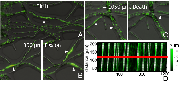
Filamentous fungi form branched and multiconnected networks of cells (called hyphae) that act as transport conduits for nutrients, fluid and organelles Lew (2005). Hyphal networks adapt to minimize transport costs Bebber et al. (2007); *heaton2010growth and also to physically mix the cytoplasmic contents Roper et al. (2013). Typically in the model filamentous fungus Neurospora crassa, nuclei are uniformly dispersed through each hypha Roper et al. (2013); Abadeh and Lew (2013), but in narrow fast flowing hyphae, nuclei have been recently reported to travel together as dense groups Hickey and Read (2009) (Fig. 1). The causes of group formation have not previously been reported. In this Letter we show groups of nuclei are fast moving solitons that form due to anti-congestion within the hyphal network: nuclei travel faster in denser groups than they can individually. In contrast to traffic jams which grow by assimilating cars from the rear Lighthill and Whitham (1955), solitons are created by anti-jamming, that is by overtaking other nuclei. Nuclei continuously transition between transported and stationary phases; anti-congestion results from cooperation between nuclei that suppresses their transitions out of the transported phase. Scheduling of nuclear traffic into intermittent solitons may represent an evolutionary solution to a common problem of biological transport networks; that the links of the network must be remodeled over time without disrupting their function as transport conduits.
Following Hickey and Read (2009) we visualized the flow of nuclei in young ( 1cm diameter) hH1::gfp N. crassa mycelia Freitag et al. (2004), grown on solid Vogels minimal medium. This genetically altered strain expresses a GFP (green fluorescent protein)-tagged histone, making nuclei fluorescent. We followed dense groups of nuclei from their first emergence near the center of network through 1 mm or more of hyphae (Fig. 1 and movie S1). In agreement with Hickey and Read (2009) we found that dense groups traveled together across multiple hyphal links and could split into two separate groups where hypha branched (Fig. 1B). Nuclear groups ultimately dispersed only after traveling 1 mm or more through the network (Fig. 1C).
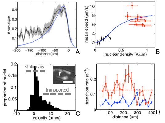
We used a hybrid particle imaging velocimetry / particle tracking method Hood et al. (2015) to track nuclear movements within and outside of dense groups. Nuclei traveling in groups were highly coherent, with no detectable difference in velocity between the front of the group and its rear (Fig 1D). Nuclear distributions were stable between different groups (Fig. 2A). Taken together, these features suggest that groups emerge represent stable traveling wave (soliton) solutions of the equations governing nuclear transport.
We compared speeds of individual and anti-jammed nuclei for over 3000 nuclei (11 individual anti-jams) in a single 400 m stretch of hypha: In general speed increased with the number density of nuclei (Fig. 2B), and groups travel faster than individual nuclei; we therefore call them anti-jams. Thus, N. crassa transport networks perform oppositely to vehicle and data networks Jacobson (1988); *treiber2000congested, in which speeds decrease congestively as traffic density increases. We measured the velocities of individual nuclei that were at least 5 m apart from other nuclei: these nuclei could be divided by Otsu’s method Otsu (1975) into two populations by their velocity stationary (ms-1) or transported (ms-1) (Fig. 2C). Velocities of nuclei propelled by bulk cytoplasmic flow are known to be nearly uniform across the entire hyphal cross-section Roper et al. (2013); Abadeh and Lew (2013); likely because cytoplasm flows like a polymer melt with a narrow slip layer along the cell membrane Roper et al. (2015). Stationary nuclei were located at the periphery of the cell, were deformed by long tubular tethers and occasionally traveled backwards (Fig. 2C and inset) suggestive of active motor-driven positioning Roux et al. (2002); *mourino2006microtubule.
Nuclei do not persist in their transported or stationary phases but freely transitioned between the two: on average nuclei remain attached to the cell membrane for 20s, and flowed with the cytoplasm was 7.8s between transitions (Fig. 2D). Attachment and detachment occurred at constant rates along the the hypha, suggesting that potential attachment sites are uniformly distributed.

To explain flow anti-congestion we model the density of transported, and stationary, , nuclei (measured in #/length of hyphae). Although we do not observe attachment sites directly, we assume that a nucleus enters the stationary phase by attaching to an available site (density ) on the cortical microtubule network. Thus our starting model (described in the Electronic Supplementary Materials); models the mass action laws for a reversible reaction system: \ce ¡=¿[k_TS][k_ST] where , are the rates of nuclear attachment and detachment respectively. We assume that transported nuclei travel with the cytoplasm, at velocity , and stationary nuclei and attachment sites remain fixed on the cell membrane. We can derive analytic results if the length scale on which and vary exceeds ; the travel distance lost by a nucleus that remains attached over the characteristic time . In this limit stationary and transported nuclei maintain a local equilibrium: , where is the (constant) density of potential attachment sites (occupied and unoccupied, so ). The total density of nuclei in both phases obeys a flux conservation equation: , where is the mean speed of transport of a mixed population of nuclei. Physically, our model predicts anti-congestion because there are finitely many available attachment sites on the cell membrane; so if density of nuclei increases then an increasing fraction are in the transported phase.
However the PDEs for and predictive dispersive, rather than free-running anti-jams (see Electronic Supporting Materials). Indeed the local equilibrium equations can be transformed to Burgers’ equation by change of variables: . Burgers’ equation has no traveling wave solutions with the same left and right boundary conditions Evans (1998). Specifically, since increases with , negative density gradients steepen with time – denser groups of nuclei travel faster than the nuclei ahead of them, becoming denser and faster. But small positive density gradients flatten over time; the sparse nuclei at the rear of an anti-jam travel slower than the denser nuclei ahead of them, so the anti-jam disperses.
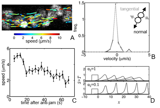
How do nuclei in anti-jams travel coherently? We wondered whether nuclei were adhering together Minke et al. (1999), so we dissected nuclear movements using high-speed (8.3 fps) confocal microscopy. Nuclei inside anti-jams rearranged continuously and fluidly (Fig 3A). We resolved the velocity fluctuations of pairs of almost touching (center-to-center distance m) nuclei into tangential and normal components. If nuclei were adhering we expected the tangential velocity component to be larger because it is easier for pairs of nuclei to rotate or slip past each other than to separate, but tangential and normal velocities were indistinguishable (Fig. 3B).
Burgers’ equation allows traveling wave solutions with different left and right boundary conditions Evans (1998), i.e. if one of our fields; , or is different ahead of and behind the anti-jam. There is no detectable difference in nuclear density on either side of an anti-jam so we hypothesized that attachment sites might be altered. Although we can not directly visualize these attachment sites, we can detect their effect on nuclear transport, by measuring velocities of nuclei in the wake of each anti-jam (Fig. 3C). Nuclei traveled faster on average immediately behind an anti-jam, dropping back to the average velocity for dilute nuclei only after a time s (Fig. 3C). Increases in nuclear velocity correspond to fewer stationary nuclei in the wake of the anti-jam. However, since (the average time for a nucleus to transition back to a stationary state), these data also require that nuclei transition more slowly to stationary state behind the anti-jam.
To explain the speed up data we assume that attachment sites are remodeled, or even lost (and then regenerated) following nuclear attachment. Thus, after a nucleus detaches from the cell periphery, an attachment site is not immediately available to accept another nucleus. We must therefore consider three attachment site densities: , available, , attached (equal to the number of attached nuclei), and , unattached but unable to accept nuclei. We assume that potential attachment sites are uniformly distributed along the hypha, with density . When written in dimensionless form (see Electronic Supplementary Materials) densities of nuclei and attachment sites are describe by a set of equations:
| (1) | |||||
Our equations include two dimensionless rate constants: (i) ; the ratio of the nuclear transition rates and (ii) ; the ratio of the time a nucleus spends attached to the time before an attachment site can accept more nuclei. In the limit we obtain the limit of the equations analyzed previously, with .

Finite-difference numerical solutions of Eq.(1) (see ESM) evolve to solitons for sufficiently small values of but dispersed for larger values of (Fig. 3D). Since small values of seem to be necessary for traveling waves to be generated, we analyzed Eq.(1) asymptotically in the limit. In this limit, attachment sites are monotonically depleted as the anti-jam completely passes. Nuclei therefore cooperate to eliminate all attachment sites, allowing anti-jams to travel through the network without dispersing. Soliton solutions exist, and the nuclear distribution within a soliton can be calculated analytically. For soliton solutions of Eq.(1) with speed , we may combine the PDEs for and to give: . We can solve for the distribution of nuclei within the anti-jam by using (the monotonically decreasing density of available attachment sites) as a dependent variable, and integrating:
| (2) |
These equation possesses a family of soliton solutions which can be parameterized either by the total number of nuclei or by the dimensionless velocity . In the wake of the anti-jam the predicted nuclear density becomes exponentially small and the population returns to its far upstream value (see ESM).
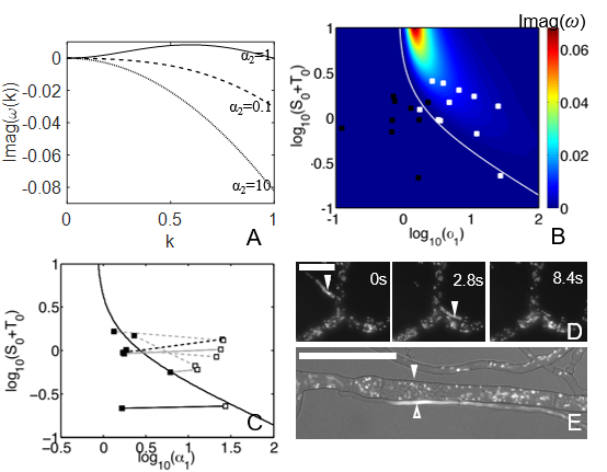
Although our simulations and analytic results show that Eq.(1) supports solitons, they are silent about whether solitons emerge spontaneously or must be created by modulating the distribution of nuclei. We analyzed the stability of uniform flow , , to infinitesimal disturbances of form: for some wavenumber and frequency , with similar representations for and . Linearizing Eqn.(1), we find that uniform flow is unstable to at most a finite range of -values, and that at large values of becomes absolutely stable (see ESM). Nonlinear simulations then showed that linearly unstable disturbances could grow into anti-jams (see ESM).
Since depends on the rate of nuclear/attachment site encounters, it is proportion to the cytoplasmic velocity. The predicted stability boundary when nuclear density and were both varied separated hyphae carrying anti-jams and hyphae that did not (Fig. 4B). We further probed the role of linear stability in anti-jam formation and eventual dispersal by examining flows into and out of network junctions in which anti-jams occurred only on one side of the junction: each junction approximately crossed the predicted stability boundary (Fig. 4C-E, movies S3 and S4). To further confirm that the parameters of our model drive anti-jam formation we redirected flows by damaging hyphae with deionized water. Rerouting of flow from a large diameter, slow hypha, to a small diameter fast hypha, generated anti-jams, as predicted by the linear stability analysis (Fig. 4E).

In summary, nuclei within high traffic links of the fungal network self-organize into stationary and anti-jammed phases. Indeed since nuclei are likely exposed to the same proteins from their shared cytoplasm Gladfelter et al. (2006); Roper et al. (2015), self-organization may provide a more robust mechanism for directing nuclear behaviors than signaling between nuclei.
Engineering of transport on the hyphal network to produce anti-congestion may provide two adaptive benefits: (1) A gain function for transport, that allows nuclei to be delivered faster in response to increases in demand, reducing the amount of redundant network links needed Corson (2010). (2) Links in the hyphal network must grow over time to minimize transport costs Bebber et al. (2007). But, similar to repair work on an open freeway, it is likely that the continuous traffic of nuclei and other organelles will disrupt this growth. Anti-jams organize nuclear transport into very short pulses separated by long ( 100s) intervals in which most () nuclei are retained at the cell periphery and stationary. The intervals between anti-jams may allow growth and other functions to be performed, controlled by stationary nuclei. In particular, given protein translation rates of 8 amino acids/s Karpinets et al. (2006), and the close agreement of translation and transcription times Milo and Phillips (2015), the residence time of a nucleus at the cell membrane () agrees well with the time needed to transcribe the key cell wall proteins RHO1 and RHO2 (195 and 200 amino acids respectively Richthammer et al. (2012)).
This work was supported by the Alfred P. Sloan Foundation and NSF grant DMS-1351860. We thank Inwon Kim, Louise Glass’ group and Nick Read for discussions.
References
- Jacobson (1988) V. Jacobson, Comput. Commun. Rev. 18, 314 (1988).
- Treiber et al. (2000) M. Treiber, A. Hennecke, and D. Helbing, Phys. Rev. E 62, 1805 (2000).
- Chiu and Jain (1989) D.-M. Chiu and R. Jain, Comput. Networks ISDN 17, 1 (1989).
- Yang and H. Bell (1998) H. Yang and M. G. H. Bell, Transport Rev. 18, 257 (1998).
- Banavar et al. (2000) J. R. Banavar, F. Colaiori, A. Flammini, A. Maritan, and A. Rinaldo, Phys. Rev. Lett. 84, 4745 (2000).
- Durand (2006) M. Durand, Phys. Rev. E 73, 016116 (2006).
- Tero et al. (2010) A. Tero, S. Takagi, T. Saigusa, K. Ito, D. P. Bebber, M. D. Fricker, K. Yumiki, R. Kobayashi, and T. Nakagaki, Science 327, 439 (2010).
- Hu and Cai (2013) D. Hu and D. Cai, Phys. Rev. Lett. 111, 138701 (2013).
- Corson (2010) F. Corson, Phys. Rev. Lett. 104, 048703 (2010).
- Katifori et al. (2010) E. Katifori, G. J. Szollosi, and M. O. Magnasco, Phys. Rev. Lett. 104 (2010).
- Chang et al. (2015) S.-S. Chang, S. Tu, Y.-H. Liu, V. Savage, S.-P. Hwang, and M. Roper, (2015), arXiv:1512.04184 .
- Alim et al. (2013) K. Alim, G. Amselem, F. Peaudecerf, M. P. Brenner, and A. Pringle, Proc. Nat. Acad. Sci. USA 110, 13306 (2013).
- Hu et al. (2012) D. Hu, D. Cai, and A. V. Rangan, PLoS ONE 7, e45444 (2012).
- Lew (2005) R. Lew, Microbiology 151, 2685 (2005).
- Bebber et al. (2007) D. Bebber, J. Hynes, P. Darrah, L. Boddy, and M. Fricker, Proc. Roy. Soc. Lond. Ser. B 274, 2307 (2007).
- Heaton et al. (2010) L. L. Heaton, E. López, P. K. Maini, M. D. Fricker, and N. S. Jones, Proc. Roy. Soc. Lond. B , rspb20100735 (2010).
- Roper et al. (2013) M. Roper, A. Simonin, P. C. Hickey, A. Leeder, and N. L. Glass, Proc. Nat. Acad. Sci. USA 110, 12875 (2013).
- Abadeh and Lew (2013) A. Abadeh and R. R. Lew, Microbiology 159, 2386 (2013).
- Hickey and Read (2009) P. C. Hickey and N. Read, Fungal Genet. Reports 56S, 306 (2009).
- Lighthill and Whitham (1955) M. J. Lighthill and G. B. Whitham, Proc. Roy. Soc. Lond. A 229, 317 (1955).
- Freitag et al. (2004) M. Freitag, P. C. Hickey, N. B. Raju, E. U. Selker, and N. D. Read, Fungal Genet. Biol. 41, 897 (2004).
- Hood et al. (2015) K. Hood, S. Kahkeshani, D. Di Carlo, and M. Roper, (2015), arXiv:1509.01643 .
- Otsu (1975) N. Otsu, Automatica 11, 23 (1975).
- Roper et al. (2015) M. Roper, C. Lee, P. C. Hickey, and A. S. Gladfelter, Curr. Opin. Microbiol. 26, 116 (2015).
- Roux et al. (2002) A. Roux, G. Cappello, J. Cartaud, J. Prost, B. Goud, and P. Bassereau, Proc. Nat. Acad. Sci. USA 99, 5394 (2002).
- Mouriño-Pérez et al. (2006) R. R. Mouriño-Pérez, R. W. Roberson, and S. Bartnicki-García, Fungal Genet. Biol. 43, 389 (2006).
- Evans (1998) L. C. Evans, Partial differential equations (Providence, Rhode Land: American Mathematical Society, 1998).
- Minke et al. (1999) P. F. Minke, I. H. Lee, and M. Plamann, Fungal Genet. Biol. 28, 55 (1999).
- Gladfelter et al. (2006) A. S. Gladfelter, A. K. Hungerbuehler, and P. Philippsen, J. Cell Biol. 172, 347 (2006).
- Karpinets et al. (2006) T. V. Karpinets, D. J. Greenwood, C. E. Sams, and J. T. Ammons, BMC Bio. 4, 30 (2006).
- Milo and Phillips (2015) R. Milo and R. Phillips, Cell biology by the numbers (Garland Science, 2015).
- Richthammer et al. (2012) C. Richthammer, M. Enseleit, E. Sanchez-Leon, S. März, Y. Heilig, M. Riquelme, and S. Seiler, Mol. Microbiol. 85, 716 (2012).