Importance of non-affine viscoelastic response in disordered fibre networks
Abstract
Disordered fibre networks are ubiquitous in nature and have a wide range of industrial applications as novel biomaterials. Predicting their viscoelastic response is straightforward for affine deformations that are uniform over all length scales, but when affinity fails, as has been observed experimentally, modelling becomes challenging. Here we introduce a numerical methodology to predict the steady-state viscoelastic spectra and degree of affinity for disordered fibre networks driven at arbitrary frequencies. Applying this method to a peptide gel model reveals a monotonic increase of the shear modulus as the soft, non-affine normal modes are successively suppressed as the driving frequency increases. In addition to being dominated by fibril bending, these low frequency network modes are also shown to be delocalised. The presented methodology provides insights into the importance of non-affinity in the viscoelastic response of peptide gels, and is easily extendible to all types of fibre networks.
pacs:
87.14.em, 87.10.Hk, 87.10.PqFibrous assemblies represent an important class of materials with many industrial applications including scaffolds for tissue engineering Burdick and Mauck (2011) and enamel remineralization Brunton et al. (2013), nonwoven fabrics for medical textiles and industrial filters Russell (2006), carbon nanotube composites Hall et al. (2008), paper and felt Alava and Niskanen (2006). Nature employs protein fibre networks in the multi-functional cellular cytoskeleton Alberts et al. (2008); Bray (2001). The mechanical stiffness of fibre networks is often central to their function, and although static properties come under most scrutiny, they often exist in dynamic environments subject to temporally-varying mechanical loads, including the cytoskeleton of motile cells Bray (2001), and scaffolds for tendon and ligament regeneration, where habitual loading propagating through the network influences the viability of embedded stem cells Burdick and Vunjak-Novakovic (2009); Raïf and Seedhom (2005); Appelman et al. (2011). Understanding the dynamical network response is essential to design novel materials with properties suited for such situations.
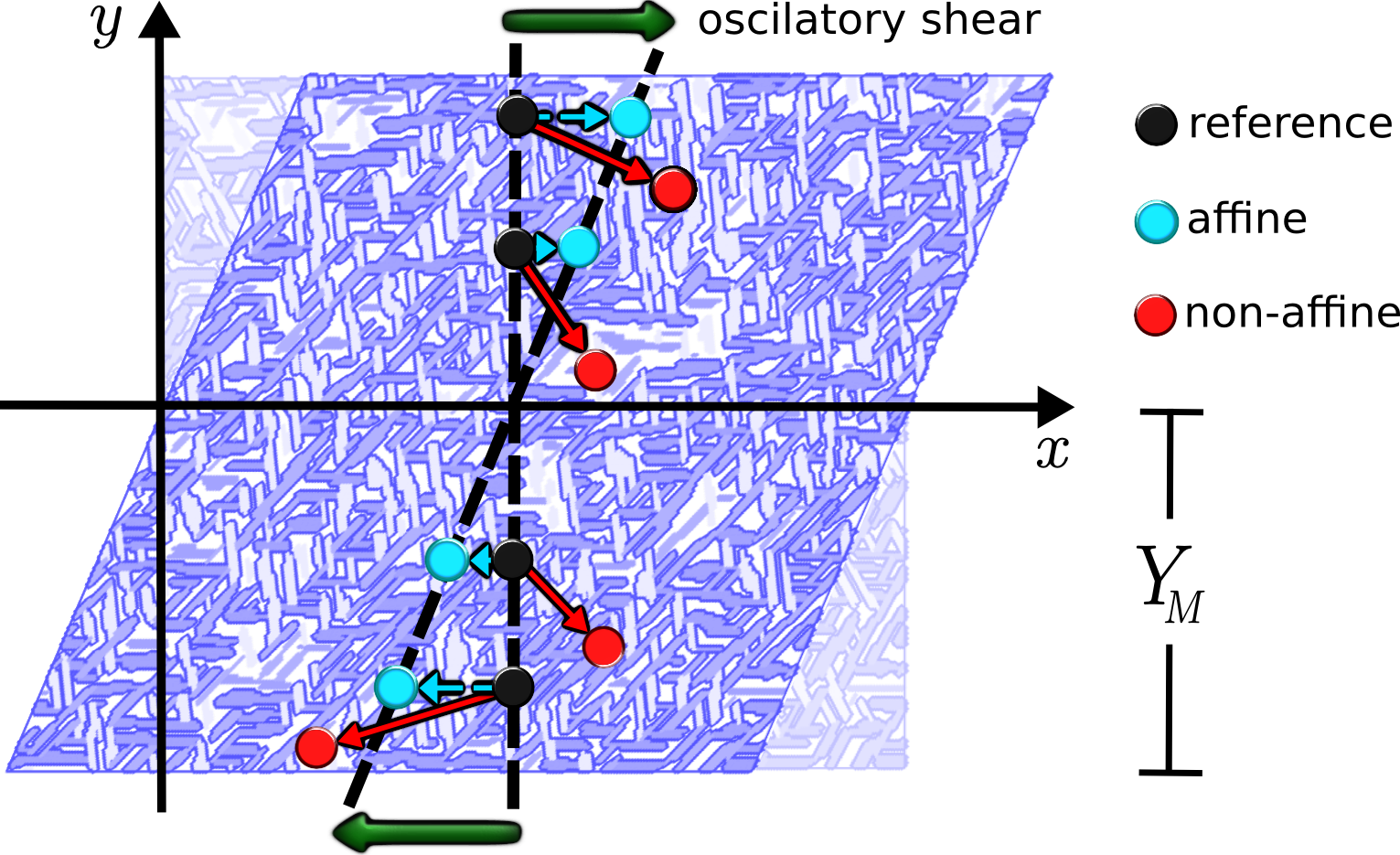
A key modelling challenge is to determine the degree to which the deformation is affine Wen et al. (2012), i.e. uniform over all relevant length scales; see Fig. 1. If affinity holds, extrapolating the macroscopic response from a putative microstructure is straightforward, and a range of thermal and athermal affine models for fibre networks have been developed Broedersz and Mackintosh (2014); Pritchard et al. (2014). When affinity fails, however, as experimentally observed over broad parameter ranges Gardel et al. (2004); Liu et al. (2007); Piechocka et al. (2011); Atakhorrami et al. (2014), it is necessary to determine the microscopic deformation field, which typically requires numerical solution for explicit network realisations. This has thus far been limited to the elastic plateau amenable to energy minimization algorithms Head et al. (2003); Wilhelm and Frey (2003); Buxton and Clarke (2007); Ström et al. (2008), or computationally–intensive particle methods that only access short times Kim et al. (2009); Huisman et al. (2010). Without a more general understanding of fibre networks dynamics, we lack the capability to predict potentially large changes in viscoelastic properties over experimentally relevant time scales.
Here we present a methodology which allows the numerical calculation of the viscoelastic spectra for any type of disordered fibre network driven at arbitrary oscillation frequencies. The method is based on normal modes which ensures linear response, and since no thermal effects or crosslink dynamics are included by construction, all measured variation in affinity and viscoelasticity can be ascribed with certainty to network properties. We demonstrate the efficacy of this method by applying it to a model of peptide gels, and reveal a rich interplay between viscoelasticity, affinity, and mode localisation that derives from the successive suppression of network modes as the driving frequency increases.
Our considerations apply to crosslinked networks of slender elastic fibres immersed in a Newtonian fluid with viscosity . To simplify the network-fluid interaction, all fibre mass is regarded as being concentrated on network nodes in the form of a spherical bead with radius and corresponding Stoke’s drag coefficient . Hydrodynamic interactions between beads are neglected. Taking the overdamped regime relevant to the intended applications, the force balance equation in terms of the node/bead displacement is
| (1) |
where is the dynamical (Hessian) matrix with components in terms of the total elastic energy of a given configuration, and is the vector amplitude of the force applied to this node. The left hand side of (1) couples fluid friction to internal forces generated by network elasticity, and these are balanced with the external force on the right hand side, here assumed to be oscillatory. A stress-controlled shear protocol is assumed where the force is applied only to boundary nodes, so that for the internal nodes, on upper boundary nodes, and on lower boundary nodes, where all on each surface sum to give the required stress. All node displacements are indexed into a single vector , which could be ordered e.g. () for two dimensional (2D) networks. All node displacements can then be written in terms of the eigenvectors of the Hessian as
| (2) |
where the sum is over all modes .
By substituting the expansion above into (1) we obtain exact expressions for the in-phase and out-of-phase components of the coefficients in steady state,
| (3) |
and
| (4) |
where are the coefficients of the expansion for the external force on all nodes, and is the eigenvalue of mode . The eigenvalues are usually related to frequencies, but because we consider the overdamped limit they are instead related to relaxation times . Note that floppy modes correspond to null eigenvalues and undefined relaxation times. We identify these using singular value decomposition Press et al. (2007), and assign to each the coefficients and corresponding to in (1). By considering the amplitudes in (3) and (4), one can use (2) to relate the displacements to the local strain in the -th bead as , where is the middle height line of the system (see Fig. 1). In order to avoid numerical instabilities due to those beads near the middle line (i.e. ), we take the mean value averaged only over beads placed at the upper and bottom boundaries. Finally, the in-phase () and out-of-phase () strains are used to compute the shear moduli of the fibre network, i.e. both the storage modulus and the loss modulus .
In practice, the numerical determination of the viscoelastic spectra of a disordered fibre network requires (i) the construction of the Hessian matrix for an explicit network realisation and a chosen model for single-fibril elasticity, and the determination of its eigenvectors and eigenvalues , (ii) the determination of the coefficients in the expansion of the external force on the network nodes in terms of the eigenvectors, (iii) knowledge of , and allows determination of the in-phase and out-phase response and from (3) and (4), which in turn allows determination of the actual displacement from (2) as a function of the frequency , (iv) from it is straightforward to determine the local strains , and the shear moduli , of the fibre network from the above formulae.
Our test system is a recently developed 2D model for peptide gels, where peptide monomers are explicitly considered in the formation of the fibre network Rizzi et al. (2015), which generalises a lattice-based elastic network model Broedersz et al. (2011) to permit variations in fibre thickness. The interactions between peptide monomers are characterized by their anisotropy ratio , where and are the strengths of strong directional hydrogen bonds and weak isotropic hydrophobicity-mediated bonds Rizzi and Auer (2013), respectively. The anisotropy in the interactions between peptide monomers enables their assembly into crosslinked networks that exhibit a universal time-dependent behaviour in their microstructural geometry (i.e. fibre thickness, fibre length, crosslink separation). Furthermore, the same time-scaling function was found to collapse the plateau value of the corresponding shear modulus and crosslink connectivities Rizzi et al. (2015) .
Unless otherwise stated, all results presented below are for networks generated from monomers with anisotropy and a coverage (mean lattice occupation) , obtained at two different simulation times measured in Monte Carlo steps (MCS). All measurements correspond to averages over 25 independent simulations. Results are reported in experimental units assuming a Young’s modulus for the fibrous material to be Pa, all beads having the same radius nm, and the fluid viscosity Pa s is that of water at 20C. Simulation relaxation times and frequencies are converted to experimental units as per and , with units of s and s-1 respectively. In addition, the viscoelastic spectra and have been normalised to the frequency-independent affine shear modulus corresponding to the storage modulus at zero frequency.
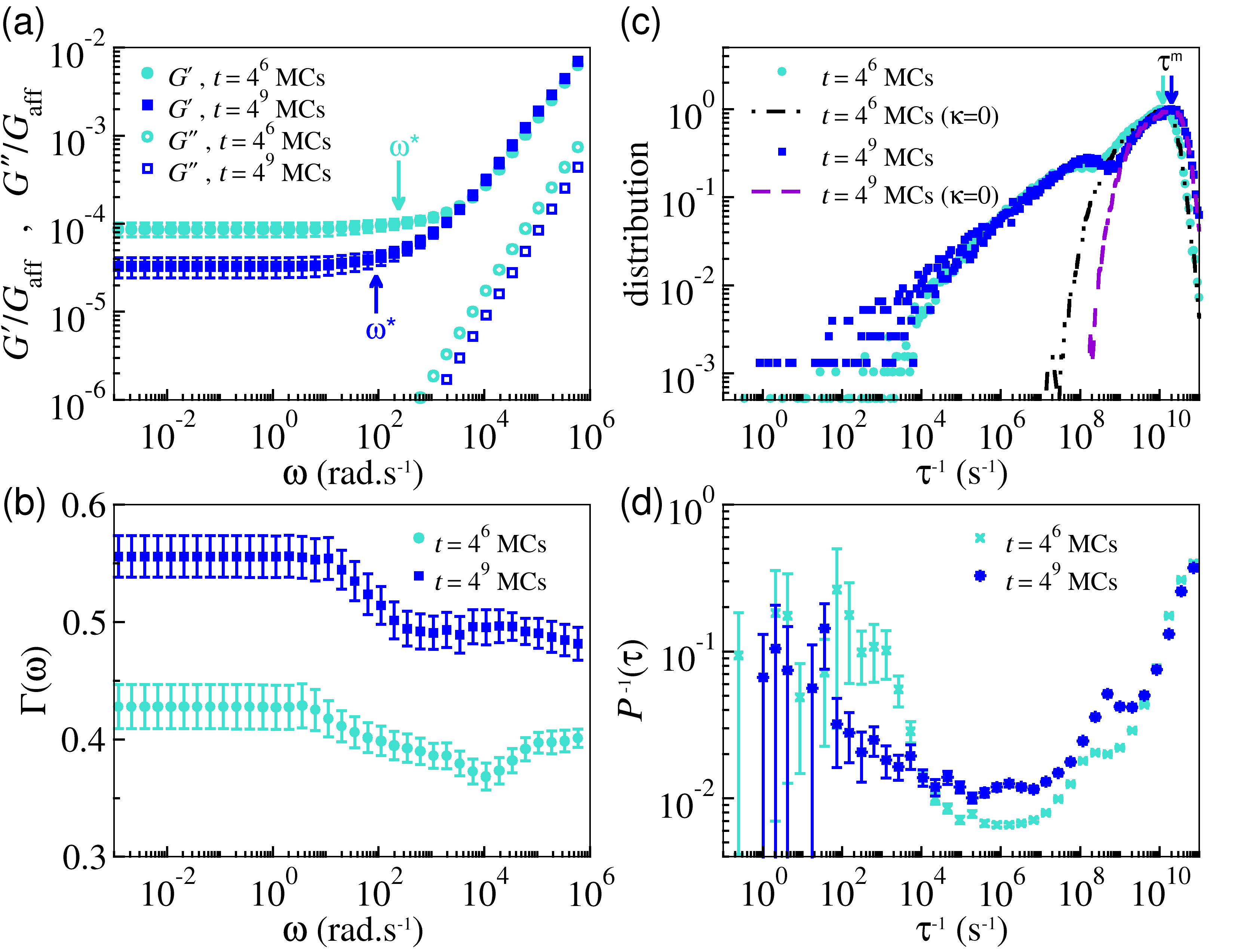
Figure 2(a) demonstrates that the storage modulus presents a plateau regime for low frequencies, and then smoothly increases above some threshold frequency here denoted . This behaviour can be rationalised in terms of the frequency cut-offs, i.e. the factors in the denominators of (3) and (4), leading to a reduction in the amplitude of mode as increases beyond this mode’s natural relaxation time . Without this mode’s contribution, the strain is reduced, so the system stiffens. At high frequencies, the increase of the storage modulus can be described by a power-law with in the range to for all values of and assayed. This range includes the value measured for fibrillar networks using passive microrheology Corrigan and Donald (2009). An exponent of 0.5 due to crosslink unbinding dynamics has been observed in experiments Lieleg and Bausch (2007) and confirmed theoretically Broedersz et al. (2010), but as our model includes no such relaxation mechanism this cannot be the origin of our . Similarly the exponent for the wormlike chain model Gittes and MacKintosh (1998) requires thermal undulations that are not present in our athermal, elastic fibres.
At the low frequencies, our networks deform in a highly non-affine manner as evident in the low values of . This non-affine response is independently confirmed by simultaneously plotting the non-affinity parameter , which is zero for affine deformations. As seen in Fig. 2(b), increases with decreasing frequency. Our 2D results can be compared to 3D experiments by scaling according to the affine predictions for each dimension, i.e. , with the inter-crosslink length nm Rizzi et al. (2015). This yields values for the storage modulus at the plateau regime equal to Pa for rad.s-1 and Pa for rad.s-1 at MCs and MCs, respectively. These values are comparable to measurements for peptide gels such as amyloid tapes Aggeli et al. (1997); Greenfield et al. (2010); Tang et al. (2011) and spider silk Rammensee et al. (2006); Gong et al. (2010). Fig. 2(a) also demonstrates our networks soften with age, which has also been observed for crosslinked actin Lieleg et al. (2011) and can be related here to the increase in non-affinity, itself due to the reduced network connectivity as shown elsewhere Rizzi et al. (2015).
In Fig. 2(c) we show the distribution of relaxation times , which confirms that the broad range over which decreases is related to a broad range of following a bimodal distribution. Previous work at zero frequency identified the fast and slow relaxation peaks with fibre stretching and bending modes, respectively Huisman and Lubensky (2011), and we can confirm this holds for finite frequency by setting the fibre bending modulus to zero in , which removes the slow relaxation modes without significantly altering the fast ones as shown in the figure. In addition, the fast stretching modes move to shorter relaxation times as the simulation time increases, in contrast to the slow bending modes which remain fixed, lending insight into the mechanism underlying the observed softening with age. The slow bending modes are also delocalised, in contrast to the localised fast stretching modes, as shown in Fig. 2(d) where is displayed the inverse participation ratio , which is high for delocalised and low for localised modes Huisman and Lubensky (2011); Silbert et al. (2009). This trend is consistent with intuitive assertions made in recent vimentin experiments Head et al. (2014).
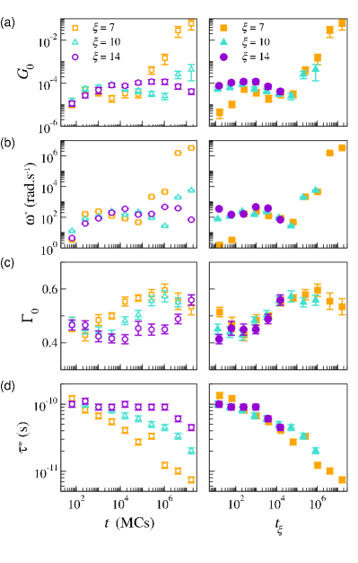
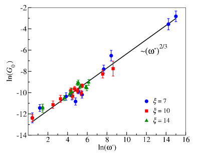
The picture just described holds for other values of the anisotropy parameter and network formation time considered. Shown in Fig. 3 are the trends as and are varied for the zero-frequency elastic modulus , the zero-frequency non-affinity , the threshold frequency and the modal relaxation time . In addition to the unscaled behaviour given as a function of simulation time (open symbols and left panels), we also plot the same quantities against the -dependent rescaled time (filled symbols and right panels) which generates data collapse at zero frequency Rizzi et al. (2015). As illustrated in Figs. 3(a) and (b), and exhibit a similar non-monotonic behaviour, while the data for in Fig. 3(c) demonstrates an increase in non-affinity with time. Figure 3(d) confirms that the trend mentioned above, i.e. that shifts to shorter relaxation times with network age, is general. We also observe a power-law behaviour which appears to be independent of , as shown in Fig. 4, but currently have no explanation for this apparently robust phenomenon. Finally, we can infer from the data collapse under the same rescaled time as Rizzi et al. (2015) that these dynamic quantities correlate to microstructural geometric quantities (fibre length and thicknesses, crosslink separation), suggesting the ultimate origin of the observed frequency dependence of our fibre networks is geometric.
In summary, we have introduced an efficient numerical scheme to extract the linear finite-frequency viscoelastic response of fibre networks, and applied it to model peptide gels to observe a power-law increase of the storage modulus with frequency . Our method precludes the possibility that this stiffening is related to dynamic crosslink unbinding Lieleg and Bausch (2007); Broedersz et al. (2010) or frequency-dependent single fibre response Gittes and MacKintosh (1998), but instead demonstrates it is due to an underlying decrease in non-affinity as shown in Fig. 2. This prediction is in principle experimentally testable Liu et al. (2007). That the transition from affine to non-affine response is gradual is consistent with Brownian dynamics Huisman et al. (2010) and elastic spring networks Yucht et al. (2013), although our results include fibre bending and are unambiguously steady state. The loss modulus never strongly deviated from the purely viscous response , in contrast to the clearly sublinear variation observed in many fibrous materials Roberts et al. (2012); Rombouts et al. (2013); Lin et al. (2010); Broedersz and Mackintosh (2014). This deviation may be due hydrodynamic interactions, which could be incorporated into this framework by including interaction terms via Oseen tensors Doi and Edwards (1986) in (1) to give a dense matrix equation. Finally, we note that even though we have applied this methodology to peptide gels in 2D, we expect our method and core findings to be applicable to fibre networks in general, including in three dimensions. Our methodology also allows a way to approach the complex and largely unexplored problem of hydrodynamic interactions in fibre networks.
We thank D. Mizuno for early discussions regarding the procedure detailed here. L.G.R. acknowledges support from the Brazilian agency CNPq (Grant N 245412/2012-3). D.A.H. acknowledges support from the Biomedical Health Research Centre, University of Leeds, UK.
References
- Burdick and Mauck (2011) J. A. Burdick and R. L. Mauck, Biomaterials for Tissue Engineering Applications (SpringerWienNewYork, 2011).
- Brunton et al. (2013) P. A. Brunton, R. P. W. Davies, J. L. Burke, A. Smith, A. Aggeli, S. J. Brookes, and J. Kirkham, British Dental Journal 215, E6 (2013).
- Russell (2006) S. Russell, Handbook of Nonwovens (Woodhead Publishing, 2006).
- Hall et al. (2008) L. J. Hall, V. R. Coluci, D. S. G. ao, M. E. Kozlov, M. Zhang, S. O. Dantas, and R. H. Baughman, Science 320, 504 (2008).
- Alava and Niskanen (2006) M. Alava and K. Niskanen, Rep. Prog. Phys. 69, 669 (2006).
- Alberts et al. (2008) B. Alberts, A. Johnson, J. Lewis, M. Raff, K. Roberts, and P. Walter, Molecular Biology of the Cell (Garland Science, 2008).
- Bray (2001) D. Bray, Cell Movements: From Molecules to Motility (Garland, 2001).
- Burdick and Vunjak-Novakovic (2009) J. A. Burdick and G. Vunjak-Novakovic, Tissue Eng. A 15, 205 (2009).
- Raïf and Seedhom (2005) E. M. Raïf and B. Seedhom, Bone 36, 433 (2005).
- Appelman et al. (2011) T. P. Appelman, J. Mizrahi, J. H. Elisseeff, and D. Seliktar, Biomat. 32, 1508 (2011).
- Wen et al. (2012) Q. Wen, A. Basu, P. A. Janmey, and A. G. Yodhb, Soft Matter 8, 8039 (2012).
- Broedersz and Mackintosh (2014) C. P. Broedersz and F. C. Mackintosh, Rev. Mod. Phys. 86, 995 (2014).
- Pritchard et al. (2014) R. H. Pritchard, Y. Y. S. Huang, and E. M. Terentjev, Soft Matter 10, 1864 (2014).
- Gardel et al. (2004) M. L. Gardel, J. Shin, F. C. Mackintosh, L. Mahadevan, P. Matsudaira, and D. A. Weitz, Science 304, 1301 (2004).
- Liu et al. (2007) J. Liu, G. Koenderink, K. E. Kasza, F. C. Mackintosh, and D. A. Weitz, Phys. Rev. Lett. 98, 198304 (2007).
- Piechocka et al. (2011) I. K. Piechocka, A. S. G. van Oosten, R. G. M. Breuls, and G. Koenderink, Biomacromolecules 12, 2797 (2011).
- Atakhorrami et al. (2014) M. Atakhorrami, G. Koenderink, J. F. Palierne, F. C. Mackintosh, and C. F. Schmidt, Phys. Rev. Lett. 112, 088101 (2014).
- Head et al. (2003) D. A. Head, A. J. Levine, and F. C. Mackintosh, Phys. Rev. Lett. 91, 108102 (2003).
- Wilhelm and Frey (2003) J. Wilhelm and E. Frey, Phys. Rev. Lett. 91, 108103 (2003).
- Buxton and Clarke (2007) G. A. Buxton and N. Clarke, Phys. Rev. Lett. 98, 238103 (2007).
- Ström et al. (2008) J. A. A. Ström, P. B. S. Kumar, I. Vattulainen, and M. Karttunen, Phys. Rev. E 77, 051913 (2008).
- Kim et al. (2009) T. Kim, W. Hwang, H. Lee, and R. D. Kamm, PLoS Comp. Biol. 5, e1000439 (2009).
- Huisman et al. (2010) E. M. Huisman, C. Storm, and G. T. Barkema, Phys. Rev. E 82, 061902 (2010).
- Rizzi and Auer (2013) L. G. Rizzi and S. Auer, J. Phys. Chem. B 119, 14631 (2013).
- Rizzi et al. (2015) L. G. Rizzi, D. A. Head, and S. Auer, Phys. Rev. Lett. 114, 078102 (2015).
- Press et al. (2007) W. H. Press, S. A. Teukolsky, W. T. Vetterling, and B. P. Flannery, Numerical Recipers, 3rd ed. (Cambridge University Press, Cambridge, 2007).
- Broedersz et al. (2011) C. P. Broedersz, X. Mao, T. C. Lubensky, and F. C. MacKintosh, Nature Phys. 7, 983 (2011).
- Corrigan and Donald (2009) A. M. Corrigan and A. M. Donald, Langmuir 25, 8599 (2009).
- Lieleg and Bausch (2007) O. Lieleg and A. R. Bausch, Phys. Rev. Lett. 99, 158105 (2007).
- Broedersz et al. (2010) C. P. Broedersz, M. Depken, N. Y. Yao, M. R. Poliak, D. A. Weitz, and F. C. MacKintosh, Phys. Rev. Lett. 105, 238101 (2010).
- Gittes and MacKintosh (1998) F. Gittes and F. C. MacKintosh, Phys. Rev. E 58, R1241 (1998).
- Aggeli et al. (1997) A. Aggeli, M. Bell, N. Boden, J. N. Keen, P. F. Knowles, T. C. B. McLeish, M. Pitkeath, and S. E. Radford, Nature (1997).
- Greenfield et al. (2010) M. A. Greenfield, J. R. Hoffman, M. O. de la Cruz, and S. I. Stupp, Langmuir 26, 3641 (2010).
- Tang et al. (2011) C. Tang, R. Ulijn, and A. Saiani, Langmuir 27, 14438 (2011).
- Rammensee et al. (2006) S. Rammensee, D. Huemmerich, K. Hermanson, T. Scheibel, and A. R. Bausch, Appl. Phys. A 82, 261 (2006).
- Gong et al. (2010) Z. Gong, Y. Yang, L. Huang, X. Chena, and Z. Shao, Soft Matter 6, 1217 (2010).
- Lieleg et al. (2011) O. Lieleg, J. Kayser, G. Brambilla, L. Cipelletti, and A. R. Bausch, Nat. Mater. 10, 237 (2011).
- Huisman and Lubensky (2011) E. M. Huisman and T. C. Lubensky, Phys. Rev. Lett. 106 (2011).
- Silbert et al. (2009) L. E. Silbert, A. J. Liu, and S. R. Nagel, Phys. Rev. E 79, 021308 (2009).
- Head et al. (2014) D. A. Head, E. Ikebe, A. Nakamasu, P. Zhang, L. G. Villaruz, S. Kinoshita, S. Ando, and D. Mizuno, Phys. Rev. E 89, 042711 (2014).
- Yucht et al. (2013) M. G. Yucht, M. Sheinman, and C. P. Broedersz, Soft Matter 9, 7000 (2013).
- Roberts et al. (2012) D. Roberts, C. Rochas, A. Saiani, and A. F. Miller, Langmuir 28, 16196 (2012).
- Rombouts et al. (2013) W. H. Rombouts, M. Colomb-Delsuc, M. W. T. Werten, S. Otto, F. A. de Wolf, and J. van der Gucht, Soft Matter 9, 6936 (2013).
- Lin et al. (2010) Y. C. Lin, N. Y. Yao, C. P. Broedersz, H. Herrmann, F. C. MacKintosh, and D. A. Weitz, Phys. Rev. Lett. 104, 058101 (2010).
- Doi and Edwards (1986) M. Doi and S. F. Edwards, The Theory of Polymer Dynamics (Oxford Science Publications, Oxford, 1986).