Patterns, Shapes and Growth of a Spherical Superabsorvent Polymers
Abstract
Combining experimental procedures and theoretical modeling, we studied patterns, shapes, and growth of a special type of SuperAbsorbent Polymers (SAP). At the initial stage, embed in water, we have a polygonal pattern over a spherical form, that evolves into a wrinkled deformed shape, which is then followed by an oblate ellipsoid with some of wrinkles over the surface. The final step is a slowly growing sphere. We measured the weights in function of time during the growing process. The results can be represented by an exponential function, which is derived from the general continuity equation.
pacs:
61.41.+e, 05.70.Ln, 68.10.-m, 82.70.GgIn the present letter, we report an integrated research in patterns, shapes and growth, as suggested by D’Arcy Thompson in his celebrated book “On the Growth and Form ”Thompson . This growth is mathematically analyzed with the continuity equation as we embed in water, a very complex network of cross linked hydrophilic polymers called SuperAbsorbent Polymer(SAP).
In general, the SAPs are commonly made from the polymerization of acrylic acid blended with sodium hydroxide. As we know the SAP is kind of gel studied Hajime Tanaka ,Suematsu in similar scenery decades ago. Due to the their high absorbency, the SAPs have been used in diapers as well as water reservoir for agriculture iranianos and horticulture and the latest accomplishment in this field is the confection of a superabsorbent membrane called Imec Imec . There is also proposals to be applied in high-performance concrete concret and petroleum petroleum .
In the early years of the present decade. SAP was retooled and sold world wide as a tiny hard spheres, with a diameter of around 3.5 mm, while the original one has an irregular shape, similar to grains or even a white powder.
One of these SAP spheres or Crystal Gel is immersed in water for a few seconds. Removing and looking at it through a microscopy, we see a polygonal pattern as shown in Fig.1(a). The origin of this pattern can be traced back to the interaction between the water dipole with the hydrophilic polymer. This guess can be verified using the following procedure.

SAP spheres were left randomly in a small container. Then, few second in the water, as noted in Fig. 1(b) the SAP spheres become a hexagonal pattern, and the equidistance between them shows the propagation of the dipole interaction through the water. The polygonal irregularity is mainly due to the shielding of the dipole-dipole interaction by the medium. We need to recall that a Buckyball effect has some role in this non-uniformity Buckyball .
Aside of polygonal pattern another fact can be seen under the microscope. Out of water, the polygons coalesces, as can be observed in a photo sequence in Fig. 2. In the middle part, we notice a labyrinthine pattern similar to the surface of our brains. This phenomenon seems to be analogous to the work on surface pattern of fruit dehydration surface , where they examined the elastic interaction between the skin and the fruit pulp. The smoothing of surface, noticed in the Fig.2, is due to the homogenization through the water diffusion over the entire surface.

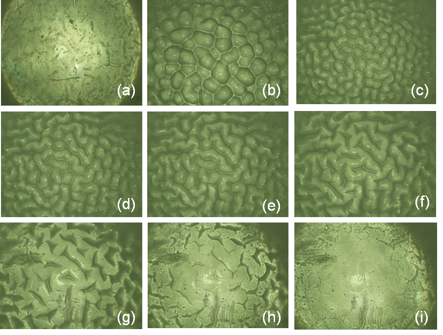
In order to look for different forms and patterns, the SAP sphere was submerged, sequentially, in a distilled water with acidity around pH 4.0. For this purpose, we built an automated system, supporting the ball with very fine needle and, taking it out time to time, in order to take a picture. This step was needed, since the water refraction does not allow a good picture. The dives were three minutes inside water and ten seconds out of it. This procedure ensures the surface to be wet during the photography stage. Fig.3 shows different shapes of SAP sphere were only the relevant pictures is displayed.

Polygonal patterns over spherical form can be seen in Fig.3 for around the first twenty minutes with water diffusing through the skin. After that, as the water enters little deeper into the polymer and the shape transformation occurs. The Crystal Gel becomes highly deformed and the surface pattern changes, it smooths and a scar or an elastic folding appears over the surface.
If we take out the highly deformed Crystal Gel from the water, the surface self smooth and after some time will take a spherical shape. In this point another experiment was performed, but not displayed. Using a razor we cut the deformed Crystal Gel in two parts, both twists considerably and after some time they can be re-assembled into a single sphere.
Before reaching the ultimate spherical form, an oblate ellipsoidal shape with a cross and semi-cross elastic folding can be noticed. Eight hours after, the Crystal Gel is spherical and the growing slows and finally a saturated phase is reached after around thirty hours.
All these experiments shows, the diffusion character of these phenomena, the search for homogenization. In the final stages, the concentration of water inside and outside the sphere is practically the same, with a dynamic equilibrium state.
Since, the Crystal Gel spheres are commercialized in different colors, we experimentally noted, that the overall behavior does not depend on the pigment material used to color these spheres.
The next step is to measure the growing process. We perform an experiment by selecting carefully, five sets with ten tiny balls and weighting them with a balance with a scale of hundredth grams of precision and, we show them in the first row of Tab.1. With the help of a sieve, each set was dived multiple times into a water with same acidity earlier mentioned and the weight was measured .
| Set | 1 | 2 | 3 | 4 | 5 |
|---|---|---|---|---|---|
| Multiple dives | 0,46 | 0,48 | 0,46 | 0,47 | 0.46 |
| Single dive | 0,45 | 0,46 | 0.44 | 0,43 | 0,45 |
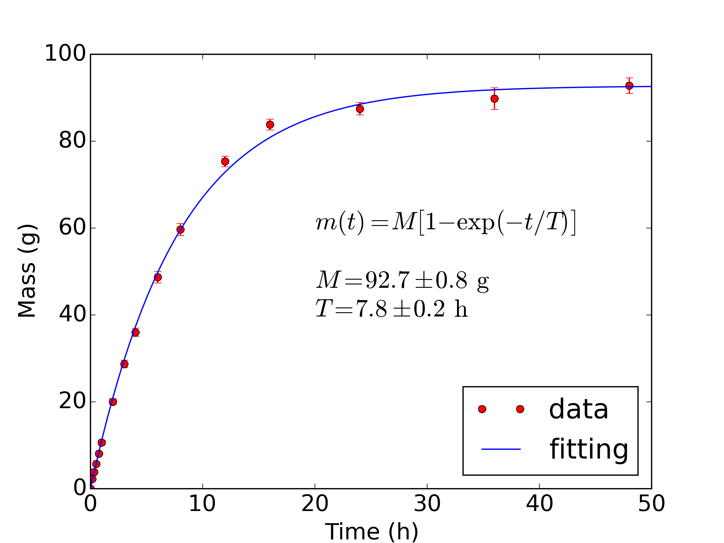
Results are displayed as a dots in the Fig.4. Time variation was measured with Nikon DX5100 camera clock and simultaneously the weight measurement was taken. Ending this experiment, we weight the individual spheres and the results will be discussed in Fig.5.

As you can notice, an exponential curve fits, with the experimental data, the error bar cannot be seen in the beginning of the curve, however it becomes larger and larger. Most likely the multiple dives, scratches the surface, allowing more water in the final steps.
To test this hypothesis, a second measurement of the final weight was done using a different batch of 5 sets of spheres, see the second row of Tab.1. But this time the spheres were dived only once and no measurement was made as they were growing. In Fig.5 shows the mass distribution of all the final spheres weight. Blue column represents one dive and green color, multiple dives. As expected in one dive, the dispersion is lower compared to multiple dives.

In the present experiment the flux of water, is going after, as we already mentioned, for homogenization. Therefore we have, only, a local form of conservation law which is governed by the general second Fick’s Law equation. Here written in the integral form:
| (1) |
where represents the density or the concentration of water inside the Crystal Gel, which increases due to absorption. The is the flux of water going towards the center of the sphere and is defined by the following expression:
| (2) |
The function is the Diffusion Coefficient. Physically, for each radius it may describe the permeability of the surface. An assemble of these surfaces is a collection of tortuous micro pipes with different diameters. As the Crystal Gel swallows the water, these micro pipes enlarges and we have in each of them, a different concentration of water.
The streaming of water inside the Crystal Gel is described by the gradient of concentration . As mentioned, experimentally in the first twenty to twenty five minutes, it seems to be a diffusive spherical field. After this time, as the streams of water overcame the skin or the first layer and the water dipoles penetrates deep in the second complex cross linked polymer network, and two types of elasticity seems to appear, one in the skin another in the pulp and the interaction between them may explain the overall aspect of the large deformations displayed Fig.3.
At this moment, it is not easy to guess a reliable density dependent diffusion coefficient. They should incorporate the permeability and the elasticity of the gel and, simultaneously, the shape deformation evolution. Further, it should also incorporate the leveling of gel surface.
Additionally, if one remembers the differential form of Eq.1, this coefficient is coupled through the divergent operator, with the gradient of water concentration, leading to non linear Differential Equation, and by solving it, someone will describe the concentration of water evolution, as it is displayed in the Fig. 3.
However, we do not have enough material to pursue this ambitious and interesting research program. Fortunately, as a first step, we can interpret the second term of the Eq.2 as the flux of water into the surface. Then, let us assume, that is always possible to transform the complicated shapes observed in the Fig.3 into spheres and the water diffuses radially. In others words we have field similar to a central field and is:
| (3) |
Replacing this term into the second term of the Eq. 1 and performing the surface integral we get,
| (4) |
The radial component of the gradient in the above Eq.4 can be interpreted as the flux of water penetrating into the surface with radius . Consequently the density of water decreases from outside to inside of the sphere in a radial and uniform way. Mathematically this is expressed by,
| (5) |
Let us now discuss the Diffusion Coefficient from Eq.4. From our experimental data, we can notice that the water flux is at maximum when polymer is placed in water and as the time evolves, the absorbency capacity diminishes and finally reaches the some kind of steady stage. The amount of water going in and out is practically the same, a dynamical equilibrium.
This dynamical situation can be abridged considering a net permeability. In our case, it can be represented by an association with both, the thickness and the inverse of spherical surface i.e., . Furthermore, an additional constant coefficient is needed to classify, simultaneously, the characteristic of Crystal Gel and the embedding medium.
Thus we suggest, rename as effective diffusion coefficient and we make a following educated guess,
| (6) |
| (7) |
Now we need to perform the calculation about the volume integral in this equation. As we do not know the variation of the of water inside the sphere, let us assume, that for each time interval, the water drips on a fixed volume, instead of adopting a Crystal Gel expansion, a real physical situation.
So the evolution of the density of water will be independent of the radius of the sphere, but there will be variation of concentration. We will also adopt the fixed volume as the maximum volume reached when saturation occurs or . Thus variation of water concentration is,
| (8) |
Replacing it, on the first term of the continuity equation Eq.7 and performing the integration over the volume, we obtain the well known differential equation,
| (9) |
which results in:
| (10) |
This function describes perfectly our measurement displayed in Fig.4, reassuring that all the spherical approximations made in the integral form of the second Fick’s Law, is robust and reliable. We can further blend it with fitted data displayed in Fig.4 to get and h/ml, where we assumed g/ml.
However, as we mentioned, the real picture is much more complicated. Additionally, the inclusion of the angular terms of the spherical coordinated is needed and also, a more realistic Diffusion Coefficient.
These additions will furnish a non-linear Differential Equation and, its solution will reveal a complete interpretation of the evolution of patterns, shapes and growth displayed throughout the pictures of present paper. Furthermore, this approach will also reveals the water evolution inside the Crystal Gel which can be subjected to an experimental scrutiny by cutting the Crystal Gels in different stages of its evolution.
Finally, as we mentioned, we developed a methodology to investigate in an integrated manner, patterns, shapes and growth of SAP spheres. Since, our approach results in the core of the Ontogenetic Growth Model, ontogenetic , which fits the grow of several animals, where the Energy is the diffusing material. Then, it is tempting to suggest that present procedure could be used to derive their basic differential equation from the general Continuity Equation.
References
- (1)
- (2) D. W. Thompson, On Growth and Form,(Cambridge University Press, 1992).
- (3) H. Tanaka, H. Tomita, A. Takasu, T. Hayashi, and T. Nishi, Phys.Rev.Lett., 68, 2794 (1992).
- (4) N. Suematsu, K. Sekimoto and K. Kawasaki, Phys.Rev A - Rapid Communications, 41, 5751 (1990).
- (5) J. M. Zohuriaan-Mehr and K. Kabiri, Iranian Polymer Journal, 17, 6, 451 (2008).
- (6) Y. Mori, Reactive and Functional Polymers, 73, 7, 936 (2013).
- (7) O. M. Jensen, Concrete International, 35, 1, 48 (2013).
- (8) J. Alaei, S.H. Boroojerdi and Z. Rabiel, Petroleum and Coal 47, 32 (2005).
- (9) H. W. Kroto, J. R. Heath, S. C. O’Brian, R. F. Curl and R. E. Smalley Nature, 318, 6042 (1985).
- (10) B. Li, F. Jia, Y.P. Cao, X. Q. Feng, H. Gao Phys.Rev.Lett., 106, 234301 (2011).
- (11) G. B. West, J. H. Brown and B. J. Enquist, Nature, 413, 628 (2001).