Binding of bivalent transcription factors to active and inactive regions folds human chromosomes into loops, rosettes and domains
Abstract
Biophysicists are modeling conformations of interphase chromosomes, often basing the strengths of interactions between segments distant on the genetic map on contact frequencies determined experimentally. Here, instead, we develop a fitting-free, minimal model: bivalent red and green “transcription factors” bind to cognate sites in runs of beads (“chromatin”) to form molecular bridges stabilizing loops. In the absence of additional explicit forces, molecular dynamic simulations reveal that bound “factors” spontaneously cluster – red with red, green with green, but rarely red with green – to give structures reminiscent of transcription factories. Binding of just two transcription factors (or proteins) to active and inactive regions of human chromosomes yields rosettes, topological domains, and contact maps much like those seen experimentally. This emergent “bridging-induced attraction” proves to be a robust, simple, and generic force able to organize interphase chromosomes at all scales.
The conformations adopted by human chromosomes in 3D nuclear space are currently an important focus in genome biology, as they underlie gene activity in health, aging, and disease Cavalli2013 . Chromosome conformation capture (3C) and high-throughput derivatives like “Hi-C” allow contacts between different chromatin segments to be mapped Lieberman-Aiden2009 . Inspection of the resulting contact maps reveals some general principles, including: (i) Each chromosome folds into distinct “topological domains” during interphase (but not mitosis when transcription ceases); domains contain 0.1-2 Mbp, active and inactive regions tend to form separate domains, and sequences within a domain contact each other more often than those in different domains Lieberman-Aiden2009 ; Dixon2012 ; Nora2012 ; Sexton2012 ; Naumova2013 ; Rao2014 ; Sexton2015 . (ii) Domains seem to be specified locally, as the same 20-Mbp region in a chromosomal fragment or the intact chromosome make much the same contacts Rao2014 . (iii) Bound transcription factors like CTCF (the CCCTC-binding factor) and active transcription units are enriched at domain “boundaries” Dixon2012 ; Rao2014 . (iv) Factors bound to promoters and enhances stabilize loops Simonis2006 ; Li2012 ; Jin2013 ; Zhang2013 ; Heidari2014 ; Rao2014 ; Mifsud2015 . (v) Co-regulated genes utilizing the same factors often contact each other when transcribed Fullwood2009 ; Schoenfelder2010 ; Yaffe2011 ; Li2012 ; Papantonis2012 . (vi) Single-cell analyses show no two cells in the same population share exactly the same contacts, but the organization is non-random as certain contacts are seen more often than others Nagano2013 . (vii) This organization is conserved; in budding yeast Hsieh2015 and Caulobacter crescentus Le2013 , “chromosomal interaction domains” (CIDs) are separated by strong promoters, and the bacterial ones are eliminated by inhibiting transcription. These principles point to central roles for transcription orchestrating this organization, with transcription factors providing the required specificity.
Biophysicists are attempting to model this organization Marenduzzo2006 ; Rosa2008 ; Nicodemi2008 ; Lieberman-Aiden2009 ; Duan2010 ; Junier2010 ; deVries2011 ; Kalhor2011 ; Rousseau2011 ; Barbieri2012 ; Bau2012 ; Brackley2013 ; Le2013 ; Naumova2013 ; Nagano2013 ; Benedetti2014 ; Jost2014 ; Tark-Dame2014 ; Trieu2014 ; Cheng2015 ; Hofmann2015 ; Johnson2015 ; Junier2015 ; Trussart2015 ; Zhang2015 , often basing the strength of interactions between segments distant in 1D sequence space on contact frequencies determined using Hi-C Lieberman-Aiden2009 ; Duan2010 ; Kalhor2011 ; Rousseau2011 ; Nagano2013 ; Trieu2014 ; Giorgetti2014 ; Junier2015 ; Trussart2015 ; Zhang2015 ; Dekker2013 ; Serra2015 . To understand the principles underlying the organization, we use a minimal model without such fitting that was originally developed to analyze non-specific binding of proteins like histones to DNA Brackley2013 ; Johnson2015 ; here, we adapt it to include specific binding. Thus, spheres (representing transcription factors) bind briefly to cognate sites in runs of beads (representing chromatin) before dissociating. These factors provide an obvious connection with transcription, as they often associate with RNA polymerase (which can remain tightly bound to the template for 10 min as it transcribes the average human gene – a binding that is also specific in the sense it occurs throughout a transcription unit but not elsewhere). [However, here, we only model transient binding.] Like many transcription factors (or complexes made up of several of these factors), ours are “bivalent”; they can bind simultaneously to two or more segments of one fiber, to create molecular “bridges” that stabilize loops. More generally, our spheres could represent any bivalent DNA-binding complex that binds specifically.
In contrast to previous work, our model is fitting free. Instead of beginning with experimentally-determined Hi-C data, we start with 1D information (i.e., whether a particular genomic region is transcriptionally active or not) and use it to generate a population of possible chromosome structures (considering fibers with more subunits than those used previously); only then, do we compare the resulting contacts with those seen experimentally. Remarkably, our coarse-grained molecular dynamic (MD) simulations show fibers spontaneously fold into structures possessing the key features outlined above. We uncover an emergent force that can act through the binding of just two (or more) types of transcription factor to their cognate sites that is able to organize interphase chromosomes locally and globally – all without inclusion of any explicit attractive force between distant segments, or between factors.
Results
Chromatin fibers spontaneously assemble into imperfect rosettes
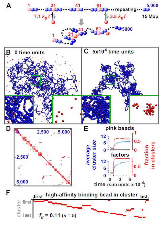
Our MD simulations use the LAMMPS software package (Large-scale Atomic/Molecular Massively Parallel Simulator Plimpton1995 ) run in Brownian dynamics (BD) mode (see Supporting Information for more details). We begin with a “chromatin fiber” of 5,000 30-nm beads – representing 15-Mbp – diffusing amongst 30-nm “transcription factors” (Fig. 1A). Initially, “transcription factors” (hereafter factors) have no affinity for any bead in the fiber (which follows a self-avoiding random walk), but then binding is “switched” on so they now have a high affinity for every 20th bead (pink), and a low affinity for all others (emulating the tight binding of transcription factors to cognate sites and non-specific binding elsewhere). Importantly, factors can bind to two (or more) beads, and affinities are just large enough to favor binding. Consequently, a factor often binds to a low-affinity site, dissociates, and rebinds nearby. As this process repeats, the factor may reach a high-affinity site and remain bound long enough to stabilize a loop (Fig. 1A); bound factors now spontaneously cluster (Fig. 1B, C; Movies S1 and S2). The force driving analogous clustering after non-specific binding was dubbed the “bridging-induced attraction”; it operates even though no explicit attraction between factors or between beads was specified, and it was not seen with monovalent factors or irreversible binding Brackley2013 . Earlier work also shows such clustering occurs with 20-nm beads Brackley2013 , so the results we now present should also apply to chromatin fibers of this (or different) size.

We next examine some properties of the system. As binding compacts the fiber, and as beads in/around each cluster make many contacts, blocks of red pixels are seen along the diagonal in the resulting contact map (Fig. 1D) – as in Hi-C data Lieberman-Aiden2009 . After switching on binding, clusters form in 1 min (one simulation time unit is 0.6 ms, calculated assuming a nuclear viscosity of 10 cP), and the fraction of pink beads in clusters increases rapidly (Fig. 1E). [We define two beads to be in the same cluster if centers lie within 90 nm.] Small clusters then slowly grow to reach a steady-state size with 12 factors/cluster (Fig. 1E), when the entropic cost of gathering loops together (which scales nonlinearly with loop number Duplantier1989 ) limits further growth. It is likely that such “coarsening” is also dynamically hindered, as merging two clusters of loops (even when thermodynamically favored), would require passage over a free-energy barrier due to inter-loop interactions. Similar growth rates are found with all fibers described, and are not discussed further.
As “rosettes” of loops are often found in models of chromosomes Pienta1984 ; Cook1995 , we developed a suitable plot – a “rosettogram” – to assess how many existed in our simulations (Fig. S1P). In a rosettogram, there is a row for every cluster, and a column for every high-affinity bead in a cluster (other beads are not shown); then, a red pixel marks the presence of a binding bead in a cluster, and increasing numbers of abutting red pixels in a row reflect increasing numbers of loops (“petals”) involving near-neighbour high-affinity sites. In Figure 1F, the first cluster includes beads from both ends and two internal segments. However, the second organizes a “perfect” rosette with 14 petals around high-affinity beads 21, 41, 61, , 281; here, contacts display “transitivity” Rao2014 , with one loop running from bead 21 to 41, another from 41 to 61, and a third from 21 to 61 via 41. In contrast, “overlapping loops” (of the type running directly from bead 21 to 61, and from 41 to 81) are rarely seen here or in Hi-C data Rao2014 . As most rows contain adjacent pixels, and as 80% pink beads share a cluster with a nearest-neighbour pink bead, a measure of the disorganization (i.e., the disorganized fraction, ) is low (Fig. S1; Table S1). In other words, rosettes and local contacts are common.
Clusters form if low-affinity sites are omitted (Fig. S2A); therefore, “sliding” from low- to high-affinity sites is not required for clustering. However, the contact map, rosettogram, and all point to a more disorganized structure. Randomly scattering the same number of high-affinity sites along a fiber creates more regular strings of rosettes (Fig. S2B), presumably because gaps between successive binding sites are exponentially distributed so that binding sites are naturally clustered nearer together in 1D genomic space (this is the so-called “Poisson clumping”). Clusters and rosettes also form with a higher concentration of chromatin (i.e., in the “semi-dilute” regime Doi1986 , see also below and Fig. S5).
Different transcription factors form into different clusters
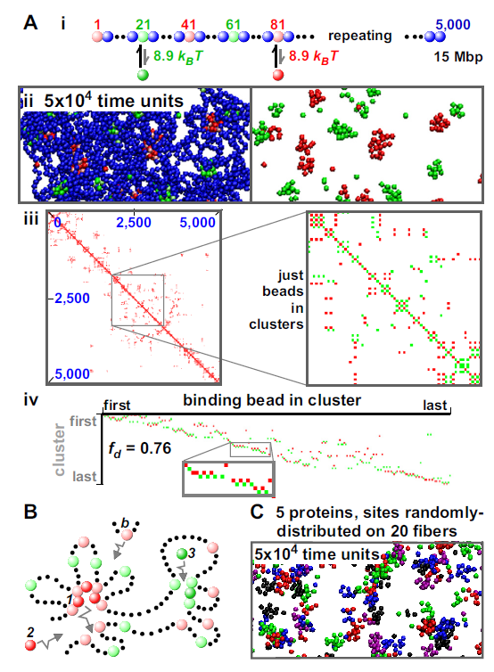
Binding of different factors to different beads was now analyzed. In Figure 2Ai, green factors interact only with light-green beads, and red ones only with pink beads. Again, no attraction is specified between factors, or between beads. Remarkably, clusters now contain only red factors – or only green ones – but rarely both (mixed clusters are not seen at the end of this simulation; Fig. 2Aii, Movies S3, S4). As before, clusters reach a steady state, but now with only 8.1 bound factors/cluster; the regular alternation of green and red binding sites renders cluster merging entropically more costly. Moreover, the contact map, rosettogram, and all point to a more disorganized structure than those seen previously; for example, there are now many “overlapping” loops where the fiber passes back and forth between a cluster of red factors to another with green ones (Fig. 2Aiii, iv). As expected, mixed clusters result if factors share binding sites (Fig. S3).

Such self-organization into structures rich in certain DNA-binding proteins but not others is commonplace in nuclear biology (see Discussion). But what might drive this extraordinary self-assembly into “specialized” clusters in the absence of any explicit interaction between factors, or between beads? We suggest there are both entropic and kinetic drivers Brackley2013 , and that the following one is important. Thus, early during the simulation in Figure 2A, a structure like that in Figure 2B might arise. Red protein is tightly bound to two pink beads, and so will rarely dissociate from the cluster; however, if it does it is likely to bind to a nearby pink bead (as there are so many). Further, as red protein and binding bead diffuse by, both are likely to join the same cluster (again because of the high local concentration of appropriate binding sites and factors). We are now in a positive feedback loop: the local concentration of red factors and pink beads makes it unlikely either will escape, and likely that more of both will be caught as they diffuse by. For the same reason, green protein is likely to bind to the right-hand cluster, and this cluster will tend to grow as other green factors and light-green sites are caught. Red and green clusters will inevitably be separate in 3D space because their cognate binding sites are separate in 1D sequence space, and cluster growth is limited when the entropic costs of bringing together ever-more loops becomes prohibitive.
In Figure 2A, red and green binding beads alternate, and overlapping loops pass back and forth between red and green clusters. Rosettes with many transitive loops result if 200-bead blocks containing 10 light-green beads (spaced every 20 beads) alternate with similar blocks containing pink beads (Fig. S4). As there are fewer binding beads of one color per block than the 12 often found in a cluster in the analogous simulation in Figure 1, successive blocks can form successive red and green clusters. Unsurprisingly, the 1D organization determines rosette and loop type.
Distinct clusters also form if more factors and more fibers are introduced. In Figures 2C and S5, 5 differently-colored factors bind to distinct cognate sites scattered randomly along 20 fibers. Distinct clusters again form; 53% contain factors of only one color, and in 80% more than 80% binding beads have the same color (Fig. S5v). Such clustering could underlie the high number of contacts seen between co-regulated genes that utilize the same factors Fullwood2009 ; Schoenfelder2010 ; Yaffe2011 ; Li2012 ; Papantonis2012 . Inter-fiber contacts are rare (Fig. S5iv), as in Hi-C data Lieberman-Aiden2009 . However, they constitute a higher fraction if just contacts made by binding beads in clusters are considered (Fig. S5iv). This is reminiscent of contacts made by active genomic regions; for example, most contacts made by (active) SAMD4A are inter-chromosomal (assessed by 4C Papantonis2012 ), as are most (active) sites binding estrogen receptor Fullwood2009 . [Figure S5 gives effects of the threshold used to define contacts on contact frequencies.]
The affinities of transcription factors for cognate sites are often tightly regulated, often by post-translational modification. Changing factor affinity was simulated using a fiber in which every 20th bead was yellow (Fig. S6). Initially, red factors bind to yellow beads, and red clusters form. Then, we switch on an attraction between green factors and yellow beads that is stronger than the red-yellow attraction; consequently, green factors compete effectively with red ones, and red/green and all-green clusters develop. This provides a precedent for how one nuclear body (e.g., a transcription factory) might evolve into another.
Forming domains
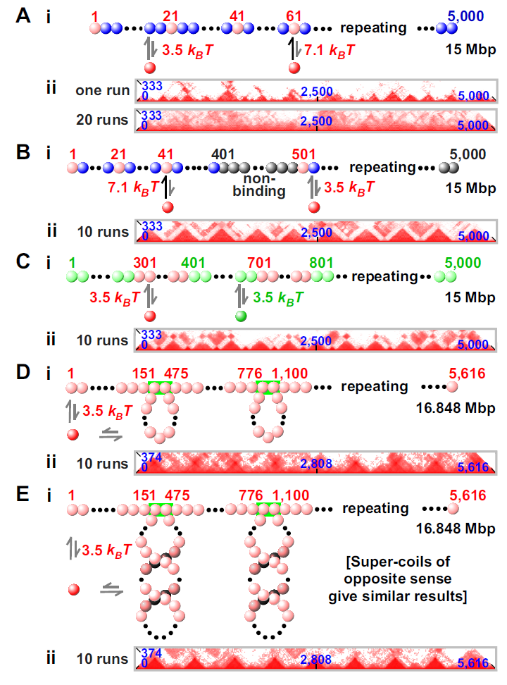
Topological domains are recognized as “pyramids” in contact maps prepared using data from Hi-C Dixon2012 ; Nora2012 ; Sexton2015 ) or simulations Barbieri2012 ; Benedetti2014 ; Jost2014 ; Trussart2015 ; Zhang2015 . Our simulations demonstrate that the pattern of binding beads in our fibers determines whether pyramids are seen. For example, Figure 3A illustrates a partial contact map obtained using data from Figure 1. As clusters appear stochastically and tend to persist, a specified bead often clusters with different partners in different simulations. Then, pyramidal patterns, which are visible in a single run (analogous to Hi-C data from a single cell), become blurred on averaging results from progressively more simulations (Fig. 3Aii). [Figure S7 gives complete contact maps for all simulations in Figure 3.]

In a homogeneous fiber, pyramids disappear on averaging because domains form stochastically; however, if the fiber is suitably patterned, domain boundaries form at specific locations. For example, if long blocks in which every 20th bead is pink (binding red factors) are interleaved with short blocks containing non-binding grey beads (representing gene-poor “deserts”), pink beads cluster but grey ones do not; then, many contacts are seen between pink beads to give pyramids sitting exactly on long segments (Fig. 3B). Here, boundaries between domains are located within grey segments. Domains are also seen if segments containing 300 successive pink beads (binding red factors) are interleaved with shorter segments containing 100 light-green beads (binding green factors); in this case, a repeating pattern of large and small pyramids is seen, with boundaries between blocks of differently-colored beads (Fig. 3C). This simulation could mimic binding of polymerizing complexes and HP1 (hetero-chromatin protein 1) to repeats of eu- and hetero-chromatin Kilic2015 . [These results confirm and extend those obtained using Monte Carlo simulations of just one segment of each type Barbieri2012 ; Hofmann2015 .] These runs of binding beads give larger clusters (i.e., 40 red factors/cluster, and 15 green factors/cluster). When the fiber is forced permanently into loops (perhaps maintained by CTCF) – and if red factors can bind to any bead – pyramids (which are more blurred than in Fig. 3B and C) tend to sit over each loop (Fig. 3D Le2013 ); this is reminiscent of Hi-C data Dixon2012 ; Le2013 ; Rao2014 . Therefore, domains can form in a non-uniform genomic landscape (Fig. 3B, C), and if there are loops (Fig. 3D). If loops are preformed into left-handed (or right-handed) inter-wound supercoils Gilbert2014 , pyramids are more sharply defined (Fig. 3E; see also Le2013 ; Benedetti2014 ).
Because our fibers form many (10) domains, we can analyze contact maps away from the diagonal: these clearly show that in all cases where domains form, inter-domain interactions are weaker than intra-domain ones (compare Fig. S7B- E) – as in Hi-C data. [Note that many domain:domain interactions seen after one run (Fig. S7F) disappear (or become fainter) after averaging data from many runs (Fig. S7B).]
Some characteristics of domains
The probability that two loci yield a Hi-C contact decreases as the number of intervening base pairs increases Lieberman-Aiden2009 , and the exponent () in the power law varies from -0.5 (in HeLa Naumova2013 ) to -1.6 (in embryonic stem cells Barbieri2012 ). [ = 1 in the fractal globule model Lieberman-Aiden2009 ; Naumova2013 .] In all simulations in Figure 3 (except for Fig. 3A), there are two regimes – below and above the domain size (the largest of the two domains appears to set the scale) with between -0.6 and -1 (strong interactions within a pyramid/domain) or close to -2 (weaker interactions between pyramids/domains; Fig. S8). Therefore our values are similar to those seen experimentally. Our results are also consistent with the exponent (seen in simulations of uniform fibers) varying with protein number and affinity Barbieri2012 .
We next used various approaches to identify domain boundaries (Fig. S9). Many current approaches are based on what we will call a Janus plot (Fig. S9), which in its simplest form quantifies all contacts that one bead makes with others to the right or the left in 1D genomic space. In Figure 3E, peaks in the two resulting plots correlate well with the left and right tethers of a loop (Fig. S9A). By subtracting signal from the two plots, we obtain a “difference plot” (i.e., the number of contacts to the right minus the number of contacts to the left). At a boundary, we expect a bead to switch its contacts, from mostly leftward to mostly rightward; consequently, boundaries are found at points where signal in the difference plot crosses zero with an upward derivative (Fig. S9B). This is essentially the method used in Dixon2012 . In the case of Figure 3E, this approach finds domains within loops, and boundaries somewhere in the linear region between them. A more accurate determination is possible by locating the peaks of the derivative of the difference plot (the “insulator” plot in Fig. S9C). This peak-finding algorithm is elegant and works well with highly-sampled contact maps (as in Fig. 3). However, it works less well with sparser data from simulations and Hi-C, where we found the “difference” plot gave better results (so we use it in what follows, aided by visual inspection to fine-tune boundary positions).
Modeling selected regions of the human genome
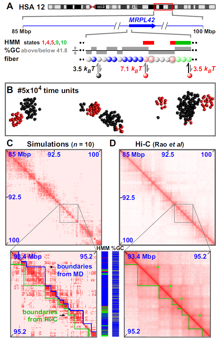
Finally, we examined whether binding of just two proteins to “active” and “inactive” beads on a 15-Mbp fiber (representing part of chromosome 12 in GM12878) could fold the genome appropriately (Fig. 4A; Movies S5 and S6). Active regions were selected using the Broad ChromHMM track on the UCSC (University of California at Santa Cruz) browser Ernst2011 , and beads (1 kbp) representing active promoters or strong enhancers (states 1, 4, 5) – and the bodies of active transcription units (states 9, 10) – were colored pink and light-green, respectively. These pink and light-green beads bind red factors (transcription factors, polymerizing complexes) with high and low affinities. Inactive heterochromatin was represented by grey beads that bind black proteins (e.g., dimers of HP1 Kilic2015 ). Heterochromatic beads were identified as those having a low GC content – an excellent and flexible marker (in principle, choice of threshold can allow any fraction of the region of interest to be classified as heterochromatin). Here, 41.8% GC was chosen as the threshold, as this led to the same percentage of heterochromatin in the 15 Mbp as that marked by state 13 (generic heterochromatin) in the HMM track. Other beads (blue) were non-binding. As before, distinct clusters of bound red or black proteins develop with 14 or 190 proteins/cluster, respectively (long runs of adjacent grey beads form the larger clusters; Fig. 4B). The resulting contact map was strikingly similar to the Hi-C one Rao2014 , with simulations correctly predicting 75% of the Hi-C domain boundaries to within 100 kbp (Figs 4C, D and S10A, B; Table S2). Boundaries were in this case found using the difference plot aided by visual inspection (Fig. S10); purely automated detection correctly locates 59% Hi-C boundaries within 100 kbp, which is still statistically significant (this is an underestimate due to algorithmic errors, some examples of which are noted in Fig. S10). Simulations also yield a more-ordered rosettogram than any seen previously (Fig. S10C); this is consistent with evolution selecting for a genetic and epigenetic sequence that gives ordered rosettes. Similar results were obtained with a 15-Mbp segment of a different chromosome (chr6) in a different cell type (Fig. S11). [Here, heterochromatic regions were defined using either %GC or HMM state 13, but fewer boundaries were correctly reproduced using the latter (Fig. S11E).]

These successes prompted us to model a whole 59-Mbp chromosome (chr19; see Fig. 5 and Movie S7). Active and inactive beads (each now representing 3 kbp) were defined as before, and heterochromatic ones using 48.4% GC (to reproduce the fraction of the chromosome bearing the state 13 mark). Now, 85% domain boundaries are correctly reproduced to within 100 kbp (Figs 5, S12A). Moreover, simulation boundaries are rich in “active” and non-binding regions and depleted of “inactive” ones (Fig. 12B). As before, the rosettogram and point to a highly-ordered structure with many local contacts (Fig. S12C). At a higher level in the organization, 3D positioning of some domains next to others – reflected by off-diagonal blocks in contact maps – is sometimes reproduced in simulations (zooms in Figs 5C and D, S12A). Our simulations of the whole chromosome further indicate that small active domains seem to be often located at the periphery of larger inactive ones; they also suggest that active domains are more dynamic and mobile than inactive ones (see Movie S7).
![[Uncaptioned image]](/html/1511.01848/assets/Fig5Bridges.png)
Discussion
These MD simulations illustrate some emergent properties of a minimalist system that involves bivalent “transcription factors” (or “proteins”) binding specifically and transiently to cognate sites in a fiber (representing “chromatin”). First, bound factors spontaneously cluster – to compact the fibers (Figs 1, 2C). This self-organization occurs in the absence of any explicit interaction between factors or between beads, and it is driven by a combination of forces dubbed the “bridging-induced attraction”. Second, and more surprisingly, factors binding to distinct sites on the fiber self-assemble into distinct (segregated) clusters. For example, bound red and green factors self-assemble into clusters that contain only red factors, or only green ones – but rarely both (Fig. 2A). These clusters arise because protein binding will inevitably yield clusters in different places in 3D space if – and only if – their cognate binding sites are spatially separated in 1D sequence space (Fig. 2B). Third, clustering organizes the loops caused by binding into higher-order rosettes, and domains (Figs 3- 5). For example, binding of just two “proteins” (transcription and HP1 complexes) to active and inactive regions in a 59-Mbp human chromosome folds the fiber to yield a contact map in which 85% of the Hi-C boundaries are both correctly placed and rich in the appropriate sequences (Figs 5 and S12, Movie S7). In other words, complexes bind locally to create loops, bound complexes cluster together with similar ones into rosettes, this folds the fiber globally into appropriate domains, and domains pack against each other – all in the expected ways. Remarkably, then, this minimalist system generates structures that possess all the key features of interphase chromosomes outlined in the Introduction. Moreover, the clusters formed are reminiscent of nuclear structures like Cajal and promyelocytic leukemia bodies, which are each rich in distinct proteins that bind to different cognate DNA sequences Sleeman2014 ; they also closely resemble nucleoplasmic transcription factories that each contain 10 active polymerizing complexes Pombo1999 ; Cook1999 ; Papantonis2013 .
The binding energy of any one factor is small (roughly comparable to that in a few H-bonds), but extended genomic regions fold simply because so many are involved. Cluster formation and protein-driven chromosome organization also occurs quickly (within minutes according to our simulations, Fig. 1E). Once clusters form, they usually persist (Fig. 1E). However, the system can evolve when new factors appear (Fig. S6), much as a transcription factory can develop into a replication factory at the beginning of S phase Hassan1994 , or into one specializing in transcribing responsive genes during the inflammatory response (when tumor necrosis factor induces nuclear influx of nuclear factor B Papantonis2012 .
Contacts made as a result of such clustering involve sites both near and far apart on the fiber. Most contacts are local, to create loops and “rosettes” like those often seen in models of chromosomes (Figs 1F, 2Aiv, see also Pienta1984 ; Cook1995 ). We suggest rosettes (with many transitive loops) are favoured over more disordered non-local structures (with many overlapping loops) partly because the entropic cost is less Marenduzzo2009 ; rosettes are also likely to be kinetically favoured when starting from knot-free structures (both in simulations and on exit from mitosis). More distant contacts also depend on patterns of binding sites in 1D genomic space, as domains separated by well-defined boundaries result if non-binding gene deserts or supercoiled loops Benedetti2014 are introduced, or if blocks of eu- and hetero-chromatin alternate (Fig. 3).
In summary, the bridging-induced attraction provides a robust, simple, and generic mechanism that can concentrate specific proteins bound to cognate sites into clusters, and fold interphase fibers in ways found in vivo. Then, the system must either spend energy to prevent the resulting clustering, or – as seems likely – it goes with the flow and uses other more or less familiar forces (charge interactions, H bonds, van der Waals, hydrophobic forces, and the depletion attraction Marenduzzo2006 ) to organize those clusters. If so, we suggest that the particular folding pattern found in any one nucleus will be largely determined by which transcription factors bind to cognate sites, and which bound factors then happen to co-cluster. We also expect that adding more proteins and fibers to our simple model will improve the concordance between contact maps obtained from simulations and Hi-C.
After the present work was completed, two studies proposed another model for formation of looping domains that is based on CTCF bridges Mirny2015 ; LibAid2015 . These involve some loop-extruding driving domain formation, and they are appealing because they can account for the recent observation that CTCF bridging depends on the orientation of the cognate binding sites Rao2014 ; CRISPCTCF . However, this model requires some as-yet undiscovered motor protein with a processivity sufficient to generate loops of hundreds of kb. On the other hand, as discussed in Ref. LibAid2015 , these studies do not address what might underlie the observed compartmentalization into active and inactive domains - which is naturally explained within our framework by binding of different factors to eu- and hetero-chromatin. Furthermore, knock-outs of CTCF have only minor effects on domain organization Zuin2014 ; Hou2010 , which again suggests that this factor cannot be the sole organizer. The results of these knock-outs are also naturally explained by our model, as the compartmentalization is driven by bivalent factors which are unrelated to CTCF. Our work and those of Refs. Mirny2015 ; LibAid2015 are therefore complementary, and it would be of interest to couple the two approaches together in the future.
Materials and Methods
Brownian dynamics (BD) simulations were run with the LAMMPS (Large-scale Atomic/Molecular Massively Parallel Simulator) code, used with a stochastic thermostat. Chromatin fibers are modeled as bead-and-spring polymers using FENE bonds (maximum extension 1.6 times bead diameter, ) and a bending potential that allows persistence length to be set. Protein:protein and chromatin:chromatin interactions involve only steric repulsion, and those between proteins and their binding sites are truncated and shifted Lennard-Jones interactions. All beads are confined within a cube with periodic boundary conditions. Additional details are listed in Figure legends and/or Supplementary Information.
Simulations of human chromosomes included two kinds of factors/proteins, one binding to active euchromatin and the other to inactive heterochromatin. Chromatin beads were colored pink or light-green if interacting strongly or weakly, respectively, with red factors (representing factors and polymerases), grey if interacting with black proteins (representing HP1), and blue if non-interacting. The Broad ChromHMM track Ernst2011 on the hg19 assembly of the UCSC Genome browser was used to determine pink/light green color: (i) if a region of 90 bp or more within one bead is labeled as an “Active Promoter” or “Strong Enhancer” (states 1,4,5) then the whole bead is colored pink, similarly if a bead contains 90 bp or more of “Transcriptional Transition” or “Transcriptional Elongation” (states 9,10) it is colored light green. Data for GC content (Figs 4, 5 and S11D) or from the HMM track (Fig. S11A, B) were used to determine grey color: (i) if GC content of the bead was below the threshold specified in each Figure legend, or (ii) if a region of 90 bp or more within one bead is labelled “Heterochromatin; low signal” (state 13). This convention allows one bead to have multiple colors. Figure legends give numbers of differently-colored beads in a simulation and affinities. Simulations included red factors (=300 in Fig. 4 and Fig. S11; =400 in Fig. 5), and black protein (=3,000 in Fig. 4 and Fig. S11; =4,000 in Fig. 5); for simplicity, protein size is equal to , and interaction range to 1.8 .
In our simulations, one bead corresponds either to 3 (Figs 1-3, 5) or 1 kbp (Figs 4 and S11). These correspond to a size of 30 nm and 20.8 nm respectively (assuming a 30-nm fiber has a packing density of 1 kbp/10 nm, and the two bead sizes contain the same DNA density). Persistence length was 3 (representing a flexible polymer), and one time unit corresponds to 0.6 or 0.2 ms (for 3 or 1 kbp beads; calculated assuming a viscosity of 10 cP, or 10-fold larger than water).
CAB and DM acknowledge support through an ERC Consolidator Grant (THREEDCELLPHYSICS, Ref.648050).
References
- (1) Cavalli, G., and Misteli, T. (2013). Functional implications of genome topology. Nat. Struct. Mol. Biol. 20:290–299.
- (2) Lieberman-Aiden, E., van Berkum, N.L., Williams, L., Imakaev, M., Ragoczy, T., Telling, A., Amit, I., Lajoie, B.R., Sabo, P.J., Dorschner, M.O., et al. (2009). Comprehensive mapping of long-range interactions reveals folding principles of the human genome. Science 326:289–293.
- (3) Dixon, J.R., Selvaraj, S., Yue, F., Kim, A., Li, Y., Shen, Y., et al. (2012). Topological domains in mammalian genomes identified by analysis of chromatin interactions. Nature 485:376–380.
- (4) Nora, E.P., Lajoie, B.R., Schulz, E.G., Giorgetti, L., Okamoto, I., Servant, N., Piolot, T., van Berkum, N.L., Meisig, J., Sedat, J., Gribnau, J., Barillot, E., Blüthgen, N., Dekker, J., and Heard, E. (2012). Spatial partitioning of the regulatory landscape of the X-inactivation centre. Nature 485:381–385.
- (5) Sexton, T., Yaffe, E., Kenigsberg, E., Bantignies, F., Leblanc, B., Hoichman, M., Parrinello, H., Tanay, A., and Cavalli, G. (2012). Three-dimensional folding and functional organization principles of the Drosophila genome. Cell 148:458–472.
- (6) Naumova, N., Imakaev, M., Fudenberg, G., Zhan, Y., Lajoie, B.R., Mirny, L.A., and Dekker, J. (2013). Organization of the mitotic chromosome. Science 342:948–953.
- (7) Rao, S.S.P., Huntley, M.H., Durand, N.C., Stamenova, E.K., Bochkov, I.D., Robinson, J.T., et al. (2014). 3D map of the human genome at kilobase resolution reveals principles of chromatin looping. Cell 159:1–16.
- (8) Sexton, T., and Cavalli, G. (2015). The role of chromosome domains in shaping the functional genome. Cell 160:1049–1059.
- (9) Simonis, M., Klous, P., Splinter, E., Moshkin, Y., Willemsen, R., de Wit, E., et al. (2006). Nuclear organization of active and inactive chromatin domains uncovered by chromosome conformation capture-on-chip (4C). Nat. Genet. 38:1348–1354.
- (10) Li, G., Ruan, X., Auerbach, R.K., Sandhu, K.S., Zheng, M., Wang, P., Poh, H.M., Goh, Y., Lim, J., Zhang, J. et al. (2012). Extensive promoter-centered chromatin interactions provide a topological basis for transcription regulation. Cell 148:84–98.
- (11) Jin F., Li Y., Dixon JR., Selvaraj S., Ye Z., Lee AY., Yen CA., Schmitt AD., Espinoza CA., and Ren B. (2013). A high-resolution map of the three-dimensional chromatin interactome in human cells. Nature 503:290–294.
- (12) Zhang, Y., Wong, C.H., Birnbaum, R.Y., Li, G., Favaro, R., Ngan, C.Y., et al. (2013). Chromatin connectivity maps reveal dynamic promoter-enhancer long-range associations. Nature 503:290–294.
- (13) Heidari, N., Phanstiel, D.H., He, C., Grubert, F., Jahanbani, F., Kasowski, M., et al. (2014). Genome-wide map of regulatory interactions in the human genome. Genome Res. 24:1905–1917.
- (14) Mifsud, B. et al. (2015). Mapping long-range promoter contacts in human cells with high-resolution capture Hi-C. Nat. Gen. 47:598–606.
- (15) Fullwood, M.J., Liu, M.H., Pan, Y.F., Liu, J., Xu, H., Bin Mohamed, Y., et al. (2009). An oestrogen-receptor-alpha-bound human chromatin interactome. Nature 462, 58–64.
- (16) Schoenfelder, S., Sexton, T., Chakalova, L., Cope, N.F., Horton, A., Andrews, S., et al. (2010). Preferential associations between co-regulated genes reveal a transcriptional interactome in erythroid cells. Nat. Genet. 42:53–61.
- (17) Yaffe, E., and Tanay, A. (2011). Probabilistic modeling of Hi-C contact maps eliminates systematic biases to characterize global chromosomal architecture. Nat. Genet. 43:1059–1065.
- (18) Papantonis, A., Kohro, T., Baboo, S., Larkin, J., Deng, B., Short, P., Tsutsumi, T., Taylor, S., Kanki, Y., Kobayashi, M., Li, G., Poh, H.-M., Ruan, X., Aburatani, H., Ruan, Y., Kodama, T., Wada, Y., and Cook, P.R. (2012). TNF signals through specialized factories where responsive coding and micro-RNA genes are transcribed. EMBO J. 31:4404–4414.
- (19) Nagano, T., Lubling, Y., Stevens, T.J., Schoenfelder, S., Yaffe, E., Dean, W., et al. (2013). Single-cell Hi-C reveals cell-to-cell variability in chromosome structure. Nature 502:59–64.
- (20) Hsieh, T.-H., S., Weiner, A., Lajoie, B., Dekker, J., Friedman, N., and Rando, O.J. (2015). Mapping nucleosome resolution chromosome folding in yeast by micro-C. Cell 162:108–119.
- (21) Le, T.B., Imakaev, M.V., Mirny, L.A., and Laub, M.T. (2013). High-resolution mapping of the spatial organization of a bacterial chromosome. Science 342:731–734.
- (22) Marenduzzo, D., Micheletti, C., and Cook, P.R. (2006). Entropy-driven genome organization. Biophys. J. 90:3712–3721.
- (23) Rosa, A., and Everaers, R. (2008). Structure and dynamics of interphase chromosomes. PLoS Comput Biol 4:e1000153.
- (24) Nicodemi, M., Panning, B., and Prisco, A. (2008). The colocalization transition of homologous chromosomes at meiosis. Phys. Rev. E Stat. Nonlin. Soft Matter Phys. 77:061913.
- (25) Duan, Z., Andronescu, M., Schutz, K., McIlwain, S., Kim, Y.J., Lee, C., Shendure, J., Fields, S., Blau, C.A., and Noble, W.S. (2010). A three-dimensional model of the yeast genome. Nature 465:363–367.
- (26) Junier I., Martin O., and Képès F. (2010). Spatial and topological organization of DNA chains induced by gene co-localization. PLoS Comput. Biol. 6:e1000678.
- (27) de Vries, R (2011). Influence of mobile DNA-protein-DNA bridges on DNA configurations: coarse-grained Monte-Carlo simulations. J. Chem. Phys. 135:125104.
- (28) Kalhor, R., Tjong, H., Jayathilaka, N., Alber, F., and Chen, L. (2011). Genome architectures revealed by tethered chromosome conformation capture and population-based modeling. Nat. Biotechnol. 30:90–98.
- (29) Rousseau, M., Fraser, J., Ferraiuolo, M.A., Dostie,J., and Blanchette,M. (2011). Three-dimensional modeling of chromatin structure from interaction frequency data using Markov chain Monte Carlo sampling. BMC Bioinformatics 12:414.
- (30) Barbieri, M., Chotalia, M., Fraser, J., Lavitas, L.M., Dostie, J., Pombo, A., and Nicodemi, M. (2012). Complexity of chromatin folding is captured by the strings and binders switch model. Proc. Natl. Acad. Sci. USA 109:16173–16178.
- (31) Baù, D., and Marti-Renom, M.A. (2012). Genome structure determination via 3C-based data integration by the Integrative Modeling Platform. Methods 58:300–306.
- (32) Brackley, C.A, Taylor, S., Papantonis, A., Cook, P.R., and Marenduzzo, D. (2013). Nonspecific bridging-induced attraction drives clustering of DNA-binding proteins and genome organization. Proc. Natl. Acad. Sci. USA 110:E3605–3611.
- (33) Benedetti F., Dorier J., Burnier Y., and Stasiak A. (2014). Models that include supercoiling of topological domains reproduce several known features of interphase chromosomes. Nucleic Acids Res 42:2848–2855.
- (34) Jost, D., Carrivain, P., Cavalli, G., and Vaillant, C. (2014). Modeling epigenome folding: formation and dynamics of topologically associated chromatin domains. Nucleic Acids Res. 42:9553–9561.
- (35) Tark-Dame, M., Jerabek, H., Manders, E.M.M., Heermann, D.W., and van Driel, R. (2014). Depletion of the chromatin looping proteins CTCF and cohesin causes chromatin compaction: insight into chromatin folding by polymer modelling. PLoS Comput. Biol. 10:e1003877.
- (36) Trieu, T., and Cheng J. (2014). Large-scale reconstruction of 3D structures of human chromosomes from chromosomal contact data. Nucleic Acids Res. 42:e52.
- (37) Cheng, T.M.K., Heeger, S., Chaleil, R.A.G., Matthews, N., Stewart, A., Wright, J., Lim, C., Bates, P.A., and Uhlmann, F. (2015). A simple biophysical model emulates budding yeast chromosome condensation. Elife 4:e05565.
- (38) Hofmann, A., and Heermann, D.W. (2015). The role of loops on the order of eukaryotes and prokaryotes. FEBS Lett., doi: 10.1016/j.febslet.2015.04.021.
- (39) Johnson, J., Brackley, C.A., Cook, P.R., and Marenduzzo, D. (2015). A simple model for DNA bridging proteins and bacterial or human genomes: bridging-induced attraction and genome compaction. J. Phys. Condens. Matter 27:064119.
- (40) Junier, I., Spill, Y.G., Marti-Renom, M.A., Beato, M., and le Dily, F. (2015). On the demultiplexing of chromosome capture conformation data. FEBS Lett., doi: 10.1016/j.febslet.2015.05.049.
- (41) Trussart, M., Serra, F., Baù, D., Junier, I., Serrano, L., and Marti-Renom, M.A. (2015). Assessing the limits of restraint-based 3D modeling of genomes and genomic domains. Nucleic Acids Res. 43:3465–3477.
- (42) Zhang, B., and Wolynes, P.G. (2015). Topology, structures, and energy landscapes of human chromosomes. Proc. Natl. Acad. Sci. USA 112:6062–6067.
- (43) Giorgetti, L., Galupa, R., Nora, E.P., Piolot, T., Lam, F., Dekker, J., Tiana, G., and Heard, E. (2014). Predictive polymer modeling reveals coupled fluctuations in chromosome conformation and transcription. Cell 157:950–963.
- (44) Dekker, J., Marti-Renom, M.A., and Mirny, L.A. (2013). Exploring the three-dimensional organization of genomes: interpreting chromatin interaction data. Nat. Rev. Genet. 14:390–403.
- (45) Serra, F., Di Stefano, M., Spill, Y.G., Cuartero, Y., Goodstadt, M., Baù, D., Marti-Renom, M.A. (2015). Restraint-based three-dimensional modeling of genomes and genomic domains. FEBS Lett., doi: 10.1016/j.febslet.2015.05.012.
- (46) Plimpton, S. (1995). Fast parallel algorithms for short-range molecular dynamics. J. Comp. Phys. 117:1–19.
- (47) Duplantier, B. (1989). Statistical mechanics of polymer networks of any topology. J. Stat. Phys. 54, 581–680.
- (48) Pienta, K.J., and Coffey, D.S. (1984). A structural analysis of the role of the nuclear matrix and DNA loops in the organization of the nucleus and chromosome. J. Cell Sci. Suppl. 1:123–135.
- (49) Cook, P.R. (1995). A chromomeric model for nuclear and chromosome structure. J. Cell Sci. 108:2927–2935.
- (50) Doi, M., and Edwards, S.F. (1986). The Theory of Polymer Dynamics. Clarendon Press (Oxford).
- (51) Kilic, S., Bachmann, A.L., Bryan, L.C., and Fierz, B. (2015). Multivalency governs HP1 association dynamics with the silent chromatin state. Nat. Commun. 6:7313.
- (52) Gilbert, N., and Allan, J. (2014). Supercoiling in DNA and chromatin. Curr. Opin. Genet. Dev. 25, 15–21.
- (53) Ernst, J., Kheradpour, P., Mikkelsen, T.S., Shoresh, N., Ward, L.D., Epstein, C.B., Zhang, X., Wang, L., Issner, R., Coyne, M. et al. (2011). Mapping and analysis of chromatin state dynamics in nine human cell types. Nature 473, 43–49.
- (54) Sleeman, J.E., and Trinkle-Mulcahy, L. (2014). Nuclear bodies: new insights into assembly/dynamics and disease relevance. Curr. Opin. Cell Biol. 28:76–83.
- (55) Pombo, A., Jackson, D.A., Hollinshead, M., Wang, Z., Roeder, R.G., and Cook, P.R. (1999). Regional specialization in human nuclei: visualization of discrete sites of transcription by RNA polymerase III. EMBO J. 18:2241–2253.
- (56) Cook, P.R. (1999). The organization of replication and transcription. Science 284:1790–1795.
- (57) Papantonis, A., and Cook, P.R. (2013). Transcription factories; genome organization and gene regulation. Chem. Rev. 113:8683–8705.
- (58) Hassan, A.B., Errington, R.J., White, N.S., Jackson, D.A., and Cook. P.R. (1994). Replication and transcription sites are colocalized in human cells. J. Cell Sci. 107, 425–434.
- (59) Marenduzzo, D. and Orlandini, E. (2009). Topological and entropic repulsion in biopolymers. J. Stat. Mech.:L09002.
- (60) Guo, Y. et al. (2015). CRISPR Inversion of CTCF Sites Alters Genome Topology and Enhancer/Promoter Function. Cell. 162:900–910.
- (61) Fudenberg, G., Imakaev, M., Lu, C., Goloborodko, A., Abdennur, N., and Mirny, L. A. (2015) Formation of Chromosomal Domains by Loop Extrusion, doi: http://dx.doi.org/10.1101/024620
- (62) Sanborn, A. L. et al. (2015). Chromatin extrusion explains key features of loop and domain formation in wild-type and engineered genomes, Proc. Natl. Acad. Sci. USA early edition, doi: 10.1073/pnas.1518552112.
- (63) Zuin, J. it et al. Cohesin and CTCF differentially affect chromatin architecture and gene expression in human cells. Proc. Natl. Acad. Sci. USA 111:996–1001 (2014).
- (64) Hou, C., Dale, R., Dean, A. (2010) Cell type specificity of chromatin organization mediated by CTCF and cohesin. Proc. Natl. Acad. Sci. USA 107:3651–3656.
Supplementary material
Appendix A Coarse grained molecular dynamics simulations
In our coarse grained molecular dynamics simulations, we represented chromatin as a bead-and-spring chain, and protein complexes as additional beads. The position of the th bead in the system changes in time according to the Langevin equation
| (1) |
where is the position of bead , is its mass, is the friction it feels due to an implicit aqueous solvent, while is a vector representing random uncorrelated noise which obeys the following relations
| (2) |
The noise is scaled by the energy of the system, given by the Boltzmann factor multiplied by the temperature of the system , taken to be 310 K for a cell. The potential is a sum of interactions between bead and all other beads, and we use phenomenological force fields as described below. For simplicity we assume that all beads in the system have the same mass and friction , and . Eq. (1) is solved in LAMMPS using a standard Velocity-Verlet algorithm.
Appendix B Force fields
For the chromatin fiber the th bead in the chain is connected to the th with a finitely extensible non-linear elastic (FENE) spring: the associated potential is given by
| (3) |
where is the separation of the beads, and the first term is the Weeks-Chandler-Andersen (WCA) potential
| (6) |
which represents a hard sphere-like steric interaction preventing adjacent beads from overlapping. In Eq. (6) is the mean of the diameters of beads and . The diameter of the chromatin beads is a natural length scale with which to parametrize the system; we denote this by , and use this to measure all other length scales. The second term in Eq. (B) gives the maximum extension of the bond, ; throughout this work we use , and set the bond energy .
The bending rigidity of the polymer is introduced via a Kratky-Porod potential for every three adjacent DNA beads
| (7) |
where is the angle between the three beads as give by
| (8) |
and is the bending energy. The persistence length in units of is given by .
Finally, steric interactions between non-adjacent DNA beads are also given by the WCA potential [Eq. (6)]. In the absence of proteins, the force field of chromatin is therefore appropriate for a biopolymer in a good solvent.
Each protein (or protein complex) we simulate is represented by a single bead; unless otherwise stated, the WCA potential is used to model steric interactions between these. Chromatin beads are labeled as binding or not-binding for each protein species according to the input data. For the interaction between proteins and the chromatin beads labeled as binding, we use a shifted, truncated Lennard-Jones potential, whose form is given by
| (9) |
with
where is a cut off distance, and and are the separation and mean diameter of the two beads respectively. This leads to an attraction between a protein and a chromatin bead if their centres are within a distance . Here is an energy scale, but due to the second term in Eq. (9) this is not the same as the minimum of the potential, which for clarity we denote as (and we refer this to as the interaction energy). For simplicity we set the diameter of the protein complexes equal to that of the chromatin beads, , and set unless otherwise stated.
The length scale , mass and energy scale give rise to a natural simulation time unit , and Eq. (1) is integrated with a constant time step , for a total of time steps or more (see main text).
Appendix C Mapping simulation units to physical units
In order to compare simulation and experimental time and length scales, it is useful here to describe how to map simulation into physical units (this is not required for energy as this was previously expressed in units of ).
Length scales are easily mapped once the value of is set in physical units. For simulations of chromatin fibers where one bead corresponds to 3 kbp, a natural choice is 30 nm, leading to a linear baseline packing of 10 nm/kbp. For the higher resolution simulations of the chr12 and chr6 regions ( Figs 4 and S11), corresponds to 1 kbp. Assuming the same chromatin density in the two models, a unit of length now corresponds to 20.8 nm.
In order to map time units, we need to recognise that there are three main time scales in the system. One is the previously defined Lennard-Jones time . A second is the inertial time (from Eq. (1)), which is the characteristic time over which a bead loses information about its velocity. A third typical time is the so-called Brownian time , which gives the order of magnitude of the time it takes for a bead to diffuse across its own diameter . Here is the diffusion constant for bead , given through the Einstein relation by . If we make the approximation that a chromatin bead will diffuse like a sphere we can then use Stokes’ law, where , with the viscosity of the fluid, and the diameter of bead . Taking realistic values for the length, mass and viscosity one finds that , with the times separated by several orders of magnitude. For numerical stability we must choose the time step smaller than all of these times, and we wish to study phenomena which will occur on times of the order ; this means that using real values for all parameters would lead to unfeasibly long run times. Instead we chose parameters such that , and map from simulation to physical time scales through the Brownian time . This assumption means that processes which occur on time-scales below the Brownian time are not resolved accurately, however this is of no practical consequence for our work as we are interested in time-scales much exceeding the Brownian time.
For simulations where chromatin beads were 30 nm in diameter (all except Figs 4 and S11), taking a viscosity of 10 cP for the nucleoplasm (10 times that of water, to account for the effective increase in viscosity due to crowding) gives a Brownian time of about 0.6 ms, so that a simulation run of time steps corresponds to about 30 s of real time. For simulations where chromatin beads correspond to 1 kbp (20.8 nm in diameter), one simulation unit of time (one Brownian time) corresponds to about 0.2 ms.
Appendix D Additional simulation details
For the simulation in Figures 3E and 3F, the force field discussed in Section B was supplemented with torsional interactions to generate results for loops with linking number Lk equal to 0 or 32. To model supercoiled or torsionally relaxed (but not nicked) loops, we use closed loops (each of contour length 324 ), which were joined to a linear backbone with a Gaussian spring. We modeled torsional interactions using spherical atoms with an associated triad of vectors, so that the Euler angles describing the relative orientation of adjacent beads allow us to track the twist as well as the bending rigidity. This scheme corresponds to model 2 described in Ref. Brackley2013 ; chromatin was modeled as a ribbon in the torsionally relaxed state, and with torsional persistence length equal to 20 .
For convenience, we also list here Lennard-Jones parameters for all attractive interactions in the simulations (the rest of the interactions are repulsive and modeled using a WCA potential, as previously mentioned). The interaction range (cut-off of Lennard-Jones interaction) was equal to 1.8 for all attractive interactions. Interaction strengths (, in units of ) were as follows: Figures 1, 3A, 3B and S2B: 7.1 (between red “transcription factors” and pink beads); 3.5 (between red factors and blue beads). Figure 2A: 8.9 (between red factors and pink beads; and between green factors and light-green beads). Figures 2C and S5: 7.1 (between each of the factors and its target binding beads). Figures 3C: 3.5 (between red factors and pink beads; and between green factors and light-green beads). Figures 3D and 3E: 3.5 (between red factors and pink beads. Figures 4, 5 and S11): 7.1 (between red factors and pink beads); 3.5 (between red factors and light-green beads; and between black “proteins” and grey beads). Figure S2A: 7.1 (between red factors and pink beads). Figure S3: 7.1 (between either red or green factors and yellow beads). Figure S4: 7.1 (between red factors and pink beads; and between green factors and light-green beads). Figure S6: initially 7.1 only between red factors and pink beads; after “switch” 7.1 (between red factors and pink beads), and 13.1 (between green factors and light-green beads).
Appendix E Initialization
Finally, as in all molecular dynamics simulations it is important to specify how the system was initialised. For all cases where a single linear polymer was modeled, chromatin fibers were first generated as random walks, and proteins randomly distributed (with uniform probability throughout the simulation box). The simulation was then run with a soft potential between all beads to remove overlaps, and with a Gaussian spring between neighboring beads (this was for at least a million time steps; in some cases it was also necessary to use a higher bending rigidity to avoid initial entanglements). After equilibration, the force field was set to the one discussed in Section B. For Figures 3E and 3F, we first equilibrated supercoiled or torsionally relaxed loops in isolation, then joined them to a linear backbone at appropriate places (see caption to Fig. 3) with Gaussian springs; the system was then allowed to equilibrate with the force field in Section B (which preserves topology and linking number as it disallows intrachain crossings). For Figure S5, we generated initial conformations for the 20 chromatin fibers as mitotic-like cylinders with random orientation, following the method described in Ref. Rosa2008 ; proteins were still distributed randomly and uniformly at the beginning of the simulations. The equilibration steps were then performed as above (with soft potential and Gaussian springs).
Appendix F Analysing contacts: contact maps, boundaries and rosettograms
An important output of both Hi-C experiments and our simulations are contact maps; in this Section we discuss how we analysed them.
The contact maps in Figures 1D, 2Aiii, S2Aiii, S2Biii, S3iii, S4iii, S5iii and S7F were obtained from a single configuration: a contact between two beads was scored if their centers were less than 150 nm (5 ) apart. We binned contacts by dividing the polymer into a number of bins (specified in each Figure Legends) to aid visualization. The colored contact maps in Figures 2Aiii, S3iii, S4iii, S5iii and S6iv were also obtained from a single configuration, by only considering the binding sites. A contact between two binding sites was scored if their centers were less than 90 nm (3 ) apart. Binding sites were colored according to the protein (or factor) which is attracted to them (i.e., pink sites are colored red as they bind red factors, etc.); in case a binding site could be the target for more than one factor (e.g., in Fig. S6iv), we colored the binding site according to the protein which was closest to them (e.g., red if the binding site was closest to a red protein, etc.). Pixels in the contact map then are colored red if they are contacts between two red pixels, etc.; mixed contacts are colored yellow if between red and green, and grey in Figure S5iii. Finally, the simulation contact maps in Figures 3, 4, 5, S7A-E and S11, were averaged over several realizations (specified in the Figure Legends, together with the binning used). In all contacts map (with the exception of colored contact maps), the entry gives the number of contacts in the bin, scaled by the maximum number of contact maps over all bins (in this way entries are between 0 and 1).
For each of the simulations in Figure 3, we plotted both the whole contact map ( Fig. S7) and just the part of it close to the diagonal (referred to as pyramid plots in the text); the latter is often used in the literature as it allows a clearer visual determination of boundaries. While the simulated contact maps are shown without any normalization, experimental contact maps for GM12878 cells ( Figs 3 and 5 in the main text) were normalized according to the square root normalization method described in Ref. Rao2014 . Experimental contact maps for Figure S11 were not normalized; these were computed from the Sequence Read Archives (SRA) data in the Gene Expression Omnibus [obtained from Ref. Dixon2012 via access number GSE35156; duplicate reads were removed].
For each of the contact maps (whether from simulation or experiments), we prepared Janus and difference plots, and computed the number of contacts (or contact probability) versus distance along the genome/simulated chromatin fibers. All contact maps were binned (the binning used varied in the different cases and in specified in Figure Legends).
The Janus forward signal for bin , , is defined as the sum over all contact map entries relative to contacts which a bead makes with other beads to the right of it: i.e., , where denotes the contact map entry relative to the -th and th bins, and is the total number of bins. The Janus backward signal for bin , , was similarly computed as .
The difference plot ( Figs S8 and S9) is the difference . When this quantity is negative, bin is making more contact to its left; when it is positive, the majority of the contacts bin makes are to its right. The difference plot is useful to get a first estimate of domain boundary locations, since boundaries are places where the pattern of contacts made by a bin changes from mostly to the left to mostly to the right (but not vice versa). Therefore, boundaries can be located at regions where crosses 0 with an upward derivative; a similar algorithm was used to locate boundaries in Ref. Dixon2012 . This is the base of the algorithm used in Figure S9 to determine boundaries automatically in the region chr12:85000000-100000000 bp ( Fig. 4 in the main text). To avoid spurious multiple nearby boundaries due to noise in the difference plot signal, we further required that either the upward trend in is common to 4 consecutive beads crossing zero, or that the upward slope at the zero crossing (forward difference where and ) is larger than a set threshold (equal to 10% or 40% of the maximum step in the function , for experiments and simulations respectively).
Another way to detect boundaries is via peaks in the derivative of (this is the insulator plot in Fig. S8): the rationale here is that we expect the relative fraction of contacts to the right should increase sharply at boundaries, however due to contacts away from the diagonal it may not necessary be that the difference plot goes through 0. In selected cases we also used the recent method described in Ref. Hsieh2015 to detect boundaries. While all methods agreed on some of the boundaries, visual inspection suggests that not all boundaries can be found by any one automated technique (see Figs 4 and S10B). While in Hi-C experiments the numerical error that these or similar algorithms make is not too important, it is much more consequential with simulation data that are noisier. Moreover, our goal is to compare experimental and simulation boundaries, rather than to estimate boundaries in either simulations or experiments with a given accuracy. Comparing boundaries in simulations and Hi-C data is a demanding task: for instance, even a two pixel error (the curve turning in an opposite direction) would lead to an artificial discrepancy of 80 kbp between the location of the same boundary in simulations and experiments, and missing out boundaries or false detection of boundaries due to noise would give an even more serious reduction in the measure of the agreement between simulations and experiments (as the number of boundaries which can be located randomly is relatively high, see text). As a result, while the automated detection of boundaries in Figure S9 shows that the agreement between simulations and experiments is statistically significant, there are errors in the boundary detection which affect this comparison. To avoid this, we resorted to locating boundaries by visual inspection (compare Fig. 4 with Fig. S9).
Finally, we discuss some details of rosettograms ( Fig. S1). To build these, we start from one configuration from the simulation, and divide the binding beads into clusters; two binding beads are defined to be in the same cluster if their separation is below a threshold (typically 90 nm, unless specified otherwise). Then clusters are numbered, starting from the first along the chromatin fiber ( Fig. S1). The rosettogram plots cluster number versus binding bead number, and for clarity we only show the binding beads which are in clusters. A string of well formed rosettes shows up as a series of continuous lines (lines made up by contiguous pixels) in the rosettogram, whereas a more disordered structure with lots of non-local contacts is characterised by breaks in the horizontal lines in the rosettogram (as binding beads in the cluster will often not be contiguous along the polymer chain). To quantify how disordered (i.e., how far from an ideal string of rosettes) the loop network of a chromatin fiber is, we compute the fraction disorganised, or . To define , we count the number of steps (upwards or downwards) in the rosettogram. In an ideal string of rosettes there will be steps, so if denotes cluster number, we subtract from the number of steps: this gives the number of “errors”, i.e., of non-local loops in the interaction network. The fraction disorganised is then defined as the number of errors per pixel (i.e., the number of errors divided by the number of pixels in the rosettogram). From this definition, it is apparent that a small value of indicates a structure akin to a regular string of rosettes, whereas a large value indicates a disordered structure with many non-local contacts.
Appendix G Analysis of bioinformatic data
Here, we explain how beads were colored using bioinformatic data in the chromosome simulations ( Fig. 4, for chr12:85000000-100000000 bp, Fig. S11 for chr6:5000000-20000000 bp, and Fig. 5 for the whole of chr19).
Beads in the simulations can interact either with (black) “proteins” binding to heterochromatin (when they are colored grey) or with (red) “transcription factors” binging to euchromatin (when they are colored pink or light-green according to binding affinity), or with both (indicated in cartoons by the surrounding halo), or with neither (when they are colored blue).
Data from the Broad ChromHMM track on the hg19 assembly of the UCSC Genome browser were used to determine pink/light green coloring as follows: (i) if a region of 90 bp or more within one bead (representing 1 kbp, Figs 4 and S11, or 3 kbp, Fig. 5) is labeled as an “Active Promoter” or “Strong Enhancer” (states 1,4,5 on the Broad ChromHMM track), then the whole bead is colored pink (so it binds with high affinity to red factors representing transcription factor/polymerase complexes); (ii) if a region of 90 bp or more within the sequence covered by one bead is labeled “Transcriptional Transition” or “Transcriptional Elongation” (states 9 and 10), then that bead is colored light green (and binds red factors with low affinity).
To determine whether a bead should be colored grey (i.e., labeled as heterochromatin) we used one of the following two methods. Either we directly used the Broad ChromHMM data (in Fig. S11): if 90 bp or more within the bead is classified as state 13, then the whole bead is classified as grey. Alternatively (in Figs 4, 5 and S11) GC content data from the UCSC Genome Browser were used to color beads by setting a threshold GC content percentage and coloring beads grey if they fell below this. Here, the rationale behind this is that heterochromatin and gene poor regions are known to correlate with low GC content (they are rich in AT). The threshold was set, in each case, so as to end up with the same overall number of heterochromatic beads as one would obtain if beads were colored grey according to the Broad ChromHMM track. As a result, the %GC content threshold used was 43.4% for chr6, 41.8% for chr12 and 48.4% for chr19. For chr19, some of the telomeric sequences are missing for hg19; we have assumed these are not binding to black proteins. We note that our coloring scheme (both when only using the HMM track and when also using GC content) allows a bead to be of more than one color. This is sensible in view of our coarse graining (a single bead can include both a euchromatic and a heterchromatic region), and also become some genomic regions can be targets for competing chromatin-associating proteins.
| Row | Fig. | pattern | low aff. sites | fd | n pixels | n clusters | % rosettes |
|---|---|---|---|---|---|---|---|
| 1 | 1F | every 20 | + | 0.13 0.02 | 173 2.4 | 21 0.8 | 0.37 0.04 |
| 2 | S2Aiv | every 20 | - | 0.2 0.02 | 146 3.6 | 20 0.7 | 0.24 0.03 |
| 3 | S2Biv | random | + | 0.06 0.002 | 201 1.4 | 17 1.0 | 0.5 0.03 |
| 4 | S2Biv | random 20 | - | 0.12 0.01 | 193 3.1 | 20 0.8 | 0.35 0.07 |
| 5 | 2Aiv | alt. every 20 | - | 0.51 0.02 | 92 5.4 | 19 1.0 | 0.18 0.03 |
| 6 | S4iv | alt. every 20 | - | 0.13 0.01 | 186 2.2 | 25 0.8 | 0.38 0.06 |
| row | thr (kbp) | p chr12 | p chr12 (auto) | p chr6 (GC) | p chr6 (state) | p chr19 |
|---|---|---|---|---|---|---|
| 1 | 20 | 0.0011 | 0.01 | 0.35 | 0.61 | 2.6e-11 |
| 2 | 50 | 0.0024 | 0.003 | 0.017 | 0.14 | 1e-11 |
| 3 | 100 | 0.000061 | 0.02 | 0.0011 | 0.13 | e-11 |
| 4 | 150 | 0.00048 | 0.37 | 0.000032 | 0.15 | e-11 |
| 5 | 200 | 0.0027 | 0.28 | 0.00015 | 0.045 | 4.6e-11 |
![[Uncaptioned image]](/html/1511.01848/assets/FigS1.png)
![[Uncaptioned image]](/html/1511.01848/assets/FigS2.png)
![[Uncaptioned image]](/html/1511.01848/assets/FigS3.png)
![[Uncaptioned image]](/html/1511.01848/assets/FigS4.png)
![[Uncaptioned image]](/html/1511.01848/assets/FigS5.png)
![[Uncaptioned image]](/html/1511.01848/assets/FigS6.png)
![[Uncaptioned image]](/html/1511.01848/assets/FigS7.png)
![[Uncaptioned image]](/html/1511.01848/assets/FigS8.png)
![[Uncaptioned image]](/html/1511.01848/assets/FigS9.png)
![[Uncaptioned image]](/html/1511.01848/assets/FigS10.png)
![[Uncaptioned image]](/html/1511.01848/assets/FigS11.png)
![[Uncaptioned image]](/html/1511.01848/assets/FigS12.png)