Investigating the Selectivity of KcsA Channel by an Image Charge Solvation Method (ICSM) in Molecular Dynamics Simulations
Abstract
In this paper, we study the selectivity of the potassium channel KcsA by a recently developed image-charge solvation method(ICSM) combined with molecular dynamics simulations. The hybrid solvation model in the ICSM is able to demonstrate atomistically the function of the selectivity filter of the KcsA channel when potassium and sodium ions are considered and their distributions inside the filter are simulated. Our study also shows that the reaction field effect, explicitly accounted for through image charge approximation in the ICSM model, is necessary in reproducing the correct selectivity property of the potassium channels.
wcai@uncc.edu (W. Cai)
1 Introduction
Ion channels are membrane-spanning proteins that form a pathway for the movement of ions through the cell membrane and they play significant roles in a wide variety of biological processes. Some examples of their many functions include the control of secretion of hormones into the bloodstream, generating electrical impulses that establish information transfer in the nervous system, and controlling the pace of the heart and other muscles [27]. The idea for assuming the existence of a means for transporting ions from the exterior of a cell to the interior was proposed 63 years ago with Hodgkin and Huxleyś study of the electrical activity in squid giant axon [20, 17]. They showed that both sodium and potassium ions contributed to the ionic current and that their fluxes were in opposite directions. Twenty years later Hladky and Haydon used small antibiotic gramicidins to actually prove the existence of an ionic pathway [20, 16]. Properties of ion channels and their functions in manipulating electric currents by conducting different ionic species heavily depend on their molecular structure in the presence of a complicated surrounding solvent environment. In past decades, great technical strides in many diverse areas of science culminated in the completion of x-ray crystal structures of ion channels. Meanwhile, for theoretical studies, a hierarchy of multi-scale mathematical models, from molecular dynamics [28, 25], Brownian dynamics [10], and Poisson-Nernst-Planck (PNP) theories [9, 11], were developed to study functions of ion channels.
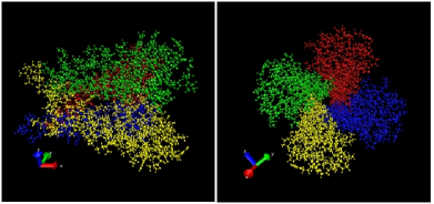
Potassium channels are specialized proteins able to facilitate and regulate the conduction of ions, ions in particular, through cell membranes [27, 13]. In 1998, MacKinnon et al.[20, 17] successfully obtained the crystal structure of KcsA (potassium crystallographically sited activation) channel at a resolution of 2.0 Å, allowing a direct laboratory observation of the selectivity filter and binding sites of ions. KcsA is comprised of around 560 residues (Fig. 1) which form four identical subunits (Fig. 2), each containing two alpha-helices connected by a loop of approximately 30 amino acids. These proteins combine to form three primary sections of the channel: the opening pore on the cytoplasmic side of the cell interior, a small cavity (of 5Å in radius) filled with water and a mix of sodium () and potassium () ions, and the selectivity filter. The selectivity filter, could be as narrow as 2Å in radius, comprised of four specific cation binding sites and formed by the backbone carbonyl groups of conserved residues threonine (T), valine (V), glycine (G), and tyrosine (Y), allows fast conduction of while being highly selective for potassium ions over sodium ions (Fig. 3) [13].


The relatively complete functioning components (gating, selectivity, conductance) and available high resolution structure of KcsA channel makes it the most interesting case attracting investigation in biological studies and mathematical modeling. However, modeling the selectivity of KcsA channel is extremely challenging due to the complicated ion-water-protein interactions. Counterintuitively, the ion has the same ionic valence as the ion does but with a smaller ionic radius, nevertheless it is the one that is rejected by the narrow selectivity filter. In [12], it was suggested that the small diameter of the selectivity filter required dehydration of the cations entering the filter. To compensate for the cost of the dehydration, the carbonyl oxygen atoms from the amino acids in the filter take the place of the water oxygen atoms. The relative rigidity of the filter precludes this action in the case of the ions with a smaller radius but stronger binding of water shell. However, a later study suggests that the selectivity can be explained by the fact that the smaller does not bind to the sites in a thermodynamically favorable way [31].
To understand the selectivity mechanism of the filter, molecular dynamics (MD), being an explicit atomistic model, is a naturally suitable method to model these characteristics but the computation remains very expensive even with current computer powers due to the large number of degree of freedom and the necessarily small time step ( seconds) versus the ion permeation time scale ( seconds) [10]. The mean-field theory, such as the PNP model, has successfully enhanced the model efficiency and reproduced the channel conductance with carefully calibrated parameters. Further, the PNP theory was strengthened recently to adopt the capacity of modeling selectivity, by taking into account the ion-size effect and nonlocal dielectric property of the solvent [4, 18, 36, 33, 22, 15]. Therefore, a highly efficient model with the ability to retain the ion-water-protein interactions is indispensable to investigate the properties, especially the selectivity, of the KcsA channel.
In this paper, we will conduct a study of the selectivity filter in the KcsA channel with our recently developed Image Charge Solvation Method (ICSM), implemented in the open-source Tinker MD package [29]. The ICSM is a hybrid explicit/implicit solvation model developed to accurately account for the reaction field of the solute-solvent environment. In contrast to the traditional full MD simulations [3, 30, 2], most of the solvent in the system outside a designated spherical region is modeled as a dielectric continuum, while only a limited number of particles (protein, water molecules, and ions) inside the sphere are given an atomistic description. The reaction field effect on the permanent charges of the protein and mobile ions, due to the solvent/membrane surroundings, are accounted for by a multiple image charge approximation [5, 7, 8]. Therefore, in the ICSM method, the treatment of electrostatic interaction does not use the periodic condition as in the EWALD approach where infinitely many artificial periodic copies of a simulated system, the ion-channel filter in this case, will occur. The effect of the dielectric exterior to the simulation sphere is accounted for easily by the image-charge method. Moreover, there is no requirement of system neutrality in the hybrid ICSM model, not like EWALD-sum based MD simulations. The efficiency, robustness, and capability of the ICSM have been tested for homogeneous water system and solvation of ions [24].
Here in this paper, we will use this hybrid solvation model to investigate the positioning of sodium ions and potassium ions inside the selectivity filter of the KcsA channel, to evaluate its likelihood of conducting the ions, and thus the selective functions of this potassium channel. In order to balance the accuracy and efficiency, cell membrane and intra/extra cellular solvents are modeled as a dielectric continuum, most of the permanent charges on the channel proteins are assumed as rigid, while the molecular dynamics is applied to the selectivity filter, as well as ions and waters in the cavity chamber. First of all, the fundamental physical properties of the KcsA channel, the electrostatic landscape is obtained by the ICSM and compared to existing results in literatures; four binding sites for are identified. Then, different combinations of and in the selectivity filter and cavity chamber are tested to show that the movement of the ion in the cavity in fact depends on which ions are in the selectivity filter, which will in turn determine what ions will eventually be transported through the whole channel. Along with these investigations, the inclusion of the reaction field in the hybrid ICSM will also be examined and will be shown to be indispensable for accurate portrayal of the ion conduction process.
2 ICSM - Image Charge Solvation Method
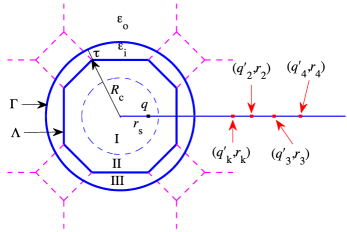
The original setup of the ICSM is shown in a schematic of Figure 4. A regular truncated octahedron (TO) with a size (the distance between its center and farthest corner) is employed as the main simulation box. Inside a spherical domain (dashed circle with radius ) labeled as region I, the solute molecule is placed in the central area of the TO. Region II is the remainder of the TO box with Region I excluded, and this region contains the solvent. Both the solute and solvent in regions I and II are described explicitly. In order to reduce the possible surface effects produced by the boundary of regions of atomistic and continuum descriptions, a buffer layer of thickness is constructed by adding a larger spherical domain containing the TO box. The area outside the TO box but inside the larger sphere is denoted as region III and it is filled with periodic images of the solvent molecules from Region II. Finally, the remaining solvent outside the larger spherical cavity of radius is modeled implicitly as a dielectric continuum, whose reaction field effect on any physical charge inside the larger sphere is approximated by multiple image charges (located outside the larger sphere) and the later will be included in the calculation of electrostatic interactions with the physical charges inside the spherical cavity of radius .
The multiple image charge approximation of the reaction field inside an dielectric sphere extends the Kelvin image concept [19] for a conducting sphere. To simulate the solvation of a protein inside a dielectric sphere containing water molecules, both the source charges and the field points are inside the sphere. For each source charge, in addition to an image point charge at the Kelvin image inversion point, there will be other image charges distributed along a ray starting from the inversion point [5] and a short summary of the multiple image charge approximation of the reaction field is given below.
Given a local volume of a spherical shape of radius in our case, and dielectric permittivity embedded in an infinite solvent of dielectric permittivity , the total electrostatic potential satisfies the Poisson and Poisson-Boltzmann equation:
| (2.1a) | ||||
| (2.1b) | ||||
| where the charge distribution inside the solute domain , contains all explicit charges of the solute and solvent molecules. And for the implicit region , is the inverse Debye-Hückel screening length [24] and if the medium is considered as simple dielectric with no mobile ion density as in this work. Here denotes the Dirac delta function. The two equations are accompanied with the following interface and boundary conditions: | ||||
| (2.1c) | ||||
| where and represent the limiting value of when approaching to the interface from two sides, and is the unit normal direction vector. | ||||
Due to the principle of superposition, we only need to consider the case of one single source charge in , i.e. , and the total potential can be written as where is the primary field that results from the source charge at and is the reaction field from the exterior dielectric medium with dielectric constant and the inverse Debye-Hückel screening length , respectively [1, 24]. The reaction field can be approximated by a set of discrete image charges based on Gauss-Radau quadratures as [5]:
| (2.2) |
where the subscript is for the Kelvin image and represents the remainder of the discrete image charges, , and for all
3 Simulation Results
In the present study, we use the KcsA channel with the PDB ID 2A9H [35]. The data from the PDB was converted to a Tinker input file using the built-in program pdbxyz.x, with the associated toxin removed and five water molecules added to the central cavity.
3.1 Simulation setup for the selectivity filter
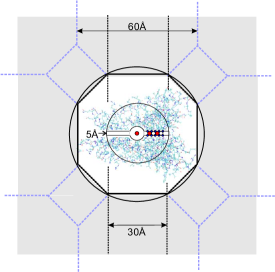
The ICSM is modified to simulate the selectivity filter inside the KcsA channel. The channel pore of the KcsA, including the cavity, selectivity filter, and residential ions are of greatest interest, so they are placed inside Region I in the ICSM schematic of Fig. 4, along with a small portion of the protein structure (see Fig.5). Meanwhile, most of the channel protein, some of the cell membrane and surrounding solvent are located in Region II. The dielectric permittivity inside the TO box is denoted as while is the dielectric constant outside the larger sphere of radius . As the difference between the two dielectric constants (and therefore the surface effect) is small, the buffer layer thickness is set to be zero.
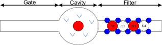
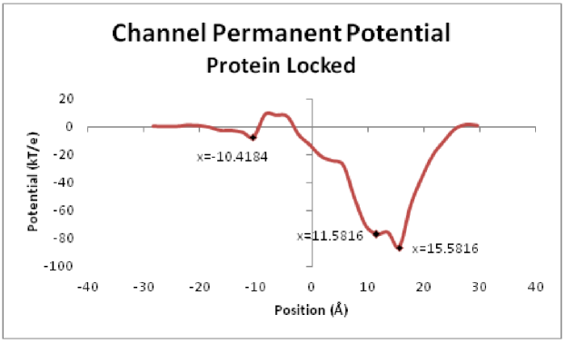
Figure 6 gives a zoomed-in view of the channel pore of the KcsA. The channel is set to run along the x-axis and its total length is taken to be 60Å. It contains three regions: the channel gate, a cavity chamber, and a selectivity filter. In our coordinates, the channel starts from -30Å and ends at 30Å . The positions of four standard binding sites for the ions in the selectivity filter (labeled S1 - S4), which are located between the carbonyl oxygen of the amino acids that make up the filter (shown in blue).
The system is first minimized with all particles allowed to move freely using the minimize program in Tinker and then run for 200ps for an initial equilibrium. For subsequent simulations, we use the ACTIVE keyword in the key file to lock down all atoms of the channel except the (sixteen) amino acids making up the selection filter, the waters, and the ions. The parameters contained in the Amber99 force field [32, 34], also included in Tinker, will be used for the MD simulations. The Velocity Verlet algorithm was chosen for the time integration with a Nose-Hoover bath set at 300K. The time step is taken to be 2 fs, and the trajectory is recorded every ps for analysis.
3.2 Fundamental characteristics
Before the selectivity of the KcsA channel is investigated, we use the ICSM to simulate some fundamental characteristics of the channel, comparing with the results in the literature.
Case 1. Profile of electrostatic landscape of the channel: We first compute the channel permanent potential using the program analyze.x of the Tinker package. This routine was modified to output the potential energy acting on a particular ion as the latter moves through the channel beginning at Åand ending at Å. The ion was moved Åat a time and the potential energy was calculated on the ion at that position. The results for the channel prior to minimization and equilibration are shown in Figure 7, which closely resembles the channel permanent potential used by Jung, et al. for their studies on ERINP [21]. In our calculation we found energy minima at -10.4 Å, 11.5Å, and 15.6Å, respectively. Next, we allowed all atoms to move freely in the system and minimized the system using the minimization program in Tinker. The final configuration will be then used as the initial input for the subsequent numerical simulations in this paper. The interval in the channel is considered as the filter, while , , , and are regarded as site S1, S2, S3 and S4, respectively.
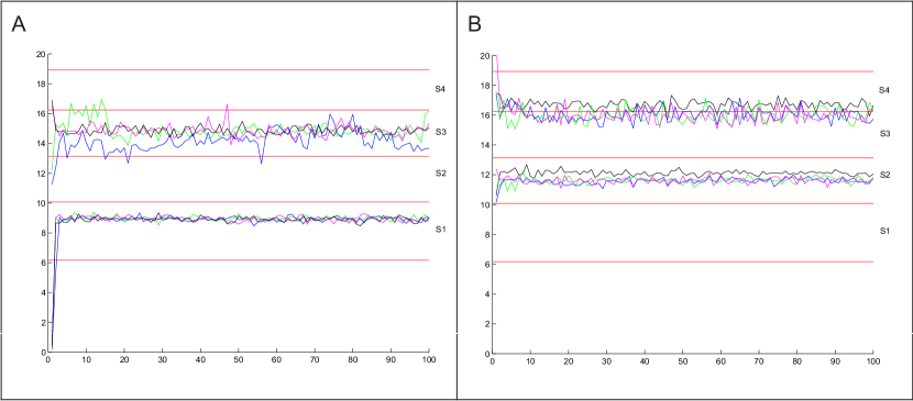
Case 2. Preferred positioning for two inside the channel: It was reported in [37] that two ions would stay at site 1 (S1) and site 3 (S3) when a third ion is far away. Otherwise they will occupy sites S2 and S4 when the third ion is relatively close to the filter entrance. To verify this conclusion by the ICSM, we run 100 ps simulations for several permutations of the positions of only two potassium ions in the channel. One ion is located in the water cavity (around Å ) and the other is placed in different positions (10-19Å ) in the selectivity filter. The detailed positions of the ions we tested are summarized in Table 1. The starting positions for the ions in the filter were set at the midpoint between the carbonyl oxygen of the filter amino acids. Regardless of their starting positions in the filter, the two potassium ions come to rest at S1 (interval [6Å, 11Å]) and S3 (interval [14Å, 16Å]) , and the relaxation period for the ions is very short. By approximately ps, the ions and waters in the filter reach their approximate final positions. An example of the trajectories for four of these runs is plotted in Figure 8 (top). In this figure and all remaining figures, the red horizontal lines indicate the position of the oxygens for the Threonine (T), Valine (V), Glycine (G), Tyrosine (Y), and Glycine (G). The intervals between TV, VG, GY, and YG are considered as S1, S2, S3, and S4, respectively. In this instance, both of the ions move into the filter and come to rest at positions S1 and S3. And this configuration will be a transitional phase between an ion exiting the channel and another one entering when three ions are present in the channel for transport.
Case 3. Preferred positioning for three inside the channel: In this case, we add a third potassium ion into the filter (interval [10Å, 16Å]) in addition to the two at the various places as in the previous study. The detailed positions of the three ions are listed in Table 2. After a similar relaxation period, two of the ions rest at site S2 (interval [11Å, 14Å]) and site S4 (interval[16Å, 19Å]), and another ion stays in the water cavity but close to the entrance of the selectivity filter (around Å). Figure 8 (bottom) shows the trajectories of the four permutations of the positions of the three potassium ions listed in Table 2. In the final states, two ions stay between V-G and between Y-G, respectively. The three ion configuration can be considered to be a “steady” or normal state for the potassium channel and we note that the final positions for the two ions in the selectivity filter are in fact S2 and S4, which are very close to the energy minima found in the empty channel in Case 1 above.
| Position(Å) | Position(Å) | Position(Å) | Position(Å) | |||||
| Initial | Final | Initial | Final | Initial | Final | Initial | Final | |
| K1 | 0.418 | 8.635 | 0.418 | 9.049 | 0.418 | 8.957 | 0.418 | 8.923 |
| K2 | 18.544 | 14.739 | 15.800 | 14.717 | 13.324 | 15.387 | 10.347 | 13.451 |
| Position(Å) | Position(Å) | Position(Å) | Position(Å) | |||||
| Initial | Final | Initial | Final | Initial | Final | Initial | Final | |
| K1 | 0.418 | 5.310 | 0.418 | 5.404 | 0.418 | 8.562 | 0.418 | 5.092 |
| K2 | 13.324 | 11.808 | 10.347 | 11.813 | 10.347 | 12.047 | 15.800 | 11.695 |
| K3 | 18.544 | 16.280 | 15.800 | 15.727 | 18.544 | 16.656 | 18.544 | 16.385 |
3.3 Selectivity and positioning of potassium versus sodium ions
Our simulations in Section 3.2 indicate that in the “normal state” of three potassium ions in the channel, two of them are located at site S2 and site S4, while the third one stays in the area around Å, which is at the entrance to the selectivity filter. This is consistent with the results in [3, 30, 2], and this location corresponds to the favorable location for a ion formed by the P helix dipoles [3, 26].
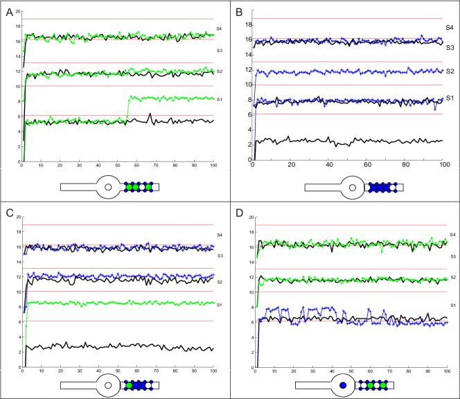
The KcsA channel dominantly conducts potassium over sodium ions, although the two type of ions have the same charge. To investigate the ability of the ICSM system to distinguish between ions and ions, we repeat the three ion tests with sodium ions in the selectivity filter instead of potassium ions, in order to simulate the preferred positionings of ions and / mixtures.
Case 1: Preferred positioning of three inside the channel: Figure 9A shows the simulation results if three ions are placed in the channel. In contrast to the case of three- (black curves), the two ions (dashed grey) in the selectivity filter rest at the sites S1 and S3 (instead of S2 and S4 as for the case of ), while the third ion stays around Å in the cavity, which is toward the center of the cavity and relatively away from the filter entrance. This simulation suggests that when two are in the filter, they are relatively away from the channel exit, meanwhile the third has difficulty approaching the filter entrance.
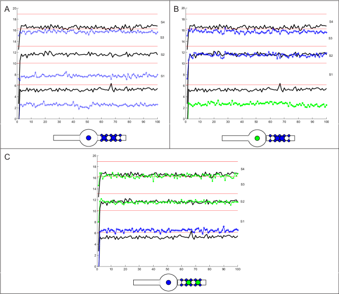
Case 2: One in cavity and two ions in filter: With Case 1 in mind, we replace the in the cavity by a and keep the two in the filter. From Fig. 9B we can clearly see that the in the cavity pushes the at site S1 in Case 1 into site S2, hence the two ions in the filter take sites S2 and S3. On the other hand, the itself stays in the cavity around , which is relatively away from the filter entrance.
Case 3. One in cavity and two ions in filter: As shown in Figure 9 C, in this case, two potassium ions are already situated in the selectivity filter and a sodium ion is located on the cellular side of the water-filled cavity. The two ions in the filter take the sites S2 and S4, as for the three- configuration. Comparing to the in the cavity in the three- system, the position of the ion is closer to the entrance of the filter and on the other side of the carbonyl oxygen for the tyrosine.
Case 4. and mixtures in filter: Figure 10 shows the permutation of and mixtures in the filter region, while in the cavity we have either or . Figs. 10A-D list the situations of , , , and , respectively. The positions of the ions are ordered from the cavity to the exit of the filter. It can be seen from the simulations that no ion can occupy the site S4 in any of the ion combinations.
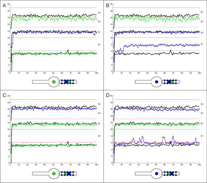
From the above simulations it can be summarized that in a three- system, two sodium ions are occupying in the filter region and stay at sites S1 and S3 positions, while the third ion in the cavity is not able to move toward the proximity of the filter. The at site S1 is able to proceed to site S2 if the sodium ion in the cavity is replaced by a potassium ion but the other in the filter stays at site S3. Alternatively, when a in the water cavity faces two in the selectivity filter, it is closer to the filter entrance than the position of the in the cavity in a three system. Therefore, we conjecture that ions are able to approach the filter entrance and translocate from site S1 to S2, but no evidence shows that they are able to move to site S4. This may be one reason for the reduced possibility of ion conducting through the channel, consistent with the fact that the KcsA channel is designed preferentially for three potassium ions in the setting for potassium transporting through the channel as shown in Section 3.2.
3.4 Effect of reaction field in ICSM
We also investigated the importance of the long range reaction field by running same configurations with the image charges and without image charges.
Case 1. Simulation of the three-system without the reaction field: We first consider the stable configuration of three potassium ions in the channel without taking into account the reaction field. For comparisons, the corresponding simulation results with reaction field are also provided. Fig. 11 A show that without the reaction field, the two ions in the selectivity filter are still occupying sites S2 and S4. However, the final resting position for the third ion, which is initially in the water cavity, is approximately at Å when the reaction field is neglected. In other words, this potassium ion is able to move closer to entrance of the filter and eventually takes site S1(between the residues T and V). Therefore, comparing to the results with reaction field for the three- system where two ions take sites S2 and S4 in the filter while one stays in the water cavity at Å. Without the reaction field the three potassium ions will take sites S1, S2, and S4; this is against the current biological conclusions.
Case 2. Simulation of the three- system without the reaction field: The positions of three ions in the channel are plotted in Fig.11B when the simulation is implemented without the reaction field. For a better comparison, the approximate final positions of these ions are shown in Table 3. Recall the simulations with the reaction field, two sodium ions in the selectivity filter take the sites S1 and S3, while the third ion stays in the cavity around Å. But without the reaction field, all three sodium ions are able to enter the filter region and they occupy the sites S1, S2, and S3, respectively. Specifically, the ion in the cavity (sodium 1) moves toward the filter and rests at Å if the reaction field is absent.
| Sodium 1 | Sodium 2 | Sodium 3 | |
|---|---|---|---|
| With RF | 2.59Å | 8.00Å | 15.66Å |
| No RF | 8.18Å | 11.99Å | 15.74Å |
Case 3. Simulation of mixtures in the channel without the reaction field: For a further investigation of the effect of the reaction field, the case of two in the selectivity filter and a in the cavity is considered. In section 3.3, it is observed that the two in the filter take sites S2 and S3, and the ion stays in the cavity around the position Å. While ignoring the reaction field, the ion enters the filter and takes the position at site S1. Thus, in this case, the selectivity filter will contain three residential ions as occupies S1 and the two ions occupy sites S2 and S3, as shown in Fig.11C. On the other hand, for the case of two in the filter and one in the cavity, the results without the reaction field are very close to the ones with the reaction field, however, the position of the in the cavity fluctuates considerably more (see Fig.11D).
From the above studies it can be summarized that the cavity ion, no matter or , is able to move into the filter on the other side of the threonine to position Å if the reaction field is neglected. This fact implies that there will be three ions in the selectivity filter, and this does not agree with the biological observations. Therefore, we can see clearly that the reaction field is critical for the fidelity of the ICSM to reproduce the physical mechanism of the KcsA channel in its selectivity and positioning of the ions.
4 Conclusions
In this paper, we have shown that the Image-Charge Solvation Method (ICSM) is applicable to study the selectivity filter of a KcsA channel where only a local spherical region around the filter section of the channel is modeled in atomistic details and the effect of the rest of the channel and membrane and solvents can be modeled as a continuum dielectric, whose reaction field is approximated by a simple multiple image charge representation.
Our numerical simulations have produced the following results:
-
•
The preferred positioning for two potassium ions inside the channel are in sites S1 and S3.
-
•
The preferred positioning for three potassium ions inside the channel are one in the cavity and two at sites S2 and S4, which is the characteristic positioning of three ions for conducting the potassium ions by the KcsA channel.
-
•
The ICSM algorithm is able to distinguish sodium and potassium ions when investigating the selectivity of the KcsA channel. In contrast to a three- system, the two ions in the selectivity filter occupy sites S1 and S3 instead of S2 and S4, and the third in the water cavity stays relatively away from the filter entrance. Among all the permutations of 3 ions, except for the case of 3 potassium ions, no ion was found to be able to move to site S4, this is consistent with the fact that the KcsA channel is indeed a potassium channel designed to transport potassium ions.
-
•
The reaction field from the dielectric environment outside the atomistic region containing the filter is critical in the accurate representation of the selectivity function of the filter and the correct predictions of the ion positioning inside the channel.
In future research, the following issues will be addressed: the layered dielectric media outside the simulation region comprised of membrane and ionic solvents as for the time being the latter is simply ignored; the I-V curve of the KcsA channel calculated by ICSM molecular dynamics simulations.
Acknowledgement
The authors acknowledge the support of the US Army Office of Research (Grant No. W911NF-14-1-0297), the National Science Foundation (DMS-1315128), and the NSFC (grant number 91230105) for the work in this paper. Authors also thank the insightful comments from Drs. Xiaolin Cheng and Chun Liu on various aspects of the simulations for KcsA channels.
References
- [1] Baker, K., Baumketner, A., Lin, Y., Deng, S., Jacobs, D., and Cai, W. ICSM: An order n method for calculating electrostatic interactions added to TINKER. Comp. Phys. Comm., 184:19–26, 2013.
- [2] Berneche, S and Roux, B. Molecular dynamics of the kcsa channel in a bilayer membrane. Biophysical Journal, 78:2900–2917, 2000.
- [3] Biggin, P. C., Smith, G. R., Shrivastava, I., Choe, S., and Sansom, M.S. P. Potassium and sodium ions in a potassium channel studied by molecular dynamics simulations. Biochim. Biophys. Acta, 1510.
- [4] Burger, M., Eisenberg, R.S., and Heinz, E. Inverse Problems Related to Ion Channel Selectivity. SIAM J Applied Math, 67(4):960–989, 2007.
- [5] Cai, W., Deng, S., and Jacobs, D. Extending the fast multipole method to charges inside or outside a dielectric sphere. J. Comput. Phys., 223:846–864, 2007.
- [6] Cai, W., Deng, S., and Xu, Z. Image charge approximations of reaction fields in solvents with arbitrary ionic strength. Journal of Computational Physics, 228:2092–2099, 2009.
- [7] Deng, S. Cai, W. Discrete image approximations of ionic solvent induced reaction field to charges. Commun. Comput. Phys., 2:1007–1026, 2007.
- [8] Deng, S. Cai,W. Extending the fast multipole method for charges inside a dielectric sphere in an ionic solvent: High-order image approximations for reaction fields. J. Comput. Phys., 227, 2007.
- [9] Chen, D.P., Lear, J., and Eisenberg, B. Permeation through an open channel: Poisson-Nernst-Planck theory of a synthetic ionic channel. Biophys J., 72(1):97–116, 1997.
- [10] Chung, S.H. and Kuyucak, S. Recent advances in ion channel research. Biochimica et Biophysica Acta, 1565:267–286, 2002.
- [11] Coalson, R. D. and Kurnikova, M. G. Poisson-Nernst-Planck theory approach to the calculation of current through biological ion channels. IEEE Trans Nanobioscience, 4(1):81–93, 2005.
- [12] Doyle, D.A., Cabral, J. M., Pfuetzner, R. A., Kuo, A., Gulbis, J. M., Cohen S.L., Chait, B.T., and MacKinnon, R. The structure of the potassium channel: Molecular basis of K conduction and selectivity. Science 280, 280:69–77, 1998.
- [13] Egwolf, B. and Roux, B. Ion selectivity of the kcsa channel: A perspective from multi-ion free energy landscapes. J. Chem. Phys., 401:831–842, 2010.
- [14] Gautschi, W. Algorithm 726; ORTHPOL-a package of routines for generating orthogonal polynomials and Gauss-type quadrature rules. ACM Trans. Math. Softw., 20:21–62, 1994.
- [15] Gillespie, D., Nonner, W., and Eisenberg, R.S. Density functional theory of charged, hard-sphere fluids. Phys Rev E, 68:1–10, 2003.
- [16] Hladky, S.B. and Haydon, D.A. Ion transfer across lipid membranes in the presence of gramicidin a. i. studies of the unit conductance channel. Biochim. Biophys. Acta, 274:294–312, 1972.
- [17] Hodgkin, A.L. and Huxley,A.F. A quantitative description of membrane current and its application to conduction and excitation in nerve. J. Physiol., 117:500–544, 1952.
- [18] Hyon, Y., Eisenberg, B., and Liu, C. A mathematical model of the hard sphere repulsion in ionic solutions. Communications in Mathematical Sciences, 9:459–475, 2011.
- [19] Jackson, J.D. Classical Electrodynamics. New York : John Wiley, 1999.
- [20] Jordan,P.C. Fifty years of progress in ion channel research. IEEE TRANSACTIONS ON NANOBIOSCIENCE, 4:3–9, 2005.
- [21] Jung, Y., Lu, B., and Mascagni, M. A computational study of ion conductance in the KcsA channel using a Nernst-Planck model with explicit resident ions. J. Chem. Phys., page 215101, 2009.
- [22] Li, H. and Lu, B. An ionic concentration and size dependent dielectric permittivity Poisson-Boltzmann model for biomolecular solvation studies. J. Chem. Phys., 141:024115, 2014.
- [23] Liang, Y., Xu, Z., and Xing, X. A Multi-scale Monte Carlo Method for Electrolytes. arXiv:1504.00454, 2015.
- [24] Lin, Y., Baumketner, A., Deng, S., Xu, Z., Jacobs, D., and Cai, W. An image-based reaction field method for electrostatic interactions in molecular dynamics simulations of aqueous solutions. J. Chem. Phys., 131, 2009.
- [25] MacKerell, A. D. Jr., Bashford, D., Bellot, M., Dunbrack, R. L. Jr., Evanseck, J. D., Field, M. J., Fischer, S., Gao, J., Guo, H., Ha, S., Joseph-McCarthy, D., Kuchnir, L., Kuczera, K., Lau, F. T. K., Mattos, C., Michnick, S., Ngo, T., Nguyen, D. T., Prodhom, B., Reiher, W. E. , III, Roux, B., Schlenkrich, M., Smith, J. C., Stote, R., Straub, J., Watanabe, M., Wiorkiewicz-Kuczera, J., Yin, D., and Karplus, M. All-atom empirical potential for molecular modeling and dynamics studies of proteins. Journal of Physical Chemistry B, 102(18):3586–3616, 1998.
- [26] Roux, B. MacKinnon, R. The cavity and pore helices in the kcsa k+ channel: electrostatic stabilization of monovalent cations. Science, 285(5424):100–102, 1999.
- [27] University of Illinois at Urbana-Champaign. Kcsa potassium channel. Online.
- [28] Perlman, D.A., Case, D.A., Caldwell, J.W., Ross, W.S., Cheatham, T.E., Debolt, S., Ferguson, D., Seibel, G., and Kollman, P. Amber, a package of computer programs for applying molecular mechanics, normal mode analysis, molecular dynamics and free energy calculations to simulate the structural and energetic properties of molecules. Comp. Phys. Commun., 91:1–41, 1995.
- [29] Ponder, J. W. Tinker - software tools for molecular design. http://dasher.wustl.edu/ffe/downloads/guide.pdf, 2004.
- [30] Shrivastava, I.H., Tieleman, D.P., Biggin, P. C., and Sansaom, M.S.P. versus ions in a K channel selectivity filter: A simulation study. Biophysical Journal, 83:633–645, 2002.
- [31] Thompson, A., Kim, I., Panosian, T., Iverson, T., Allen, T., and Nimigean, C. Mechanism of potassium-channel selectivity revealed by na+ and li+ binding sites within the kcsa pore. Nature Structural and Molecular Biology, 16:1317–1324, 2009.
- [32] Weiner, P. K. and Kollman, P. A. Amber: Assisted model-building with energy refinement.a general program for modeling molecules and their interactions. J. Comput. Chem., 2:287–303, 1981.
- [33] Xie, D., Jiang, Y., and Scott, L.R. Efficient algorithms for solving a nonlocal dielectric model for protein in ionic solvent. SIAM Journal on Scientific Computing, 38:B1267–1284, 2013.
- [34] Yang, L.J. New-generation amber united-atom force field. J. Phys.Chem. B, 110:13166–13176, 2006.
- [35] Yu, L., Sun, C., Song, D., Shen, J., Xu, N., Gunasekera, A., Hajduk, P. J., and Olejniczak, E. T. Nuclear magnetic resonance structural studies of a potassium channel-charybdotoxin complex. Biochemistry, pages 15834–15841, 2005.
- [36] Zhou, S., Wang, Z., and Li, B. Mean-field description of ionic size effects with non-uniform ionic sizes: A numerical approach. Phys. Rev. E, 84:021901, 2011.
- [37] Zhou, Y., Morais-Cabra, J., Kaufman, A., and Mackinnon, R. Chemistry of ion coordination and hydration revealed by a channel-Fab complex at 2.0 Å resolution. Nature, 414:43–48, 2001.