Function changing mutations in glucocorticoid receptor evolution correlate with their relevance to mode coupling
Abstract
Nonlinear effects in protein dynamics are expected to play role in function, particularly of allosteric nature, by facilitating energy transfer between vibrational modes. A recently proposed method focusing on the non-Gaussian shape of the population near equilibrium projects this information onto real space in order to identify the aminoacids relevant to function. We here apply this method to three ancestral proteins in glucocorticoid receptor (GR) family and show that the mutations that restrict functional activity during GR evolution correlate significantly with locations that are highlighted by the nonlinear contribution to the near-native configurational distribution. Our findings demonstrate that nonlinear effects are not only indispensible for understanding functionality in proteins, but they can also be harnessed into a predictive tool for functional site determination.
.1 Introduction
Mechanisms of information transfer and function in proteins continue to be challenging problems where different points of view compete. The ensemble view, i.e., that a ligand binding event triggers allostery by modifying the free energy landscape is now a commonly recognized paradigm Kumar et al. (2000); Tsai et al. (1999); Okazaki and Takada (2008). The so called “population shift” picture is helpful in understanding allostery without shape change and finds support from recent NMR studies Kar et al. (2010). Models which focus on mechanistic aspects, such as the suppression of a certain vibrational mode or energy transfer between two modes Brooks and Karplus (1983); Haliloglu et al. (1997); Yogurtcu et al. (2009); Ma (2005); Piazza and Sanejouand (2009) are also widely employed, thanks to the intuitive picture they offer. Through such models, it is, for example, possible to discuss positive/negative allostery Rodgers et al. (2013).
A recently proposed approach that rests on dynamical simulations around equilibrium delivers a tool for predicting locations relevant to mode coupling Kabakçıoğlu et al. (2010); Varol et al. (2014). The central idea of the method is to quantify the nonlinear contribution (required for energy transfer between vibrational modes) to the near-native configurational probability distribution function and identify the residues on which it has the highest impact. Present work is an application of this method to a set of ancestral glucocorticoid receptor (GR) proteins and observes a statistically significant correlation between locations underlined by mode coupling and function changing mutations that took place during the evolution of the GR proteins. Multiple molecular dynamics (MD) trajectories of for each protein further allow us to discuss the robustness of the method between independent MD runs, as well as the sensitivity of our findings to the simulation length.
.2 Extracting information on mode coupling
Consider a protein composed of aminoacids. Let the Cartesian coordinates of carbon-alpha () atoms be stored in the vector of length , encoding a coarse representation of the protein’s spatial arrangement, or configuration. The configurational probability distribution, can be derived numerically from an real-valued matrix, where is the number of snapshots acquired by sampling sufficiently long MD trajectories. This configurational distribution is then used to determine fluctuations around the mean structure, , where indicates averaging over time and multiple MD trajectories generated using different random seeds.
Within the framework of elastic network models, the configurational distribution is most conveniently expressed in terms of “modal fluctuations”
| (1) |
where is the covariance matrix associated with real-space fluctuations. A general analytical expression for in terms of Hermite tensor polynomials was originally proposed by Flory Flory and Yoon (1974):
| (2) |
where denotes the Hermite tensor polynomial of rank , and the coefficients follow from the orthogonality relation .
In basis, all expansion coefficients in Eq.(2) vanish for a perfectly elastic network. As a result, the configurational distribution of such a linear system is separable into identical Gaussian functions with unit standard deviation and zero mean:
| (3) |
The superscript (g) implies the Gaussian product form of . All vibrational modes of the linear system are represented in an identical fashion in this normalized form of the distribution. Yet, their physical difference is evident from and encoded in the corresponding eigenvalues and eigenvectors of .
Nonlinearity can be introduced into this picture without coupling the vibrational modes, by simply adding higher-order terms to the corresponding harmonic oscillator Hamiltonians:
| (4) |
where and are the effective mass and the spring constant for the mode and indicate the strength of higher-order terms in appropriate units, all of which can in principle be derived from the underlying dynamics. The resulting configurational distribution function can then be expressed as
| (5) |
where
| (6) |
Note that, above is now the ordinary Hermite polynomial of rank . Eqs.(5,6) describe the most general separable distribution for the variables , hence the superscript (s).
An arbitrary configurational distribution can be expressed in a similar fashion, starting from Eq.(2) Kabakçıoğlu et al. (2010):
| (7) |
Eq.(7) allows one to address all nonlinear corrections to the Gaussian description of near-native fluctuations in a methodical, order-by-order fashion. For the type of nonlinearity which yields Eq.(5) in a suitably chosen basis we propose the term “marginal anharmonicity”, because it is fully characterized by marginal distributions . In other words, all coefficients and their higher-order counterparts in Eq.(7) can be derived in this case from the coefficients of “one-body” terms . For example, . On the other hand, the diagonalization problem, that is, determining the transformation for an arbitrary marginally anharmonic system is significantly harder than that for a linear one. (This problem arises naturally in signal processing field and is known as “Independent Component Analysis” or ICA Hyvärinen et al. (2004).)
Deviations in from the separable form in Eq.(5) are, by construction, due to coupling between (possibly anharmonic) vibrational modes. In fact, when such coupling is strong, the solutions of the nonlinear system will depart significantly from those found in the noninteracting limit in Eq.(5); so that, describing the dynamics around the vibrational “modes” in Eq.(4) may no more be justified. However, numerous earlier studies suggest that a quasi-elastic treatment of protein dynamics is an adequate and fruitful course at physiological temperatures Brooks and Karplus (1983); Karplus and McCammon (1983); Levy et al. (1982); Levitt et al. (1985); Doruker et al. (2000). Therefore, in the following we assume that the eigenvectors of in Eq.(1) continue to serve as a meaningful basis in which calculated along the eigen-directions yield a good approximation for the best marginally anharmonic description of the system. Our results reported below corroborate this expectation.
While the degree of coupling between mode pairs (also triplets, quartets and so on) can be discussed by investigating corresponding high-order terms in Eq.(7), it was shown earlier that a cumulative treatment of all coupling corrections to Eq.(5) is not only possible but also easier Varol et al. (2014). This is done by quantifying the difference between the near-native conformational distribution obtained from MD (which, in principle, includes effects at all orders) and the best description of the data in terms independent modes, as given by Eq.(5).
A motivation for studying mode coupling is to gain insight about the mechanism and regulation of function in allosteric proteins. Accordingly, one is typically interested in locations on the structure where mutations or immobilization by means of ligand binding alter protein’s activity. After isolating multi-mode contributions to fluctuations as described above, the next step is therefore to project this information onto the protein structure in order to identify regions which are critical for mediating the coupling between relevant modes. To this end, we first map the distribution back onto Cartesian space by
| (8) |
Next, the difference between and is measured along each Cartesian component of the position vector of residue by means of the Jensen-Shannon (JS) divergence defined as Manning and Schütze (1999):
| (9) |
where and is the Kullback-Leibler divergence that is given by
| (10) |
An advantage of using JS divergence is that it is symmetric, therefore immune to the possibility that one of the two distributions may vanish at certain points (Kullback-Leibler divergence yields infinity for such instances).
Following the recipe above, we calculate the mode-coupling score for each amino acid in a protein, as the sum of JS divergences calculated along the components of . Note that, comparing and aminoacid by aminoacid rather than in their full domain not only yields a testable output (locations relevant to mode coupling in the protein), but is also more robust to stochastic fluctuations inherent to the method, simply because of reduced dimensionality. That is, histograms for spatial fluctuations of single aminoacids can be represented with much fewer bins compared to joint distributions or for the whole protein, therefore they are sufficiently well sampled along the MD trajectories. Variations in the output as a function of the simulation length as well as between independent MD runs are investigated in Section .4.
An earlier version of this prescription was applied to motor protein myosin II with encouraging results Varol et al. (2014). We here consider the ancestral chain of GR proteins and show that the outlined analysis reveals a significant bias in mode-coupling scores for experimentally validated function-altering mutations in the family.
.3 Glucocortocoid receptors and evolutionary data
Glucocorticoid receptors are a class of endogenous steroid hormones that regulate imflammatory and stress responses, growth, development, and apoptosis Kino et al. (2009); Nicolaides et al. (2010); Chrousos (2004); Clark JK (1992); Zhou and Cidlowski (2005); Barnes (1998); Sapolsky et al. (2000). GR positively regulates transcription through a process known as transactivation in which the ligand-bound GR dislocates from cytoplasm to enter cell nucleus where it activates transcription Carson et al. (2014). Its paralogous counterpart, mineralocorticoid receptor (MR) is mainly responsible for regulating electrolyte homeostasis Li et al. (2005). While GR binds glucocorticoid hormone cortisolBentley (1998), MR acts as a host for aldosterone, 11-deoxycorticosterone (DOC) and with a lesser affinity for cortisol Yang and Young (2009). Through phylogenetic analysis, the sequence and the crystal structure of their common ancestor AncCR, as well as the ancestral GR proteins in cartilaginous fish (AncGR1) and in bony vertebrates (AncGR2) have been determined Bridgham et al. (2009); Ortlund et al. (2007). It has been shown that AncCR, similar to AncGR1, indiscriminately binds to DOC, aldosterone and cortisol. On the other hand, AncGR2 exclusively binds cortisol and is not activated by aldosterone and DOC.
Considerable effort has been devoted to understanding the basis of ligand specificity in the evolution of GR Ortlund et al. (2007); Bridgham et al. (2009); Harms and Thornton (2014). Structural variations are minute, with RMSD difference between AncGR1 and AncGR2 Glembo et al. (2012). Among the 36 mutations that transform AncGR1 to AncGR2, it has been shown that two strictly conserved mutations, S106P and L111Q (group X), are sufficient to increase cortisol specifity. S106P changes the architecture of ligand binding pocket and allows L111 to be located at a closer position to the ligand. The effect of L111Q is biochemical rather than mechanical, since it creates an additional hydrogen bond between 111Q and the cortisol, which lacks in DOC and aldosterone binding. Three additional mutations, L29M, F98I and S212 (group Y), wipe out the affinity towards DOC and aldosterone. However, AncGR1+X+Y structure cannot activate transcription due to the damaged hydrogen bond network which destabilizes the activation-function helix. Two further mutations, N26T and Q105L (group Z), are necessary in order to reestablish the hydrogen bond network and thereby stabilize the structure. All together, AncGR1+X+Y+Z, captures AncGR2 phenotype. A recent study found that XYZ mutations correlate with a measure based on the difference in fluctuation amplitudes of a residue in principal vibrational modes of ancestral GR proteins Glembo et al. (2012).
Besides historically occuring mutations, another study on alternative evolutionary pathways that restore AncGR2 phenotype Harms and Thornton (2014) demonstrated that, among a set of suggested alternatives, only the mutation pair Q114L/M197I recovers cortisol sensitivity similar to the historical set of permissive mutations, albeit with a loss of associated transcriptional function.
.4 Specifics on molecular dynamics simulations
Crystal structures of AncCR, AncGR1, and AncGR2 are publicly available at PDB with accession codes 2Q3Y, 3RY9, and 3GN8, respectively. MD simulations of each were carried out on Tesla K20 GPUs by means of Amber 14 Molecular Dynamics Package Case et al. (2014) using force-field Cornell et al. (1995). Ligand molecules were also included in the simulations and their parametrization were done with Antechamber using generalized Amber force field Wang et al. (2004, 2006). All simulations were performed in ensemble with explicit water solvent and with Langevin dynamics which maintained the temperature at 310 K and the pressure at 1 bar. No rigid bonds were assumed. 1 fs timesteps were used between successive frames while trajectories were captured every 1000 frames, i.e. 1 ps apart, throughout 128 ns long simulations performed for each sample. Each protein was simulated four times with the same initial condition but different random number generator seeds. Before further analysis, trajectories were aligned by means of the backbone atoms. Discarding the first 7 ns of each simulation for equilibration, this protocol resulted in snapshots, derived from long simulations of the near-native dynamics for each protein.
.5 Mode coupling in GR proteins and comparison with evolutionary data
A substantial contribution from marginal anharmonicity is observable in the amplitude distributions of the slowest modes of AncGR2 protein shown in Fig. 1. Strong deviation from Gaussian in
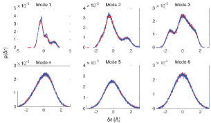
eigenmodes with large eigenvalues is typical for the whole GR family, in fact for most proteins Roh et al. (2005); Hayward et al. (1995). A comparison of the marginal distributions and Eq.(6) with a cut-off at confirms that they are represented accurately by the analytical approach in Section .2.
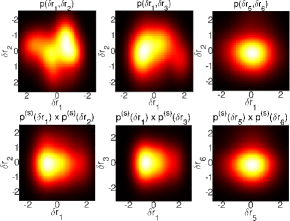
On the other hand, presence of mode coupling is evident from the difference between and , as shown in Fig. 2 by considering joint amplitude distributions for corresponding to slowest mode pairs . Contribution of mode coupling is exemplified by the difference between the two rows of Fig. 2. Information content of such deviation from marginal anharmonicity in protein dynamics and its relevance to protein’s biological function is our focus in this study.
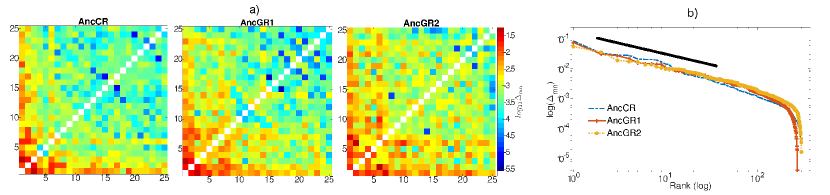
We start by asking whether certain mode pairs stand out in the above analysis. One can assess the overall impact of a mode pair by considering the (dimensionless) conformational free energy
| (11) |
calculated with and without the mode coupling contribution from the mode pair in Eq.(7). For this purpose, we approximated by the one- and two-body terms spelled out in Eq.(7). We then defined , in which the mode-coupling contribution of the pair is discarded. The difference is a measure of the impact of the interaction between modes and on protein’s behavior near equilibrium. for all pairs composed out of slowest 25 modes of each protein are given in Fig. 3. It is interesting that displays a power-law dependence with a scaling exponent over more than two decades on the rank order of the pair (Fig. 3b). An exhaustive analysis over all mode pairs was not performed due to its heavy computational cost.
We found that the highest-impact mode pairs are 1-3 for AncCR and AncGR1; and 1-7 and 2-6 for AncGR2. Spatial fluctuations associated with these mode pairs coincide with helices 7 and 10, along with the loop region preceeding helix-7. Indeed, these helices form part of the ligand binding pocket, while the loop before helix-7 is where the two X mutations are located. We additionally observed a region on helix-9 with high sequence conservation score also involved in mode coupling which, to our knowledge, has not been highlighted in earlier studies.
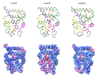
We next performed the analysis outlined in Section .2 in order to derive a mode-coupling score for each aminoacid. The resulting score vectors obtained over the full data set (four trajectories) separately for each member of the GR family are shown as a heat map superimposed onto the proteins’ three-dimensional structure in Fig. 4. We observed that the loop (100-110) preceeding helix-7 yields considerably high scores in all proteins, despite the fact that this loop and the nearby helix-7 exhibit the largest structural variability between AncGR1 and AncGR2 Ortlund et al. (2007). Furthermore, the same region also accommodates 4 of the 6 (XYZ-)mutations mentioned above. These observations hint at the relevance of mode coupling to the evolutionary history of function in the GR protein’s lineage, which we investigate below in further detail.
Note that, the location of the X-mutation S106P consistently has one of the highest scores in all proteins. Considering that S106P alone decreases activation in AncGR indepedent of the ligand type Ortlund et al. (2007) suggests that the mechanism underlying the activity loss is mechanical in origin, rather than biochemical (to which the present method is insensitive). The opposite is true for the second X mutation L111Q which recovers cortisol specificity Ortlund et al. (2007) by allowing formation of a hydrogen bond with cortisol. Mode-coupling score of location 111 shows no significant deviation from the mean. On the other hand, the synthetic mutations Q114L/M197I in AncGR1 - that also recover cortisol specificity and disrupt communication between cortisol binding and transcriptional activity - coincide with the two mode-coupling peaks in AncGR1 located on helix-7 and helix-10.
Complementing these observations, an objective evaluation of the correlation between mode-coupling scores and the AncGR1 AncGR2 mutation set is desirable. For this purpose, we use the recall analysis where mutation sites under consideration are labelled as the target set and their rankings are inspected in the full residue list sorted according to mode-coupling scores. The result is presented as a recall curve which is a plot of the fraction of the target set elements (-coordinate) observed in a given fraction (-coordinate) of the list picked from the top. In absence of correlation between the target set and the scoring function, one expects to see the recall to remain on the diagonal upto statistical fluctuations. A recall curve remaining significantly above the diagonal indicates a positive correlation, since it reflects the fact that the aminoacids in the target set come with higher-than-average scores.
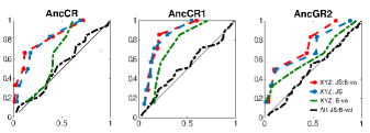
Fig. 5 shows the outcome of the recall analysis for the three proteins in the GR family. In all cases, we observe no visible correlation between mode-coupling scores and the complete set of mutations accompanying each evolutionary step. Focusing on the function changing XYZ-mutations only, we first note an overall positive correlation with B-values (variance of fluctuations around equilibrium for each atom), due to the fact that these mutations are located mostly on the loop regions. It is striking that the mode-coupling based ordering yields better recall values in all three proteins. We furthermore observe that the recall performances improve slightly when mode-coupling scores are divided by the B-values in order to factor out the bias mentioned above. This is the central result of the present work which, together with a similar observation on myosin II Varol et al. (2014), lends support to the thesis that coupling between vibrational modes is a key physical mechanism in protein function.
.6 Robustness wrt data acquisition period
Since most proteins carry out their function in time scales beyond the reach of computer simulations, it is natural to ask how sensitive above results are to the simulation time window. We investigated the robustness of our findings by re-analyzing the data in varying time intervals. To this end, we divided each trajectory into fragments of ns each () and calculated mode-coupling scores by using each fragment separately. We then compared score vectors for each interval pair with identical lengths by measuring the angle between them. This was done by evaluating the dot product of the two vectors after setting their mean to zero (by a
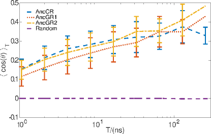
constant shift) and rescaling them to unit length. The mean and the standard devitation of the obtained dot products were recorded separately for each interval length . Results shown in Fig. 6 confirm that the analysis detailed in section .2 yields progressively more consistent results with increasing .
.7 Conclusion
While it is natural from a physical point of view to postulate that nonlinear effects mitigate energy transfer within a proteinPiazza and Sanejouand (2009), precisely how the nonlinearity observed in protein dynamics can be fruitfully exploited to yield biologically relevant predictions is unclear. Even the relevance of nonlinear effects to protein function is far from being universally acknowledged. While part of the literature (such as on discrete breathers Piazza (2014); Luccioli et al. (2011); Juanico et al. (2007); Piazza and Sanejouand (2008)) attests to its importance, there is substantial amount of past and recent work which investigate mechanisms of protein function within a linear (harmonic) framework or at the level of principal component analysis Bahar and Rader (2005); Haliloglu et al. (1997); Levy et al. (1982); Ma (2005); Rodgers et al. (2013); McLeish et al. (2015). By demonstrating that functionally critical mutations along the evolutionary descent which relates three ancestral proteins of the GR family are highlighted in an analysis of the nonlinear contribution to dynamics, the present work emphasizes the significance of nonlinearity, in particular that beyond marginal anharmonicity, to protein function.
The selective power of the mode-coupling analysis for functionally relevant sites (in GR protein family reported here and in myosin II earlier Varol et al. (2014)) is suggestive. However, it is also evident from the data that not all known functional locations come with high mode-coupling scores. Given the complexity of the system and the multitude of factors beyond protein dynamics that play role in functionality, this is only expected. Applying the analysis on carefully constructed toy nonlinear models may help clarify the mechanistic role played by the aminoacids that score high in the present analysis. Such information could be useful for characterizing the proteins on which the current approach may be expected to be successful in future.
.8 Acknowledgements
We thank B. Erman, D. Yuret, O. Keskin and A. Erdoğan for beneficial discussions, C. Atılgan for a critical reading, and S. B. Ozkan for a stimulating seminar which motivated the present work. We acknowledge support of TÜBİTAK through the grant MFAG-113F092.
References
- Kumar et al. [2000] Sandeep Kumar, Buyong Ma, Chung-Jung Tsai, Neeti Sinha, and Ruth Nussinov. Folding and binding cascades: dynamic landscapes and population shifts. Protein Science, 9(1):10–19, 2000.
- Tsai et al. [1999] Chung-Jung Tsai, Buyong Ma, and Ruth Nussinov. Folding and binding cascades: shifts in energy landscapes. Proceedings of the National Academy of Sciences, 96(18):9970–9972, 1999.
- Okazaki and Takada [2008] Kei-ichi Okazaki and Shoji Takada. Dynamic energy landscape view of coupled binding and protein conformational change: induced-fit versus population-shift mechanisms. Proceedings of the National Academy of Sciences, 105(32):11182–11187, 2008.
- Kar et al. [2010] Gozde Kar, Ozlem Keskin, Attila Gursoy, and Ruth Nussinov. Allostery and population shift in drug discovery. Current opinion in pharmacology, 10(6):715–722, 2010.
- Brooks and Karplus [1983] Bernard Brooks and Martin Karplus. Harmonic dynamics of proteins: normal modes and fluctuations in bovine pancreatic trypsin inhibitor. Proceedings of the National Academy of Sciences, 80(21):6571–6575, 1983.
- Haliloglu et al. [1997] Turkan Haliloglu, Ivet Bahar, and Burak Erman. Gaussian dynamics of folded proteins. Physical review letters, 79(16):3090, 1997.
- Yogurtcu et al. [2009] Osman N Yogurtcu, Mert Gur, and Burak Erman. Statistical thermodynamics of residue fluctuations in native proteins. The Journal of chemical physics, 130(9):095103, 2009.
- Ma [2005] Jianpeng Ma. Usefulness and limitations of normal mode analysis in modeling dynamics of biomolecular complexes. Structure, 13(3):373–380, 2005.
- Piazza and Sanejouand [2009] Francesco Piazza and Yves-Henri Sanejouand. Long-range energy transfer in proteins. Physical biology, 6(4):046014, 2009.
- Rodgers et al. [2013] Thomas L Rodgers, Philip D Townsend, David Burnell, Matthew L Jones, Shane A Richards, Tom CB McLeish, Ehmke Pohl, Mark R Wilson, and Martin J Cann. Modulation of global low-frequency motions underlies allosteric regulation: demonstration in crp/fnr family transcription factors. PLoS Biol, 11(9):e1001651, 2013.
- Kabakçıoğlu et al. [2010] A Kabakçıoğlu, D Yuret, M Gur, and B Erman. Anharmonicity, mode-coupling and entropy in a fluctuating native protein. Physical biology, 7(4):046005, 2010.
- Varol et al. [2014] Onur Varol, Deniz Yuret, Burak Erman, and Alkan Kabakçıoğlu. Mode coupling points to functionally important residues in myosin ii. Proteins: Structure, Function, and Bioinformatics, 82(9):1777–1786, 2014.
- Flory and Yoon [1974] PJ Flory and DY Yoon. Moments and distribution functions for polymer chains of finite length. i. theory. The Journal of Chemical Physics, 61(12):5358–5365, 1974.
- Hyvärinen et al. [2004] Aapo Hyvärinen, Juha Karhunen, and Erkki Oja. Independent component analysis, volume 46. John Wiley & Sons, 2004.
- Karplus and McCammon [1983] M Karplus and JA McCammon. Dynamics of proteins: elements and function. Annual review of biochemistry, 52(1):263–300, 1983.
- Levy et al. [1982] Ronald M Levy, David Perahia, and Martin Karplus. Molecular dynamics of an -helical polypeptide: temperature dependence and deviation from harmonic behavior. Proceedings of the National Academy of Sciences, 79(4):1346–1350, 1982.
- Levitt et al. [1985] Michael Levitt, Christian Sander, and Peter S Stern. Protein normal-mode dynamics: trypsin inhibitor, crambin, ribonuclease and lysozyme. Journal of molecular biology, 181(3):423–447, 1985.
- Doruker et al. [2000] Pemra Doruker, Ali Rana Atilgan, and Ivet Bahar. Dynamics of proteins predicted by molecular dynamics simulations and analytical approaches: Application to -amylase inhibitor. Proteins: Structure, Function, and Bioinformatics, 40(3):512–524, 2000.
- Manning and Schütze [1999] Christopher D Manning and Hinrich Schütze. Foundations of statistical natural language processing. MIT press, 1999.
- Kino et al. [2009] Tomoshige Kino, Irini Manoli, Sujata Kelkar, Yonghong Wang, Yan A Su, and George P Chrousos. Glucocorticoid receptor (gr) has intrinsic, gr-independent transcriptional activity. Biochemical and biophysical research communications, 381(4):671–675, 2009.
- Nicolaides et al. [2010] Nicolas C Nicolaides, Zoi Galata, Tomoshige Kino, George P Chrousos, and Evangelia Charmandari. The human glucocorticoid receptor: molecular basis of biologic function. Steroids, 75(1):1–12, 2010.
- Chrousos [2004] George P Chrousos. The glucocorticoid receptor gene, longevity, and the complex disorders of western societies. The American journal of medicine, 117(3):204–207, 2004.
- Clark JK [1992] O’Malley BW Clark JK, Schrader WT. Mechanism of steroid hormones, pages 35–90. Philedelphia: WB Sanders Co., 1992.
- Zhou and Cidlowski [2005] Junguo Zhou and John A Cidlowski. The human glucocorticoid receptor: one gene, multiple proteins and diverse responses. Steroids, 70(5):407–417, 2005.
- Barnes [1998] Peter J Barnes. Anti-inflammatory actions of glucocorticoids: molecular mechanisms. Clinical science, 94(6):557–572, 1998.
- Sapolsky et al. [2000] Robert M Sapolsky, L Michael Romero, and Allan U Munck. How do glucocorticoids influence stress responses? integrating permissive, suppressive, stimulatory, and preparative actions 1. Endocrine reviews, 21(1):55–89, 2000.
- Carson et al. [2014] Matthew W Carson, John G Luz, Chen Suen, Chahrzad Montrose, Richard Zink, Xiaoping Ruan, Christine Cheng, Harlan Cole, Mary D Adrian, Dan T Kohlman, et al. Glucocorticoid receptor modulators informed by crystallography lead to a new rationale for receptor selectivity, function, and implications for structure-based design. Journal of medicinal chemistry, 57(3):849–860, 2014.
- Li et al. [2005] Yong Li, Kelly Suino, Jennifer Daugherty, and H Eric Xu. Structural and biochemical mechanisms for the specificity of hormone binding and coactivator assembly by mineralocorticoid receptor. Molecular cell, 19(3):367–380, 2005.
- Bentley [1998] Peter John Bentley. Comparative vertebrate endocrinology. Cambridge University Press, 1998.
- Yang and Young [2009] Jun Yang and Morag J Young. The mineralocorticoid receptor and its coregulators. Journal of molecular endocrinology, 43(2):53–64, 2009.
- Bridgham et al. [2009] Jamie T Bridgham, Eric A Ortlund, and Joseph W Thornton. An epistatic ratchet constrains the direction of glucocorticoid receptor evolution. Nature, 461(7263):515–519, 2009.
- Ortlund et al. [2007] Eric A Ortlund, Jamie T Bridgham, Matthew R Redinbo, and Joseph W Thornton. Crystal structure of an ancient protein: evolution by conformational epistasis. Science, 317(5844):1544–1548, 2007.
- Harms and Thornton [2014] Michael J Harms and Joseph W Thornton. Historical contingency and its biophysical basis in glucocorticoid receptor evolution. Nature, 512(7513):203–207, 2014.
- Glembo et al. [2012] Tyler J Glembo, Daniel W Farrell, Z Nevin Gerek, MF Thorpe, and S Banu Ozkan. Collective dynamics differentiates functional divergence in protein evolution. PLoS computational biology, 8(3):e1002428, 2012.
- Case et al. [2014] DA Case, V Babin, Josh Berryman, RM Betz, Q Cai, DS Cerutti, TE Cheatham Iii, TA Darden, RE Duke, H Gohlke, et al. Amber 14. 2014.
- Cornell et al. [1995] Wendy D Cornell, Piotr Cieplak, Christopher I Bayly, Ian R Gould, Kenneth M Merz, David M Ferguson, David C Spellmeyer, Thomas Fox, James W Caldwell, and Peter A Kollman. A second generation force field for the simulation of proteins, nucleic acids, and organic molecules. Journal of the American Chemical Society, 117(19):5179–5197, 1995.
- Wang et al. [2004] Junmei Wang, Romain M Wolf, James W Caldwell, Peter A Kollman, and David A Case. Development and testing of a general amber force field. Journal of computational chemistry, 25(9):1157–1174, 2004.
- Wang et al. [2006] Junmei Wang, Wei Wang, Peter A Kollman, and David A Case. Automatic atom type and bond type perception in molecular mechanical calculations. Journal of molecular graphics and modelling, 25(2):247–260, 2006.
- Roh et al. [2005] JH Roh, VN Novikov, RB Gregory, JE Curtis, Z Chowdhuri, and AP Sokolov. Onsets of anharmonicity in protein dynamics. Physical review letters, 95(3):038101, 2005.
- Hayward et al. [1995] Steven Hayward, Akio Kitao, and Nobuhiro Gō. Harmonicity and anharmonicity in protein dynamics: a normal mode analysis and principal component analysis. Proteins: Structure, Function, and Bioinformatics, 23(2):177–186, 1995.
- Piazza [2014] Francesco Piazza. Nonlinear excitations match correlated motions unveiled by nmr in proteins: a new perspective on allosteric cross-talk. Physical biology, 11(3):036003, 2014.
- Luccioli et al. [2011] Stefano Luccioli, Alberto Imparato, Stefano Lepri, Francesco Piazza, and Alessandro Torcini. Discrete breathers in a realistic coarse-grained model of proteins. Physical biology, 8(4):046008, 2011.
- Juanico et al. [2007] Brice Juanico, Y-H Sanejouand, Francesco Piazza, and Paolo De Los Rios. Discrete breathers in nonlinear network models of proteins. Physical review letters, 99(23):238104, 2007.
- Piazza and Sanejouand [2008] Francesco Piazza and Yves-Henri Sanejouand. Discrete breathers in protein structures. Physical biology, 5(2):026001, 2008.
- Bahar and Rader [2005] Ivet Bahar and AJ Rader. Coarse-grained normal mode analysis in structural biology. Current opinion in structural biology, 15(5):586–592, 2005.
- McLeish et al. [2015] Tom CB McLeish, Martin J Cann, and Thomas L Rodgers. Dynamic transmission of protein allostery without structural change: Spatial pathways or global modes? Biophysical journal, 2015.