IR-Laser Welding and Ablation of Biotissue Stained with Metal Nanoparticles
Abstract
In the present work we have studied the possibility of laser welding and ablation of biological tissue by the using of spherical metal nanoparticles (NPs) and infrared laser irradiation which spectrally located far from plasmon resonances. YAG:Nd laser with 1064 nm wavelength, 8ns pulse duration, and operating in transverse electromagnetic modes TEM00 was used for the synthesis of metal NPs. The Au,Ti Ni and Cu as well as Au-Ag and Au-Cu hybrid metal NPs were formed in the liquid medium. Effectiveness of laser ablation in the case of the biotissue sample that stained with the metal NPs was approximately on 4-5 times larger than for the native sample. Also the scheme of a laser point welding for the deep-located biotissue layer selectively stained by the metal NPs has been demonstrated.
pacs:
79.20.Eb, 81.20.Vj, 78.67.BfKeywords: nanoparticles, biotissue, laser, ablation, welding
1 Introduction
Nanoparticles (NPs) are homogeneous and composite materials with size in a range of 1-100 nm which exhibit some unexpected optical, electromagnetic, chemical and mechanical properties not inherent to bulk materials and atomic-molecular structures due to quantum confined nature of their energy levels and surface area to volume ratio. The unique properties of NPs depend on their size, morphology, composition, uniformity, and agglomeration. These extraordinary qualities make superior the application of NPs in nanophotonics, biomedicine, environment science, engineering etc [1]-[5]. Especially, in medical applications the NPs are able to pass through biological filters inside of organism by that deliver drugs. Also, nanomaterials are used for protein detection, fluorescent biological labels, tumor destruction, tissue engineering etc. Various physical and chemical processes are currently widely used for the production of NPs, nevertheless we focus on the laser synthesis of colloidal spherical NPs by the laser ablation in liquid media [6]-[9]. This method favorably differs from other methods in its simplicity, efficiency, and it does not harm the environment. Also, by this method we can obtain nanoparticles of different sizes varying in the wide range and strongly depending on the details of experimental setup. In work the [6] we have reported on laser synthesis of GaAs and CdSe semiconductor NPs with picosecond Nd:YAG laser. The considerable blue shift of the photoluminescence was observed when picosecond laser beam with transverse electromagnetic modes TEM00 structure has been used. Particle sizes was estimated (2-3 nm) from the spectral data and the bandwidth of the luminescence spectra of about 40 nm reveals a narrow size distribution of the produced quantum dots which was achieved without any size selection methods. Such ultra fine sizes of NPs are excellently meet with the requirements for medical applications. In recent years the use of nanoparticles in biomedicine for the early detection, accurate diagnosis and treatment of cancer is extensively studied. Particularly, the possibilities for cancer photothermal therapy due to tissue heating by using metal NPs have been successfully demonstrated [10, 11, 12]. This method is based on localized surface plasmon resonance (LSPR), when nanoscale localization and amplification of electromagnetic fields occur in the vicinity of metal nanoparticles [13]. The locally amplified optical fields generate localized thermal energy due to the rapid conversion of photon energy into heat via electron–electron scattering and electron–phonon coupling [14] in the wavelengths of the plasmon resonance [15]-[18]. The plasmon resonances for gold and silver nanospheres are in the green-blue range of the visible spectrum, which can be red-shifted into the near infrared if nanoparticles shape is modified to nanorods or nanoshels. This is more practical as biological tissues are relatively transparent to near infrared light. However the strong tuning to plasmon resonance wavelength is required in medical applications of photoheating that in most cases is not provided by commercial medical laser devises.
2 Experimental results
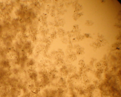
For syntheses of metal NPs in the considered experiment, the YAG:Nd laser with 1064nm wavelength, 8 ps pulse duration, repetition rate of 10Hz and operating in transverse electromagnetic modes TEM00 was used. The same scheme of laser synthesis described in the work [6] has been used. The laser beam was focused on the surface of a bulk metal target allocated in the glass cuvette with distilled water. Exposition of the laser irradiation was two hours. The Au, Ti, Ni and Cu, as well as Au-Ag and Au-Cu hybrid spherical metal NPs were formed in the liquid medium. Laser welding and ablation of biotissue stained with the metal nanoparticles has been studied on samples of chicken skin. Figure (1) shows typical photographical picture (taken by an optical microscope) of tissue stained with the metal NPs.
In the present work we have studied the possibility of tissue heating by using infrared laser irradiation at 1064 nm wavelengths. This wavelength is located far from plasmon resonances of known spherical metal NPs. Particularly, silver and gold nanoparticles exhibit strong plasmon resonance at wavelengths 405 nm and 520 nm correspondingly. The absorption properties of colloidal solutions of Ag, Au and Cu nanoparticles that were synthesized by method of laser ablation in a liquid has been measured (see Figure (2)). Obtained data are consistent with the previous publications on plasmon resonances of widely used Ag, Au spherical NPs with the sizes of about 50 nm.
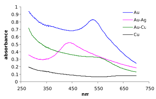
As we can see in Figure (2), the plasmon resonance of Cu nanoparticles in comparison with other considered metal NPs is located in the red range of the optical spectrum, however, its value is rather smaller. The continuous wave YAG:Nd laser with an output beam of 3mm diameter and power up to 4W was used for the biotissue welding and ablation. Two areas of sample’s surface - unstained and stained with metal nanoparticles were ablated at the same dose of the laser irradiation. Figure (3) shows the picture of skin surface after ablation procedure.
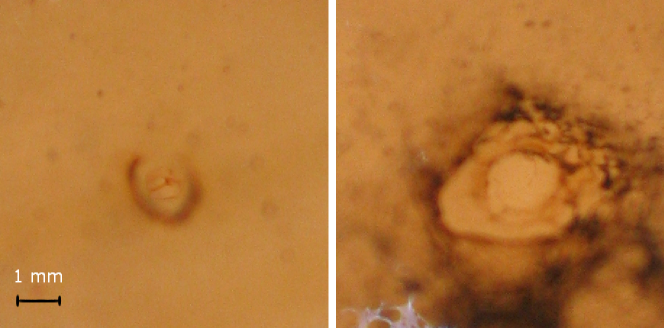
The surface of the area which has been exposed to laser ablation in the case of the tissue sample that colored with silver NPs is approximately on 4-5 times larger than that for the sample with non-colored area. Furthermore, the same result was obtained in case of several other metal nanoparticles with different wavelength of plasmon resonances, as well as with Ni and Ti nanoparticles which does not exhibit any plasmon resonance properties. Thus, we observe significant difference in photodamage for biotissue samples which are unstained or stained with the metal nanoparticles. Such difference in absorption of infrared radiation may be explained by the presence of the metal nanoparticles clusters in the tissue samples. Indeed, as we can see in the Figure (1), the coloration of biotissue is highly nonhomogeneous and one can observe the number of cluster centers colored in intense black. In the book [19] there are given experimental and numerical analysis of optical characteristics of fractal aggregates of nanoparticles. As it is shown, the absorption spectra for clusters with large amount of nanoparticles (500-10000) are shifted toward infrared range. This result demonstrates that the application of metal NPs in laser surgical procedures will lead to significant reduction of an irradiation dose even in cases when plasmonic resonance is absent or the wavelength of laser radiation is far from it. Furthermore the application of nanoparticles in laser surgery allows effectively use IR laser light that penetrates deeply in the tissue and absorbed poorly by the native tissue itself. However, this radiation may be absorbed strongly by the nanoparticles aggregates. This advantage can be used for development of the schemes of laser welding for deep-located biotissue areas selectively colored by the metal NPs. For example, we performed the point welding of two chicken skin samples with thickness of 2 mm each (see Figure (4)).

The surfaces of the samples were colored with the silver nanoparticles. The laser radiation penetrates through the first sample from the noncolored side without production of the visible damages of tissue and reaches the contacting area of samples that contain nanoparticles. In this case we observe the strong absorption of light in the area of biotissue colored with the nanoparticles and, subsequently, the local heating of the tissue. In the result, the local spot welding of two layers of the skin tissue was realized. Note that in case of the using of radiations of 405 nm or 520 nm wavelengths which are in the range of Ag and Au plasmon resonances, we would have strong absorbance by the tissue of 2 mm thickness, which will lead to the photodamage of the superficial layers. Furthermore, the application of Ag nanoparticles that exhibit strong microbicidal effect may lead to significant improvement of the postsurgical healing of the patients. It is also important as tissue photoheating could cause inflammatory process due to local antimicrobial immune resistance reduction, and this negative effect could be also minimized.
3 Acknowledgments
We would like to thank Prof. H. K. Avetissian for valuable discussions. This work was supported by State Committee of Science of RA.
References
References
- [1] Alivisatos A P 1996 J. Phys. Chem. 100 13226 –13239.
- [2] Salata O V 2004 J. Nanobiotechnology 2 3.
- [3] Jong W H Borm P J 2008 Int. J. Nanomedicine 3(2) 133-149.
- [4] Liu W T 2006 J. Bioscience and Bioengineering 102(1) 1-7.
- [5] Haider A Kang I-K 2015 Adv. Materials Science and Engineering 2015 165257.
- [6] Lalayan A 2005 Laser Phys. Lett. 2 12-15.
- [7] Svrcek V et al 2007 JLMN-Journal of Laser Micro/Nanoengineering 2 15-20.
- [8] Ganeev R A Baba M Ryasnyansky A I Suzuki M Kuroda H 2005 Appl. Phys. B 80 595–601.
- [9] Mafune F Kohno J Takeda Y Kondow T Sawabe H 2000 J. Phys. Chem. B104 9111.
- [10] Loo C et al. 2004 Technol. Cancer Res. Treat. 3 33-40.
- [11] Hirsch L R et al. 2003 Proc. Natl. Acad. Sci. U. S. A.100 13549–13554.
- [12] O’Neal D P et al. 2004 Cancer Lett. 209 171–176.
- [13] Kreibig U Vollmer M 1995 Optical Properties of Metal Clusters (Springer-Verlag) 436.
- [14] Seol Y Carpenter A E Perkins T T 2006 Opt. Lett. 31(16) 2429–2431.
- [15] Murray W A Barnes W L 2007 Adv. Mater. 19(22) 3771–3782.
- [16] Kelly K L Coronado E Zhao L L Schatz G C 2003 J. Phys. Chem. B. 107(3) 668–677.
- [17] Jain P K Huang X El-Sayed I H El-Sayad M A 2007 Plasmonics 2(3) 107–118.
- [18] Maier S A 2007 Plasmonics: Fundamental and Applications (Springer Science) 223.
- [19] Shalaev V M 2000 Nonlinear Optics of Random Media: Fractal Composites and Metal-Dielectric Films (Springer) 158.