High Resolution Angle Resolved Photoemission with Tabletop 11eV Laser
Abstract
We developed a table-top vacuum ultraviolet (VUV) laser with nm wavelength (10.897eV) and demonstrated its viability as a photon source for high resolution angle-resolved photoemission spectroscopy (ARPES). This sub-nanosecond pulsed VUV laser operates at a repetition rate of 10 MHz, provides a flux of 21012 photons/second, and enables photoemission with energy and momentum resolutions better than 2 meV and 0.012 Å-1, respectively. Space-charge induced energy shifts and spectral broadenings can be reduced below 2 meV. The setup reaches electron momenta up to 1.2 Å-1, granting full access to the first Brillouin zone of most materials. Control over the linear polarization, repetition rate, and photon flux of the VUV source facilitates ARPES investigations of a broad range of quantum materials, bridging the application gap between contemporary low energy laser-based ARPES and synchrotron-based ARPES. We describe the principles and operational characteristics of this source, and showcase its performance for rare earth metal tritellurides, high temperature cuprate superconductors and iron-based superconductors.
I INTRODUCTION
Angle-resolved photoemission spectroscopy (ARPES) directly accesses electronic band structures and electronic self-energies in a momentum-resolved manner, which makes it a powerful and unique technique in condensed matter research. This momentum resolution allows one to distinguish multiple electronic band dispersions in multiband systems, to assess the symmetry of order parameters, and to characterize electronic anisotropy in solid state systems. Recent progress in the ARPES technique has pushed energy resolutions down to meV scales, which gives unprecedented access to low energy excitations in quantum materials. With its unique combination of energy and momentum resolution, ARPES contributes significantly to the understanding of many two-dimensional quantum materials Ohta et al. (2006); Zhou et al. (2007); Johnson (2002); Kordyuk (2014), in particular high Tc superconducting cuprates Damascelli, Hussain, and Shen (2003); Ding et al. (1996), iron based superconductors Stewart (2011); Lu et al. (2012); Lee et al. (2014) and topological states of matter Hsieh et al. (2008); Chen et al. (2009).
ARPES collects photoemitted electrons as a function of kinetic energy and emission angle with respect to the sample surface normal. The conservation of energy and in-plane momentum of each electron111In general, the out-of-plane momentum is not conserved as the electron overcomes the surface potential barrier during the photoemission process. allows the calculation of the electron’s binding energy with respect to the Fermi level and its parallel momentum :Damascelli, Hussain, and Shen (2003)
| (1) |
| (2) |
Here, denotes the work function of the material and the photon energy. Eq. (1) describes how larger photon energies provide access to states with higher binding energy. Eq. (2) shows that the accessible electron momenta are limited by the electron kinetic energy, which scales monotonically with the photon energy. For typical values of = 50∘ and = 4.5 eV, the minimum photon energy required to capture the entire first Brillouin zone (BZ) of a material with 3.5 Å lattice constant is = 9.7 eV.222 = 35.0∘ (maximum sample surface rotation) + 15.0∘ (typical detector acceptance angle)
The relation between photon energy and parallel momentum resolution is given by
| (3) |
As indicated in Eq. (2-3), for a given detector angular resolution and momentum feature, higher photon energies degrade the momentum resolution due to increasing and decreasing .
When employing pulsed photon sources Coulomb repulsion of electrons photoemitted by a single pulse can lead to space charging effects, which both shift and broaden the spectrum.Graf et al. (2010); Passlack et al. (2006) The effects of space charging can be mitigated by higher repetition rates while maintaining a constant photon flux. Therefore to achieve ultimate resolution and applicability, a light source optimized for high-resolution ARPES studies of complex materials should satisfy several requirements.
- 1.
-
2.
Sufficiently low photon energy ( 20 eV) to facilitate high momentum resolution (Eq. (3))
-
3.
Sufficiently high repetition rate ( 100 kHz) to limit the loss of energy and momentum resolutions due to space-charge effects while maintaining sufficiently high signal-to-noise ratio
-
4.
Sub-meV bandwidth, high flux and long term stability
-
5.
Variable polarization for control over photoemission matrix elements
-
6.
Additional requirements may include a small ( 1 mm) beam spot and short ( 100 ps) pulse duration for time-of-flight detection schemes
While it is challenging to integrate all these properties into one single light source, existing light sources successfully capture different aspects of the requirements listed above (TABLE I). Noble gas discharge lamps, helium and xenon in particular, were among the earliest photoemission light sources with photon energies in the range of 8.4 - 41 eV.Harter et al. (2012); Zhang (2012); Souma et al. (2007); VGV (2012) These pioneering experiments established ARPES as a valuable tool for the analysis of electronic structures in solids.
Continuous advances of synchrotron technology provided bright and tunable light sources that were pivotal to the tremendous success of ARPES in the last two decades. Comparing with gas discharge lamps, synchrotron-based ARPES features small ( 0.5 mm) beam spots which improve momentum resolution, and avoids reduced sample lifetimes due to gas molecules effusing from the light source.Harter et al. (2012); Zhang (2012) Additionally, third generation synchrotron sources feature low photon energies in combination with excellent energy-momentum resolution.Strocov et al. (2010, 2014) Despite the outstanding capabilities of synchrotron-based ARPES the demand for accessible, table-top ultraviolet light sources with higher photon flux and improved energy stability remains high.
Laser-based ARPES commonly utilizes non-linear optical crystals to up-convert infrared light pulses into the UV spectral region.Baichang Wu and Chen (1996) The generated low photon energies of 6-7 eV result in the excellent energy and momentum resolution required for studies of detailed band dispersions and subtle low-energy excitations.Kiss et al. (2008); Kirchmann et al. (2008); Jiang et al. (2014); Damm et al. (2015) Yet these low photon energies limit the access to high momenta and valence bands.
Although highly desirable, frequency up-conversion to sub-170 nm wavelengths is not feasible in nonlinear optical crystals due to the re-absorption in the vacuum UV (VUV) range in any material. Instead, these short wavelengths can be generated via high-harmonic generation in polarizable gases. Tjernberg and coworkers built a 10.5 eV light source dedicated to ARPES by frequency tripling the third harmonic of a pulsed IR laser in Xe gas,Berntsen, Goetberg, and Tjernberg (2011) which, when integrated with a time-of-flight detector, for the first time made laser-based photoemission study possible at many materials’ BZ boundaries. Such non-resonant XUV generation schemes usually require MW peak power, which in turn typically limit the laser repetition rate to 1 MHz.Berntsen, Goetberg, and Tjernberg (2011) The capabilities of commonly used light sources for ARPES are summarized in TABLE I.
| \pbox20cmE | |||||||
|---|---|---|---|---|---|---|---|
| (meV) | \pbox20cmspot FWHM | ||||||
| (mm) | \pbox20mm max k∥ | ||||||
| (Å-1) | \pbox20cm max BE | ||||||
| (eV) | \pbox20cm polarization | ||||||
| control | \pbox20cmrep rate | ||||||
| (MHz) | \pbox20cm photon flux | ||||||
| (photons/second) | |||||||
| 5.8 - 8eV laserKoralek et al. (2007); Kiss et al. (2008, 2005) | 2 | 0.1 | 0.75 | 2 | Yes | 102 | 1014 |
| He/Xe discharge lampHarter et al. (2012); Zhang (2012); Souma et al. (2007) | 10 | 1 | \pbox20cm 0.82 | ||||
| 1.8 | |||||||
| 2.7 | \pbox20cm 3 (XeI) | ||||||
| 16 (HeI) | |||||||
| 36 (HeII) | No | CW | 1013 | ||||
| SynchrotronBL (5, 4); BES | 1 | 0.3 | multiple BZ | \pbox20cmup to | |||
| core level | |||||||
| (keV) | Limited | 500 | 1012 | ||||
| 11eV laser | 2 | 0.5 | 1.2 | 6 | Yes | 120 | 1013 |
In this article, we describe a table-top pulsed VUV light source optimized for high resolution ARPES studies of quantum materials. Its photon energy of 11 eV is sufficient to cover the complete BZ in most materials, while maintaining exceptional energy and momentum resolution at high photon flux. We demonstrate its capabilities for high resolution ARPES on a number of materials, including antimony, rare earth metal tritelluride, unconventional copper and iron-based high temperature superconductors and single unit cell FeSe film.
II SYSTEM OVERVIEW
II.1 Vacuum-Ultraviolet (VUV) Laser Light Source

Our coherent VUV light source utilizes three cascaded stages of nonlinear frequency conversion of a quasi-continuous wave pulsed, 1024 nm infrared (IR) solid-state laser. As shown schematically in Fig. 1, the first two conversion stages occur in birefringent nonlinear crystals; VUV flux is generated via two-photon resonant, sum-frequency generation in xenon gas using the fundamental () and fourth-harmonic (4) of this laser system.
The VUV light source is driven by a fiber-coupled wavelength-tunable (1024 nm) IR seed source with variable repetition rate and sub-nanosecond pulse duration. Its output is amplified by a Yb-doped fiber amplifier to an average power of 10 W, and a peak power of 10 kW. The amplified IR light is up-converted to the second harmonic (512 nm) using a first second harmonics crystal (SHG1) with 50% efficiency. The second harmonic light is separated from the fundamental using a dichroic beam splitter (BS) and refocused into a second nonlinear crystal (SHG2) to generate UV light (256 nm). By controlling the polarization of the 512 nm beam, the output power of the frequency quadrupled UV light can be continuously modulated. This attenuation method provides VUV (114 nm) flux control, without change to the alignment or beam profile.
Following the beam splitter, the polarization of the residual IR beam is controlled by a second half-wave plate (HWP) to achieve any given VUV beam polarization. Then the IR and UV beams are overlapped in time and space using dichroic dielectric-coated beam-combiner mirrors, and focused by a single lens into a xenon-filled gas cell. The energy-level diagram of the specific nonlinear process in atomic xenon, and the control of the final VUV polarization, are shown in Fig. 2. Two UV photons (4) with wavelengths of 256.015 nm resonantly drive the dipole forbidden - transition.Moore (1958); Gornik et al. (1981); Plimmer et al. (1989) By mixing this local atomic oscillator with a fundamental IR photon (), light at the 9th harmonic (9) is generated (10.897 eV, or 113.778 nm). By driving the two-photon transition with linearly-polarized UV light, the polarization of the VUV photon is determined by the optical polarization of the IR photon.

Traditional gas-phase nonlinear optical systems have typically required MW-scale peak-power driving lasers for efficient conversion, thereby severely limiting the achievable repetition rates. However, by operating at a two-photon resonance condition, the peak optical powers required for efficient up-conversion are reduced to the kW level, which is a necessary condition for increasing the repetition rate of the source to the MHz range,Berntsen, Goetberg, and Tjernberg (2011) and for reducing the overall source size to a table-top device.
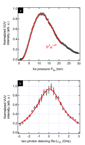
Atomic xenon is negatively dispersive for degenerate two-photon-resonant sum-frequency generation, which enables a tightly-focussed geometry for VUV generation.Vidal (1980) Fig. 3(a) demonstrates typical variation of generated VUV flux on the xenon gas pressure. The single-peaked curve, with a maximum at approximately 12 torr xenon pressure, is typical of tightly-focussed sum-frequency generation.Bjorklund (1975)
Due to the intermediate resonance step in the frequency-conversion process, the generated VUV flux is particularly sensitive to the absolute frequency of the UV beam. As shown in Fig. 3(b) for the 1 MHz repetition rate, 1 ns pulsewidth laser setup, detuning of the UV wave from the two-photon resonance results in a rapid reduction in VUV generation efficiency.Stappaerts et al. (1976) The width of this resonance profile is set by the convolution of the laser linewidth and the natural linewidth (including isotopic and Doppler broadening) of the two-photon transition. In order to maintain peak flux, the central frequency of the UV beam must be maintained to within 1 GHz; the IR frequency must therefore be stabilized to the central frequency within 250 MHz uncertainty. In comparison, the total width of the resonance curve for the case of the broader-bandwidth excitation laser pulses produced in the 10 MHz, 100 ps laser configuration reaches 8 GHz. This provides a lower bound on both the energy resolution and the absolute energy stability of the 11 eV laser of 30 eV.
II.2 VUV DISPERSION AND FOCUSING
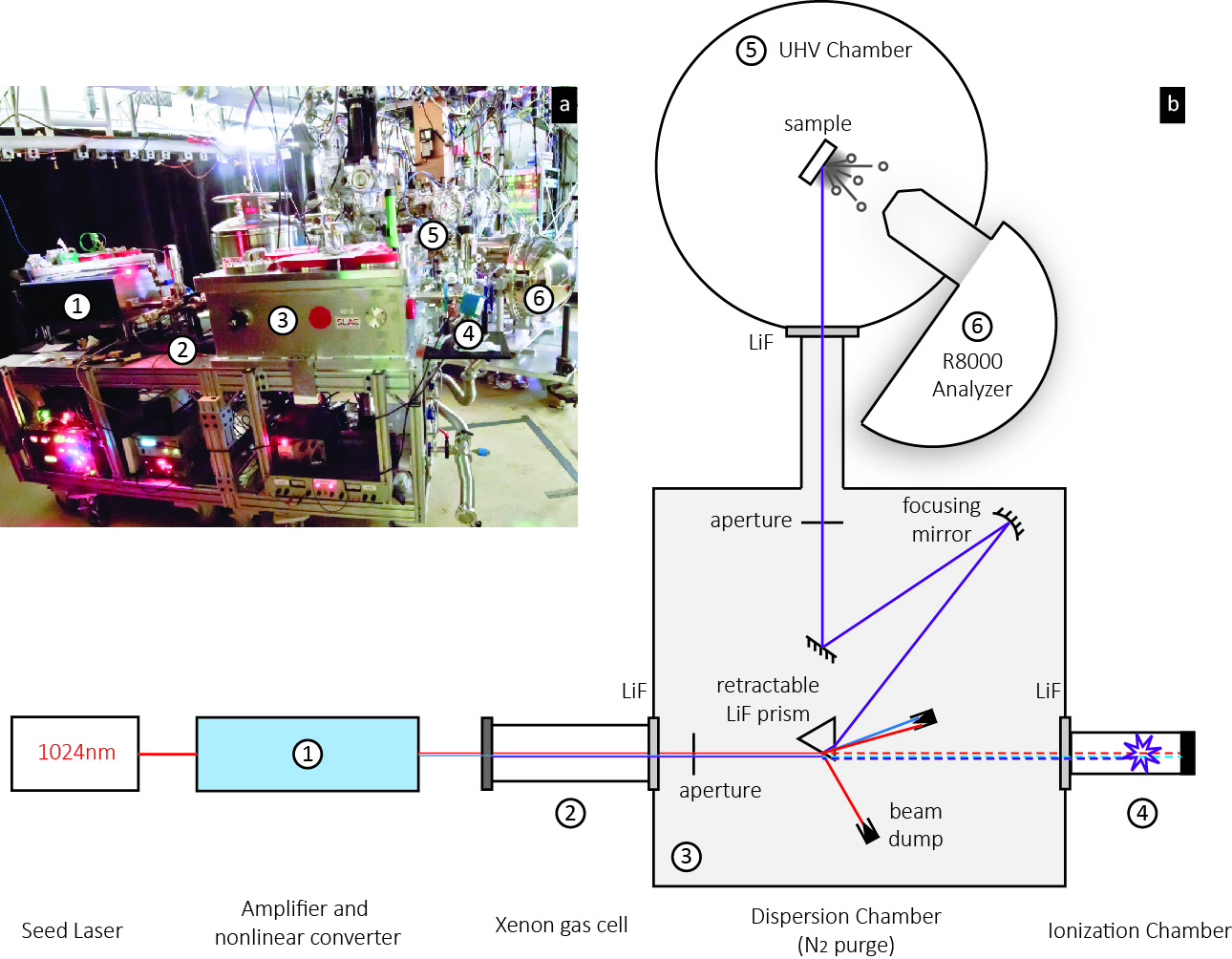
VUV light is absorbed strongly by several atmospheric constituents, most notably oxygen and water vapor.Krupenie (1972); Tennyson et al. (2009) The 1/e attenuation length for 11 eV photons in air is approximately 3 mm.Watanabe, Inn, and Zelikoff (1953) Therefore, the entire beam path for the 11 eV light must be either evacuated or purged to remove the absorbing species.
The xenon-filled gas converter is connected directly to a N2-purged dispersion chamber using an uncoated LiF window. The 11 eV light is generated inside the xenon gas cell as a nearly-diffraction-limited beam that propagates collinearly with the driving IR and UV beams. The IR and UV photons have energies of 1.21 eV and 4.84 eV, respectively, and Watt-level average powers. Illumination of the sample with these IR and UV beams can cause heating and photoemission and must be avoided. We employ an equilateral LiF prism to refractively disperse the 11 eV flux from the lower-energy photons prior to photoemission. The prism is operated in a minimum-deviation condition, so that the incidence angle is near the Brewster angle for the VUV light, thus minimizing reflective losses for horizontal-linearly polarized 11 eV flux.Laporte and Subtil (1982)
Under normal photoemission measurement conditions, the prism is positioned to refract the co-propagating beams; both the front-surface reflections, and the refracted IR and UV beams, are trapped by beam dumps to minimize scattered light. In order to measure the 11 eV source power, the prism may be automatically retracted to allow the three laser beams to propagate to the opposite port of the dispersion chamber (dashed line in Fig. 4(b)). The photocurrent produced by an ionization chamber mounted to this port (Lumeras model IC-LF-C, with a LiF entrance window and filled with isopropanol vapor) provides solar-blind measurements of the 11 eV flux.
The 11 eV light is then focused onto the ARPES sample using a single concave reflective MgF2-overcoated aluminum mirror. This mirror images the laser waists in the xenon converter onto the ARPES sample with a magnification factor of approximately 3. The 11 eV photons propagate through a second, uncoated 2¬mm-thick LiF window into the UHV chamber. Photoelectrons are collected by a Scienta R8000 hemispherical electron analyzer.
III SYSTEM CHARACTERIZATION AND BENCHMARK
III.1 Ultimate resolution
Fig. 5(a) shows the momentum-integrated spectrum of an evaporated polycrystalline gold film. The angle integrated Energy distribution curves (EDC) was fit with a Fermi-Dirac function convolved with a Gaussian instrument energy-resolution function (red lines), giving 0.9 meV as an upper bound of the experimental energy resolution for the raw spectrum (open markers). A similar analysis was carried out after correcting the spectrum for detector nonlinearity (solid markers). The correction gives an experimental resolution of 2.2 meV. The origin and correction procedure of the detector nonlinear effect were detailed in Ref[40]. As shown in Fig. 5(b), by continuously varying the laser power, the detector count rate was measured as a function of total photoemission current over a large photon flux range. To remove the nonlinearity, detector count rates were mapped to the corresponding photoemission current (incident laser power) for every pixel.Reber et al. (2014)
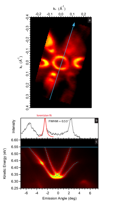
Antimony is known to host a metallic surface state on the (111) surface,Sugawara et al. (2006) which we can use to estimate the upper bound of the setup’s momentum resolution as shown in Fig. 6. A bulk Sb crystal is cleaved in-vacuo on the (111) surface with Fig. 6(c) showing the band dispersion along the -M cut. The angular distribution curve of the surface state from inner pocket at reaches a FWHM of 0.53∘, or 0.012 Å-1 when converted to parallel momentum. It should be noted that this is a combined effect from both instrument resolution and intrinsic sample-dependent electron scattering at . In comparison, high quality measurements on optimally doped Bi-2212 can yield 0.37∘ angular or 0.0039 Å-1 momentum FWHM at with 7 eV laser-ARPES.Ishizaka et al. (2008)
III.2 Space charging
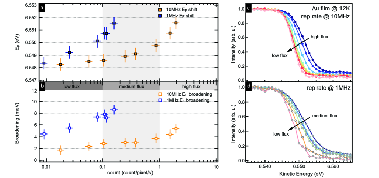
We employ two systems with comparable flux but different repetition rates to calibrate the system’s space-charging effect. For our control experiment setup, at 1 MHz repetition rate, 500 ps pulse duration and photons/second, space charging caused the measured Fermi level (on evaporated polycrystalline gold) to shift by 6 meV.
In order to mitigate the space charging induced spectral changes, we reduce the pulse duration to 100 ps while maintaining the peak pulse intensity of the IR beam, which is crucial to preserve high conversion efficiency of VUV photon generation.Hellmann et al. (2009) The repetition rate is boosted up to 10 MHz to compensate for the reduced photon count within every single pulse comparing to 1 MHz setup.
Fig. 7 shows the photon-intensity dependence of space charging for both the 1 MHz and the 10 MHz setup. The severity of space charging is quantified both by the shift in (Fig. 7(a)) as well as the broadening of the Fermi edge (Fig. 7(b)). The broadening is defined as the fitted instrument resolution as is adopted in Fig. 5(a). Electron count/pixel/second from the Scienta R8000 detector camera is used as a measure of beam intensity. We find that up to 1012 photons/sec or 1 electron count/pixel/sec, space charging can be limited to 2 meV shift and 4 meV broadening for the 10 MHz configuration. In the control experiment (blue markers), the 1 MHz setup gives worse space charging with a decade lower photon flux. In typical high resolution ARPES experiment operating at 0.1-1 count/pixel/sec, space charging effects are almost absent in the 10 MHz setup.
IV MATERIAL SYSTEM APPLICATIONS
IV.1 Rare earth metal tritelluride
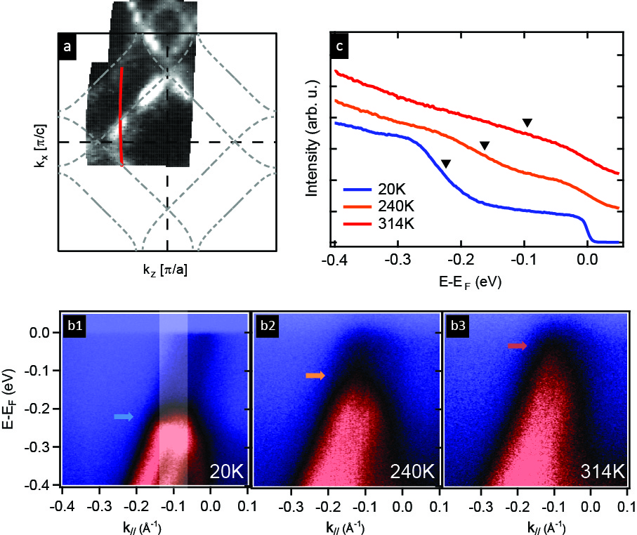
Rare earth metal tritellurides have been a model system to study charge density wave (CDW) formation, where a large CDW gap extends over the entire BZ.Moore et al. (2010); Brouet et al. (2004) 11 eV photons provide access to the entire first folded BZ (Fig. 8(a)). When the system is warmed up from 20K to 314K, the CDW order weakens. This manifests as a shrinking CDW gap below (b1-b3), which is observed in our experiment as an uplifting gap edge in the integrated EDC (c).
IV.2 High temperature cuprate superconductor
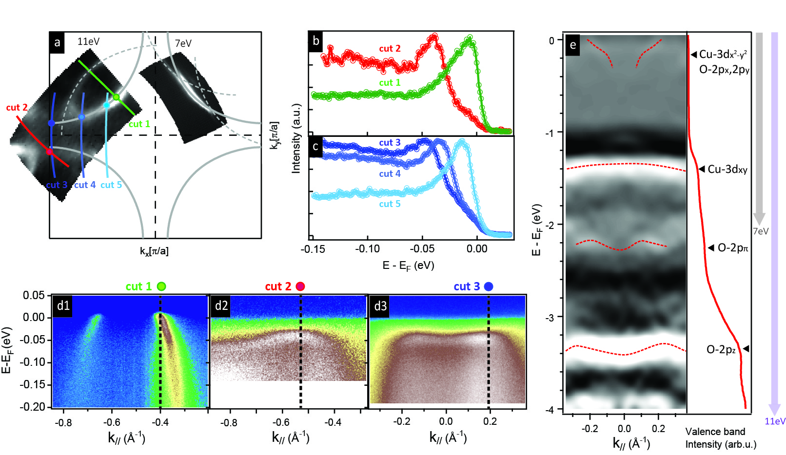
We then apply the 11 eV laser-ARPES to cuprate high temperature superconductors, namely bilayer Pb-doped Bi2Sr2CaCu2O8+δ with a Tc of 80K (overdoped). Previous laser-based ARPES studies at both 6 eV and 7 eV photon excitation energies have provided tremendous insights towards near-nodal excitation, yet the antinodal region near the BZ boundary was not accessible with these low photon energies.Peng et al. (2013); Parham et al. (2013); Kondo et al. (2011); Vishik et al. (2012) Fig. 9(a) compares the Fermi surface in the superconducting state obtained by 11 eV (upper left quadrant) and 7 eV (upper right quadrant) photoemission. The grey dashed lines are superstructures related to Bi-O sublattice distortion.Levin, Smolin, and Shepelev (1994); Shan et al. (2003)
With 11 eV photons, the entire BZ can be mapped with high energy and momentum resolution. Notably, the 11 eV system can measure an antinodal (near (,0)) spectrum (Fig. 9(d2-d3)), whereas measurements with 7 eV laser can barely reach the antinode and have poor cross section there.Hashimoto et al. (2012) EDCs with clear quasiparticle peaks from node to antinode are shown in panel (b) and (c) with two different analyzer slit orientations to account for different matrix element effects.Hashimoto et al. (2008) Here we adopted photon flux of photons/sec during data collection to ensure the absence of space-charging.
Another advantage is the ability to access valence bands (Fig. 9(e)). While a 7 eV laser cannot reach the copper 3 band (grey arrow), the 11 eV system enables access all the way down in binding energy including non-interacting oxygen band (purple arrow). This is a significant advancement compared to previous laser ARPES setups in addressing materials’ chemical potential evolution, and it provides opportunities for the application of a full 5-band treatment in cuprate superconductors.Shen et al. (2004); Rösch and Gunnarsson (2004); Kung et al. (2014)
IV.3 Iron-based superconductor
Previous laser-based ARPES studies on iron-based high temperature superconductors,Shimojima et al. (2010); Okazaki et al. (2012a, b); He2 (2011) were limited by the small cross-section of 7 eV photons when compared to higher energy synchrotron light sources.Lu et al. (2008); Shimojima et al. (2011) In addition, as a multi-band system, iron based superconductors have a rich Fermi surface structure, which has significant contributions from both the BZ center and corner, as the nesting-driven spin fluctuations are considered to govern the physics in these materials.Lu et al. (2012); Zi-Rong et al. (2013) The importance of the corner pockets has recently been highlighted in KxFe2-ySe2Zhang et al. (2011); Qian et al. (2011); Mou et al. (2011); Okazaki et al. (2012b) and the superconducting monolayer FeSe/SrTiO3 film systems, which have only Fermi surfaces at the BZ corner.Liu et al. (2012); Tan et al. (2013); Lee et al. (2014) For ARPES studies on these systems, access to the full BZ with high resolution and flux is required.
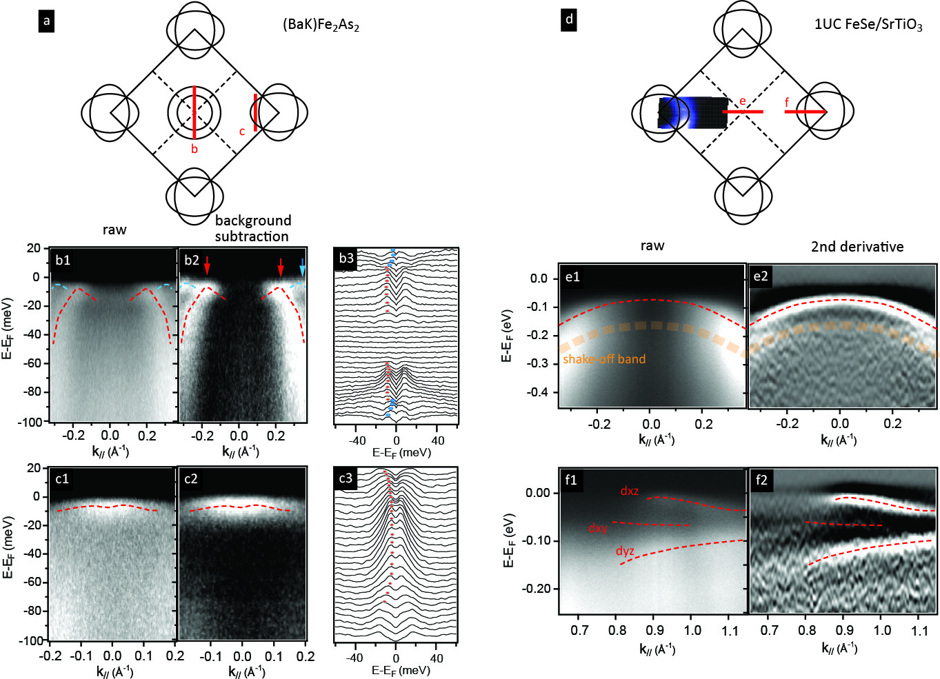
The 11 eV laser-based ARPES system is ideal to tackle these questions. Here we demonstrate the much improved cross section and extended momentum range in both (Ba,K)Fe2As2 (Tc 38K, Fig. 10(a)-(c)) bulk crystal system at 10 K and monolayer FeSe/STO film system (Tgap 65K, Fig. 10(d)-(f)) at 10 K. The VUV light is polarized along the -M direction, and is kept perpendicular to the cut direction.
In (Ba,K)Fe2As2, two hole pockets with different kF’s centered around (Fig. 10(b))Zhang et al. (2010) are identified, with the inner pocket (red arrows) showing a bigger superconducting gap (7.2 meV) than the outer pocket (blue arrows, 2.4 meV). For the two bands the Bogoliubov quasiparticle band backbending is observed in the superconducting state. For the zone corner cut (c), the dxz/dyz band shows a shallow band bottom and forms an electron-like Fermi surface, consistent with previous observations from synchrotron studies.Yi et al. (2011); Tan et al. (2013); Yi et al. (2013)
The single unit cell FeSe film is grown on Nb-doped SrTiO3 substrate, and is transferred to the ARPES measurement chamber in-situ. The Fermi surface shows that Fermi pockets only exist at the BZ corner (Fig. 10(f)), and the hole band does not cross at the point (Fig. 10(e)). The superconducting backbending of the dxz electron band, as well as its hybridization gap with dxy band are observed, consistent with previous measurements at higher photon energy.Liu et al. (2012); Zhang et al. (2015) The measurements also show evidence, although weaker than synchrotron data, of the shadow band due to the strong coupling to the substrate’s out-of-plane phonon, which could play a decisive role in boosting Tc by as much as 50% from its KxFe2-ySe2 counterpart.Lee et al. (2014) The polarization control and improved cross section of our 11 eV laser facilitates resolving gap functions with high resolution on multiple bands across the entire BZ. This is essential to understand the pairing symmetry and superconducting mechanism of this multi-band system.
V SUMMARY
We have presented a table-top 11eV laser-based ARPES system. The system utilizes the 9th harmonics of a 1024 nm IR laser to produce the 113.778 nm VUV radiation for photoemission. Combined with a Scienta R8000 hemispherical electron analyzer, we demonstrate the system’s energy resolution of 2 meV and momentum resolution of 0.012 Å-1 under realistic experimental conditions. The system is capable of reaching to Å-1 parallel momenta as well as 5 eV in binding energy (assuming 4.5 eV material work function). Space charging effects are reduced at high repetition rates of 10 MHz. On the other hand, short pulses ( 100 ps) can be utilized for time-of-flight applications where time resolution is more prioritized.
This system provides a long desired solution to bridging the gap between the existing high-resolution small-momentum-coverage laser-based ARPES and large-momentum-coverage synchrotron-based ARPES. It is capable of producing high resolution spectra in many correlated electron systems and reaching their BZ boundaries as well as valence bands, including the temperature dependent CDW gap in rare earth metal tritelluride, the antinode and oxygen 2p band spectrum in cuprate superconductor and superconducting gap measurements in iron-pnictide and iron-chalcogenide film.
VI ACKNOWLEDGEMENT
The authors acknowledge enlightening discussions with Donghui Lu and Makoto Hashimoto. Samples are kindly provided by Wei Li, Ian Fisher and Hiroshi Eisaki. A.J.M. acknowledges partial support for the VUV light source development from the National Science Foundation under SBIR grant #0848526. S.L.Y. acknowledges Stanford Graduate Fellowship for support. This work is a collaboration between Lumeras LLC and Stanford Institute for Materials and Energy Sciences (SIMES). The photoemission studies were supported by the Department of Energy, Office of Basic Energy Sciences, Division of Materials Sciences and Engineering.
References
- Ohta et al. (2006) T. Ohta, A. Bostwick, T. Seyller, K. Horn, and E. Rotenberg, “Controlling the electronic structure of bilayer graphene,” Science 313, 951–954 (2006).
- Zhou et al. (2007) S. Y. Zhou, G.-H. Gweon, A. V. Fedorov, P. N. First, W. A. de Heer, D.-H. Lee, F. Guinea, A. H. Castro Neto, and A. Lanzara, “Substrate-induced bandgap opening in epitaxial graphene,” Nat Mater 6, 770–775 (2007).
- Johnson (2002) P. D. Johnson, “Photemission and the influence of collective excitations,” Journal of Electron Spectroscopy and Related Phenomena 126, 133 – 144 (2002).
- Kordyuk (2014) A. A. Kordyuk, “ARPES experiment in fermiology of quasi-2d metals (review article),” Low Temperature Physics 40, 286–296 (2014).
- Damascelli, Hussain, and Shen (2003) A. Damascelli, Z. Hussain, and Z.-X. Shen, “Angle-resolved photoemission studies of the cuprate superconductors,” Rev. Mod. Phys. 75, 473–541 (2003).
- Ding et al. (1996) H. Ding, T. Yokoya, J. C. Campuzano, T. Takahashi, M. Randeria, M. R. Norman, T. Mochiku, K. Kadowaki, and J. Giapintzakis, “Spectroscopic evidence for a pseudogap in the normal state of underdoped high-tc superconductors,” Nature 382, 51–54 (1996).
- Stewart (2011) G. R. Stewart, “Superconductivity in iron compounds,” Rev. Mod. Phys. 83, 1589–1652 (2011).
- Lu et al. (2012) D. Lu, I. M. Vishik, M. Yi, Y. Chen, R. G. Moore, and Z.-X. Shen, “Angle-resolved photoemission studies of quantum materials,” Annual Review of Condensed Matter Physics 3, 129–167 (2012).
- Lee et al. (2014) J. J. Lee, F. T. Schmitt, R. G. Moore, S. Johnston, Y.-T. Cui, W. Li, M. Yi, Z. K. Liu, M. Hashimoto, Y. Zhang, D. H. Lu, T. P. Devereaux, D.-H. Lee, and Z.-X. Shen, “Interfacial mode coupling as the origin of the enhancement of tc in fese films on SrTiO3,” Nature 515, 245–248 (2014).
- Hsieh et al. (2008) D. Hsieh, D. Qian, L. Wray, Y. X. Y. S. Hor, R. J. Cava, and M. Z. Hasan, “A topological dirac insulator in a quantum spin hall phase,” Nature 452, 970–974 (2008).
- Chen et al. (2009) Y. L. Chen, J. G. Analytis, J.-H. Chu, Z. K. Liu, S.-K. Mo, X. L. Qi, H. J. Zhang, D. H. Lu, X. Dai, Z. Fang, S. C. Zhang, I. R. Fisher, Z. Hussain, and Z.-X. Shen, “Experimental realization of a three-dimensional topological insulator, ,” Science 325, 178–181 (2009).
- Note (1) In general, the out-of-plane momentum is not conserved as the electron overcomes the surface potential barrier during the photoemission process.
- Note (2) = 35.0∘ (maximum sample surface rotation) + 15.0∘ (typical detector acceptance angle).
- Graf et al. (2010) J. Graf, S. Hellmann, C. Jozwiak, C. L. Smallwood, Z. Hussain, R. A. Kaindl, L. Kipp, K. Rossnagel, and A. Lanzara, “Vacuum space charge effect in laser-based solid-state photoemission spectroscopy,” Journal of Applied Physics 107, 014912 (2010).
- Passlack et al. (2006) S. Passlack, S. Mathias, O. Andreyev, D. Mittnacht, M. Aeschlimann, and M. Bauer, “Space charge effects in photoemission with a low repetition, high intensity femtosecond laser source,” Journal of Applied Physics 100, 024912 (2006).
- Harter et al. (2012) J. W. Harter, P. D. C. King, E. J. Monkman, D. E. Shai, Y. Nie, M. Uchida, B. Burganov, S. Chatterjee, and K. M. Shen, “A tunable low-energy photon source for high-resolution angle-resolved photoemission spectroscopy,” Review of Scientific Instruments 83, 113103 (2012).
- Zhang (2012) W. Zhang, Photoemission Spectroscopy on High Temperature Superconductor: A Study of by Laser-Based Angle-Resolved Photoemission, Springer Theses (Springer, 2012).
- Souma et al. (2007) S. Souma, T. Sato, T. Takahashi, and P. Baltzer, “High-intensity xenon plasma discharge lamp for bulk-sensitive high-resolution photoemission spectroscopy,” Review of Scientific Instruments 78, 123104 (2007).
- VGV (2012) “High intensity extreme uv source scienta vuv 5000,” VG Scienta Technical Data Sheet (2012), rev. 4.1.
- Strocov et al. (2010) V. N. Strocov, T. Schmitt, U. Flechsig, T. Schmidt, A. Imhof, Q. Chen, J. Raabe, R. Betemps, D. Zimoch, J. Krempasky, X. Wang, M. Grioni, A. Piazzalunga, and L. Patthey, “High-resolution soft x-ray beamline adress at the swiss light source for resonant inelastic x-ray scattering and angle-resolved photoelectron spectroscopies,” Journal of Synchrotron Radiation 17, 631–643 (2010).
- Strocov et al. (2014) V. N. Strocov, X. Wang, M. Shi, M. Kobayashi, J. Krempasky, C. Hess, T. Schmitt, and L. Patthey, “Soft-X-ray ARPES facility at the ADRESS beamline of the SLS: concepts, technical realisation and scientific applications,” Journal of Synchrotron Radiation 21, 32–44 (2014).
- Baichang Wu and Chen (1996) N. Y. Baichang Wu, Dingyuen Tang and C. Chen, “Linear and nonlinear optical properties of the KBe2BO3F2 (KBBF) crystal,” Optical Materials 5, 105 – 109 (1996).
- Kiss et al. (2008) T. Kiss, T. Shimojima, K. Ishizaka, A. Chainani, T. Togashi, T. Kanai, X.-Y. Wang, C.-T. Chen, S. Watanabe, and S. Shin, “A versatile system for ultrahigh resolution, low temperature, and polarization dependent laser-angle-resolved photoemission spectroscopy,” Review of Scientific Instruments 79, 023106 (2008).
- Kirchmann et al. (2008) P. Kirchmann, L. Rettig, D. Nandi, U. Lipowski, M. Wolf, and U. Bovensiepen, “A time-of-flight spectrometer for angle-resolved detection of low energy electrons in two dimensions,” Applied Physics A 91, 211–217 (2008).
- Jiang et al. (2014) R. Jiang, D. Mou, Y. Wu, L. Huang, C. D. McMillen, J. Kolis, H. G. Giesber, J. J. Egan, and A. Kaminski, “Tunable vacuum ultraviolet laser based spectrometer for angle resolved photoemission spectroscopy,” Review of Scientific Instruments 85, 033902 (2014).
- Damm et al. (2015) A. Damm, J. Güdde, P. Feulner, A. Czasch, O. Jagutzki, H. Schmidt-Böcking, and U. Höfer, “Application of a time-of-flight spectrometer with delay-line detector for time- and angle-resolved two-photon photoemission,” Journal of Electron Spectroscopy and Related Phenomena 202, 74 – 80 (2015).
- Berntsen, Goetberg, and Tjernberg (2011) M. H. Berntsen, O. Goetberg, and O. Tjernberg, “An experimental setup for high resolution 10.5 eV laser-based angle-resolved photoelectron spectroscopy using a time-of-flight electron analyzer,” Review of Scientific Instruments 82, 095113 (2011).
- Koralek et al. (2007) J. D. Koralek, J. F. Douglas, N. C. Plumb, J. D. Griffith, S. T. Cundiff, H. C. Kapteyn, M. M. Murnane, and D. S. Dessau, “Experimental setup for low-energy laser-based angle resolved photoemission spectroscopy,” Review of Scientific Instruments 78, 053905 (2007).
- Kiss et al. (2005) T. Kiss, T. Shimojima, F. Kanetaka, K. Kanai, T. Yokoya, S. Shin, Y. Onuki, T. Togashi, C. Zhang, C. Chen, and S. Watanabe, “Ultrahigh-resolution photoemission spectroscopy of superconductors using a VUV laser,” Journal of Electron Spectroscopy and Related Phenomena 144, 953 – 956 (2005), proceeding of the Fourteenth International Conference on Vacuum Ultraviolet Radiation Physics.
- BL (5) “Experimental Station 5-4 — Stanford Synchrotron Radiation Lightsource,” http://www-ssrl.slac.stanford.edu/content/beam-lines/bl5-4, accessed: 2015-01-17.
- BL (4) “Beamline 4.0.3.” http://www-als.lbl.gov/index.php/holding/99-403.html, accessed: 2015-01-17.
- (32) “One-cubed ARPES,” https://www.helmholtz-berlin.de/pubbin/igama_output?modus=einzel&sprache=en&gid=1679&typoid=40737, accessed: 2015-05-17.
- Moore (1958) C. E. Moore, Atomic Energy Levels, National Bureau of Standards 3, 467 (1958).
- Gornik et al. (1981) W. Gornik, S. Kindt, E. Matthias, and D. Schmidt, “Two‐photon excitation of xenon atoms and dimers in the energy region of the 5p5 6p configuration,” The Journal of Chemical Physics 75, 68–74 (1981).
- Plimmer et al. (1989) M. D. Plimmer, P. E. G. Baird, C. J. Foot, D. N. Stacey, J. B. Swan, and G. K. Woodgate, “Isotope shift in xenon by Doppler-free two-photon laser spectroscopy,” Journal of Physics B: Atomic, Molecular and Optical Physics 22, L241 (1989).
- Vidal (1980) C. R. Vidal, “Coherent VUV sources for high resolution spectroscopy,” Appl. Opt. 19, 3897–3903 (1980).
- Bjorklund (1975) G. C. Bjorklund, “Effects of focusing on third-order nonlinear processes in isotropic media,” Quantum Electronics, IEEE Journal of 11, 287–296 (1975).
- Stappaerts et al. (1976) E. Stappaerts, G. Bekkers, J. Young, and S. Harris, “The effect of linewidth on the efficiency of two-photon-pumped frequency converters,” Quantum Electronics, IEEE Journal of 12, 330–333 (1976).
- Krupenie (1972) P. H. Krupenie, “The spectrum of molecular oxygen,” Journal of Physical and Chemical Reference Data 1 (1972).
- Tennyson et al. (2009) J. Tennyson, P. F. Bernath, L. R. Brown, A. Campargue, M. R. Carleer, A. G. Császár, R. R. Gamache, J. T. Hodges, A. Jenouvrier, O. V. Naumenko, O. L. Polyansky, L. S. Rothman, R. A. Toth, A. C. Vandaele, N. F. Zobov, L. Daumont, A. Z. Fazliev, T. Furtenbacher, I. E. Gordon, S. N. Mikhailenko, and S. V. Shirin, “IUPAC critical evaluation of the rotational–vibrational spectra of water vapor. part I — energy levels and transition wavenumbers for H2 17O and H2 18O,” Journal of Quantitative Spectroscopy and Radiative Transfer 110, 573 – 596 (2009).
- Watanabe, Inn, and Zelikoff (1953) K. Watanabe, E. C. Y. Inn, and M. Zelikoff, “Absorption Coefficients of Oxygen in the Vacuum Ultraviolet,” The Journal of Chemical Physics 21 (1953).
- Laporte and Subtil (1982) P. Laporte and J. L. Subtil, “Refractive index of LiF from 105 to 200 nm,” J. Opt. Soc. Am. 72, 1558–1559 (1982).
- Reber et al. (2014) T. J. Reber, N. C. Plumb, J. A. Waugh, and D. S. Dessau, “Effects, determination, and correction of count rate nonlinearity in multi-channel analog electron detectors,” Review of Scientific Instruments 85, 043907 (2014).
- Takayama et al. (2014) A. Takayama, T. Sato, S. Souma, and T. Takahashi, “Rashba effect in antimony and bismuth studied by spin-resolved ARPES,” New Journal of Physics 16, 055004 (2014).
- Sugawara et al. (2006) K. Sugawara, T. Sato, S. Souma, T. Takahashi, M. Arai, and T. Sasaki, “Fermi Surface and Anisotropic Spin-Orbit Coupling of Sb(111) Studied by Angle-Resolved Photoemission Spectroscopy,” Phys. Rev. Lett. 96, 046411 (2006).
- Ishizaka et al. (2008) K. Ishizaka, T. Kiss, S. Izumi, M. Okawa, T. Shimojima, A. Chainani, T. Togashi, S. Watanabe, C.-T. Chen, X. Y. Wang, T. Mochiku, T. Nakane, K. Hirata, and S. Shin, “Doping-dependence of nodal quasiparticle properties in high- cuprates studied by laser-excited angle-resolved photoemission spectroscopy,” Phys. Rev. B 77, 064522 (2008).
- Hellmann et al. (2009) S. Hellmann, K. Rossnagel, M. Marczynski-Bühlow, and L. Kipp, “Vacuum space-charge effects in solid-state photoemission,” Phys. Rev. B 79, 035402 (2009).
- Moore et al. (2010) R. G. Moore, V. Brouet, R. He, D. H. Lu, N. Ru, J.-H. Chu, I. R. Fisher, and Z.-X. Shen, “Fermi surface evolution across multiple charge density wave transitions in ,” Phys. Rev. B 81, 073102 (2010).
- Brouet et al. (2004) V. Brouet, W. L. Yang, X. J. Zhou, Z. Hussain, N. Ru, K. Y. Shin, I. R. Fisher, and Z. X. Shen, “Fermi surface reconstruction in the cdw state of observed by photoemission,” Phys. Rev. Lett. 93, 126405 (2004).
- Peng et al. (2013) Y. Peng, J. Meng, D. Mou, J. He, L. Zhao, Y. Wu, G. Liu, X. Dong, S. He, J. Zhang, X. Wang, Q. Peng, Z. Wang, S. Zhang, F. Yang, C. Chen, Z. Xu, T. K. Lee, and X. J. Zhou, “Disappearance of nodal gap across the insulator–superconductor transition in a copper-oxide superconductor,” Nature Communications 4, 3459 (2013).
- Parham et al. (2013) S. Parham, T. J. Reber, Y. Cao, J. A. Waugh, Z. Xu, J. Schneeloch, R. D. Zhong, G. Gu, G. Arnold, and D. S. Dessau, “Pair breaking caused by magnetic impurities in the high-temperature superconductor Bi2.1Sr1.9Ca(Cu1-xFex)2Oy,” Phys. Rev. B 87, 104501 (2013).
- Kondo et al. (2011) T. Kondo, Y. Hamaya, A. D. Palczewski, T. Takeuchi, J. S. Wen, Z. J. Xu, G. Gu, J. Schmalian, and A. Kaminski, “Disentangling cooper-pair formation above the transition temperature from the pseudogap state in the cuprates,” Nature Physics 7, 21–25 (2011).
- Vishik et al. (2012) I. M. Vishik, M. Hashimoto, R.-H. He, W.-S. Lee, F. Schmitt, D. Lu, R. G. Moore, C. Zhang, W. Meevasana, T. Sasagawa, S. Uchida, K. Fujita, S. Ishida, M. Ishikado, Y. Yoshida, H. Eisaki, Z. Hussain, T. P. Devereaux, and Z.-X. Shen, “Phase competition in trisected superconducting dome,” Proceedings of the National Academy of Sciences 109, 18332–18337 (2012).
- Levin, Smolin, and Shepelev (1994) A. A. Levin, Y. I. Smolin, and Y. F. Shepelev, “Causes of modulation and hole conductivity of the high-t c superconductor according to X-ray single-crystal data,” Journal of Physics: Condensed Matter 6, 3539 (1994).
- Shan et al. (2003) L. Shan, A. Ejov, A. Volodin, V. V. Moshchalkov, H. H. Wen, and C. T. Lin, “STM studies of the surface structure in cleaved single crystals,” EPL (Europhysics Letters) 61, 681 (2003).
- Hashimoto et al. (2012) M. Hashimoto, R.-H. He, I. M. Vishik, F. Schmitt, R. G. Moore, D. H. Lu, Y. Yoshida, H. Eisaki, Z. Hussain, T. P. Devereaux, and Z.-X. Shen, “Superconductivity distorted by the coexisting pseudogap in the antinodal region of Bi1.5Pb0.55Sr1.6La0.4CuO6+δ: A photon-energy-dependent angle-resolved photoemission study,” Phys. Rev. B 86, 094504 (2012).
- Hashimoto et al. (2008) M. Hashimoto, T. Yoshida, H. Yagi, M. Takizawa, A. Fujimori, M. Kubota, K. Ono, K. Tanaka, D. H. Lu, Z.-X. Shen, S. Ono, and Y. Ando, “Doping evolution of the electronic structure in the single-layer cuprate : Comparison with other single-layer cuprates,” Phys. Rev. B 77, 094516 (2008).
- Shen et al. (2004) K. M. Shen, F. Ronning, D. H. Lu, W. S. Lee, N. J. C. Ingle, W. Meevasana, F. Baumberger, A. Damascelli, N. P. Armitage, L. L. Miller, Y. Kohsaka, M. Azuma, M. Takano, H. Takagi, and Z.-X. Shen, “Missing quasiparticles and the chemical potential puzzle in the doping evolution of the cuprate superconductors,” Phys. Rev. Lett. 93, 267002 (2004).
- Rösch and Gunnarsson (2004) O. Rösch and O. Gunnarsson, “Electron-phonon interaction in the three-band model,” Phys. Rev. B 70, 224518 (2004).
- Kung et al. (2014) Y. F. Kung, C.-C. Chen, B. Moritz, S. Johnston, R. Thomale, and T. P. Devereaux, “Numerical exploration of spontaneous broken symmetries in multiorbital hubbard models,” Phys. Rev. B 90, 224507 (2014).
- Shimojima et al. (2010) T. Shimojima, K. Ishizaka, Y. Ishida, N. Katayama, K. Ohgushi, T. Kiss, M. Okawa, T. Togashi, X.-Y. Wang, C.-T. Chen, S. Watanabe, R. Kadota, T. Oguchi, A. Chainani, and S. Shin, “Orbital-dependent modifications of electronic structure across the magnetostructural transition in ,” Phys. Rev. Lett. 104, 057002 (2010).
- Okazaki et al. (2012a) K. Okazaki, Y. Ito, Y. Ota, Y. Kotani, T. Shimojima, T. Kiss, S. Watanabe, C. T. Chen, S. Niitaka, T. Hanaguri, H. Takagi, A. Chainani, and S. Shin, “Evidence for a modulation of the superconducting energy gap of optimally doped single crystals using laser Angle-Resolved Photoemission Spectroscopy,” Phys. Rev. Lett. 109, 237011 (2012a).
- Okazaki et al. (2012b) K. Okazaki, Y. Ota, Y. Kotani, W. Malaeb, Y. Ishida, T. Shimojima, T. Kiss, S. Watanabe, C.-T. Chen, K. Kihou, C. H. Lee, A. Iyo, H. Eisaki, T. Saito, H. Fukazawa, Y. Kohori, K. Hashimoto, T. Shibauchi, Y. Matsuda, H. Ikeda, H. Miyahara, R. Arita, A. Chainani, and S. Shin, “Octet-line node structure of superconducting order parameter in KFe2As2,” Science 337, 1314–1317 (2012b).
- He2 (2011) “The orbital characters and kz dispersions of bands in iron-pnictide NaFeAs,” Journal of Physics and Chemistry of Solids 72, 479 – 482 (2011).
- Lu et al. (2008) D. H. Lu, M. Yi, S.-K. Mo, A. S. Erickson, J. Analytis, J.-H. Chu, D. J. Singh, Z. Hussain, T. H. Geballe, I. R. Fisher, and Z.-X. Shen, “Electronic structure of the iron-based superconductor LaOFeP,” Nature 455, 81–84 (2008).
- Shimojima et al. (2011) T. Shimojima, F. Sakaguchi, K. Ishizaka, Y. Ishida, T. Kiss, M. Okawa, T. Togashi, C.-T. Chen, S. Watanabe, M. Arita, K. Shimada, H. Namatame, M. Taniguchi, K. Ohgushi, S. Kasahara, T. Terashima, T. Shibauchi, Y. Matsuda, A. Chainani, and S. Shin, “Orbital-Independent Superconducting Gaps in Iron Pnictides,” Science 332, 564–567 (2011).
- Zi-Rong et al. (2013) Y. Zi-Rong, Z. Yan, X. Bin-Ping, and F. Dong-Lai, “Angle-resolved photoemission spectroscopy study on iron-based superconductors,” Chinese Physics B 22, 087407 (2013).
- Zhang et al. (2011) Y. Zhang, L. X. Yang, M. Xu, Z. R. Ye, F. Chen, C. He, H. C. Xu, J. Jiang, B. P. Xie, J. J. Ying, X. F. Wang, X. H. Chen, J. P. Hu, M. Matsunami, S. Kimura, and D. L. Feng, “Nodeless superconducting gap in AxFe2Se2 (A=K,Cs) revealed by angle-resolved photoemission spectroscopy,” Nat Mater 10, 273–277 (2011).
- Qian et al. (2011) T. Qian, X.-P. Wang, W.-C. Jin, P. Zhang, P. Richard, G. Xu, X. Dai, Z. Fang, J.-G. Guo, X.-L. Chen, and H. Ding, “Absence of a holelike fermi surface for the iron-based superconductor revealed by Angle-Resolved Photoemission Spectroscopy,” Phys. Rev. Lett. 106, 187001 (2011).
- Mou et al. (2011) D. Mou, S. Liu, X. Jia, J. He, Y. Peng, L. Zhao, L. Yu, G. Liu, S. He, X. Dong, J. Zhang, H. Wang, C. Dong, M. Fang, X. Wang, Q. Peng, Z. Wang, S. Zhang, F. Yang, Z. Xu, C. Chen, and X. J. Zhou, “Distinct fermi surface topology and nodeless superconducting gap in a superconductor,” Phys. Rev. Lett. 106, 107001 (2011).
- Liu et al. (2012) D. Liu, W. Zhang, D. Mou, J. He, Y.-B. Ou, Q.-Y. Wang, Z. Li, L. Wang, L. Zhao, S. He, Y. Peng, X. Liu, C. Chen, L. Yu, G. Liu, X. Dong, J. Zhang, C. Chen, Z. Xu, J. Hu, X. Chen, X. Ma, Q. Xue, and X. J. Zhou, “Electronic origin of high-temperature superconductivity in single-layer FeSe superconductor,” Nat Commun 3, 931 (2012).
- Tan et al. (2013) S. Tan, Y. Zhang, M. Xia, Z. Ye, F. Chen, X. Xie, R. Peng, D. Xu, Q. Fan, H. Xu, J. Jiang, T. Zhang, X. Lai, T. Xiang, J. Hu, B. Xie, and D. Feng, “Interface-induced superconductivity and strain-dependent spin density waves in FeSe/SrTiO3 thin films,” Nature Materials 12, 634–640 (2013).
- Zhang et al. (2010) Y. Zhang, L. X. Yang, F. Chen, B. Zhou, X. F. Wang, X. H. Chen, M. Arita, K. Shimada, H. Namatame, M. Taniguchi, J. P. Hu, B. P. Xie, and D. L. Feng, “Out-of-plane momentum and symmetry-dependent energy gap of the pnictide superconductor revealed by angle-resolved photoemission spectroscopy,” Phys. Rev. Lett. 105, 117003 (2010).
- Yi et al. (2011) M. Yi, D. Lu, J.-H. Chu, J. G. Analytis, A. P. Sorini, A. F. Kemper, B. Moritz, S.-K. Mo, R. G. Moore, M. Hashimoto, W.-S. Lee, Z. Hussain, T. P. Devereaux, I. R. Fisher, and Z.-X. Shen, “Symmetry-breaking orbital anisotropy observed for detwinned above the spin density wave transition,” Proceedings of the National Academy of Sciences 108, 6878–6883 (2011).
- Yi et al. (2013) M. Yi, D. H. Lu, R. Yu, S. C. Riggs, J.-H. Chu, B. Lv, Z. K. Liu, M. Lu, Y.-T. Cui, M. Hashimoto, S.-K. Mo, Z. Hussain, C. W. Chu, I. R. Fisher, Q. Si, and Z.-X. Shen, “Observation of temperature-induced crossover to an orbital-selective mott phase in (, Rb) superconductors,” Phys. Rev. Lett. 110, 067003 (2013).
- Zhang et al. (2015) Y. Zhang, M. Yi, Z.-K. Liu, W. Li, J. J. Lee, R. G. Moore, M. Hashimoto, N. Masamichi, H. Eisaki, S.-K. Mo, Z. Hussain, T. P. Devereaux, Z.-X. Shen, and D. H. Lu, “Distinctive momentum dependence of the band reconstruction in the nematic state of FeSe thin film,” ArXiv e-prints (2015), arXiv:1503.01556 .