A critical look at methods for calculating charge transfer couplings fast and accurately
Abstract
We present here a short and subjective review of methods for calculating charge transfer couplings. Although we mostly focus on Density Functional Theory, we discuss a small subset of semiempirical methods as well as the adiabatic-to-diabatic transformation methods typically coupled with wavefunction-based electronic structure calculations. In this work, we will present the reader with a critical assessment of the regimes that can be modelled by the various methods – their strengths and weaknesses. In order to give a feeling about the practical aspects of the calculations, we also provide the reader with a practical protocol for running coupling calculations with the recently developed FDE-ET method.
1 Introduction
Charge transfer (CT) between molecular species play vital roles in processes that occur in biology such as protein communication1, 2, 3, 4, 5, respiratory systems in the mitochondria6, oxidative damage on DNA7, 8, 9, photosynthetic cycles 10, 11, as well as in materials science conduction in organic semiconductors12, 13. In order to achieve an accurate modeling of these processes in the simulations, one needs to include several levels of complexity, which in most instances lead to considering model systems featuring hundreds of atoms and an even larger number of electrons. The large system sizes preclude the use of high-level wavefunction-based quantum-chemical methods. For this reason, researchers worldwide have invested a great deal of effort in developing approximate, fast, yet still accurate methods for describing CT reactions. Methods based on Density-Functional Theory have in recent years become competitive in regards to the accuracy while still maintaining a generally low computational cost.
Marcus theory14, 15 is perhaps the most applicable theory for modeling a CT process. This theory was originally derived under three main approximations. First, a CT event is thought of in terms of a two-dimensional basis set (donor and acceptor). The interaction matrix element of the Hamiltonian is the central quantity in determining the probability of a transition in populations from the basis function representing the donor state to one representing the acceptor state. This interaction is the electronic coupling of the two electronic states involved in the CT reaction16, 17. Second, it relies on the Condon approximation18, 19, in which the electronic coupling is considered to be independent of the nuclear motion when the transfer occurs. Third, reactants and products are modeled as being enclosed by spheres on which the polarization of the solvent is represented as a dielectric continuum 20, 21, 22, 15. Marcus theory can be summarized as23:
| (1) |
where is the reorganization energy, and is the electronic coupling.
States that most resemble the initial and final states of electron transfer have been often referred to as “diabatic states” 24, 25 and their corresponding wavefunctions “diabats”. Although it is known that diabatic states have a formal definition 26, 27, it was shown 28 that charge-localized states satisfy the requirements for diabatic states for condensed phase electron transfer reactions.
Several approaches are available in the literature to generate and evaluate Hamiltonian matrix elements with wavefunctions of charge-localized, diabatic states. They differ in the level of theory used in the calculation and in the way localized electronic structures are created26, 29, 30, 31, 15, 25. When wavefunction-based quantum-chemical methods are employed, the framework of the generalized Mulliken-Hush method (GMH)29, 32, 33, 34, is particularly successful. So far, it has been used in conjunction with accurate electronic structure methods for small and medium sized systems35, 36, 37. As an alternative to GMH and other derived methods 38, 39, additional methods have been explored for their applicability in larger systems such as constrained density functional method (CDFT)40, 41, 25, 37, and fragmentation approaches42, 43, 44, 45, 46, 47, which also include the frozen density embedding (FDE) method48, 49.
So far, we have mentioned methods that produce all-electron diabatic wavefunctions and corresponding Hamiltonian matrix elements. There are two other classes of methods which simplify the quantum problem by focusing on the wavefunction of the transferred charge: such as methods making use of the frozen core approximation Fragment Orbital methods (FO), and methods that assume the charge to be localized on single atomic orbitals 50. In this work, we will also treat these computationally low-cost methods.
As our group is involved in the development of the Frozen Density Embedding (FDE) formulation of subsystem DFT, this chapter will pay particular attention to the FDE methodology. We believe FDE to be a very promising method capable of achieving a good description of the electronic coupling in CT reactions, while maintaining a low computational complexity.
This chapter is divided in two parts: the first part is devoted to the FDE method as well as other DFT-based alternatives. The second part covers more accurate methods (wavefunction-based). In each of the two parts, we discuss the numerical stability and accuracy of the methods in the generation of diabatic states with the overarching goal of obtaining reliable electronic couplings with a contained computational effort.
We will start with a description of FDE and its ability to generate diabats and to compute Hamiltonian matrix elements – the FDE-ET method (ET stands for Electron Transfer). In the subsequent section, we will present specific examples of FDE-ET computations to provide the reader with a comprehensive view of the performance and applicability of FDE-ET. After FDE has been treated, four additional methods to generate diabatic states are presented in order of accuracy: CDFT, FODFT, AOM, and Pathways. In order to output a comprehensive presentation, we also describe those methods in which wavefunctions methods can be used, in particular GMH and other adiabatic-to-diabatic diabatization methods. Finally, we provide the reader with a “protocol” for running FDE-ET calculations with the only available implementation of the method in the Amsterdam Density Functional software51. In closing, we outline our concluding remarks and our vision of what the future holds for the field of computational chemistry applyed to electron transfer.
2 DFT Based Methods
2.1 The Frozen Density Embedding formalism
The frozen density-embedding (FDE) formalism 52 developed by Wesolowski and Warshel53, 54, 52 has been applied to a plethora of chemical problems, for instance, solvent effects on different types of spectroscopy55, 56, 57, magnetic properties 58, 59, 60, 61, 62, excited states63, 64, 65, 55, 66, charge transfer states 67, 49, 68. Computationally, FDE is available for molecular systems in ADF 51, 69, Dalton 70, 71, Q-Chem 72, 73, and Turbomole 74, 75, 76 packages, as well as for molecular periodic systems in CP2K 77, 78 and fully periodic systems (although in different flavors) in CASTEP 79, 80, Quantum Espresso 81, 82, 83, and Abinit 84, 85.
FDE prescribes that the total electron density should be expressed as the sum of subsystem electron densities53, 86, 87, 88, 89, this is based on the idea that a molecular system can be more easily approached if it is subdivided into many smaller subsystems. Namely,
| (2) |
As in regular DFT calculations, the electron density of each subsystem is computed by solving selfconsistently a Kohn–Sham (KS) like equation per subsystem. These KS like equations read as:
| (3) |
Where , are the molecular orbitals and orbital energies of subsystem . In Eq. (3) we have augmented the Kohn–Sham single particle Hamiltonian by an embedding potential, , in which are encoded the interactions with the other subsystems. In the following, is the embedding potential acting on subsystem :
| (4) |
In the above, , and are kinetic and exchange–correlation energy functionals, and the nuclear charge, respectively, and is the total number of subsystems considered. In practical FDE calculations, the kinetic energy is calculated in terms of orbital free semilocal functionals. This approximation is ultimately the biggest difference between an FDE and a full KS-DFT calculation of the supersystem90, 91, 92, 93. As a consequence, the embedding potential becomes inaccurate when the subsystems feature a large overlap between their electron densities 83, 94, 95 (this is because the larger the density overlap is, the larger the magnitude of the nonadditive potentials become). In FDE, the subsystem KS equations are left to converge to selfconsistency with respect to each other. This is often achieved by employing the so–called freeze-and-thaw procedure 96, 69 (as done in ADF and other molecular codes) or via updating the embedding potential at every SCF cycle as done in CP2K 78, 97 and Quantum-Espresso 83, 81, 98. It is worth noting that FDE scales linearly with the number of subsystems provided that linear scaling methods for the solution of the electrostatic problem are employed 69.
The earliest example of diabatization by FDE was given in Ref.48. This is shown in Figure 1, where the spin densities for a pair of guanines are calculated. KS-DFT of the supersystem carried out with semilocal XC functionals fails in the prediction of the spin density. This is because the self-interaction error makes the spin density spread on both guanines against the prediction given by more accurate theoretical work35 and experimental studies99, 100, 101. On the contrary, FDE localizes the charge on a guanine of choice.
The fact that FDE was able to provide subsystem-localized electronic structures was known since its early application to systems with unpaired electrons 60, 62, 61, 102. Later, this ability of FDE was explored for the computation of diabatic states for electron transfer 48, 49, 67, 103 and to compute hyperfine coupling constants 60, 61.

The question that one can raise is why FDE calculations yield charge localized states? We provide here four reasons 49, 103.
-
1.
Orthogonality is not imposed between the molecular orbitals belonging to different subsystems.
-
2.
FDE calculations can be executed in the monomer basis set. This is known as FDE(m) method 104.
-
3.
FDE calculations are always initiated with a subsystem localized initial guess of the electron density.
-
4.
Electrons of a subsystem, remain localized also because there are repulsive walls in the region of the surrounding (frozen) fragments.
The first reason, is important because it directly removes a bias towards delocalization which results due to orthonormalization of the molecular orbitals, as already noted by Dulak and Wesolowski 105. The second and third reasons come together, the lack of basis functions on the surrounding subsystems, does not allow substantial charge transfer between the subsystems. As a consequence, the SCF is biased to converge to localized electronic structures.
The fourth reason makes reference to the approximate nature of the term (also known as nonadditive kinetic energy potential which is part of the embedding potential) in the region of the frozen fragments (e.g. in the region where with is larger than any other subsystem electron density). Approximate nonadditive kinetic energy potentials fail in canceling out the attractive potential due to the nuclear charge in the vicinity of the nucleus of the surrounding frozen subsystems 104, 106, and they do not reproduce the exact potential at intermediate regions 107, 108, 109, especially in the vicinity of an atomic shell 106, 109. In that region they cross the exact potential and large potential walls arise. A simplified depiction of this effect is devised in figure 2.

In this scenario, diabatic states can be generated with FDE by performing at least two simulations, one featuring a hole/electron on the donor while the acceptor is neutral and one calculation in which the charge hole/electron is on the acceptor. The result is two charge localized states, whose, densities and Kohn-Sham orbitals are used in a later step in order to build the diabatic Hamiltonian and overlap matrices, needed to compute the diabatic coupling matrix element.
2.1.1 FDE-ET method
FDE-ET is a methodology which computes Hamiltonian couplings from diabatic states generated by an FDE calculation. Electron transfer reaction are usually described in the basis of a two-state formalism30, taking as basis set two broken-symmetry charge-localized states. This methodology can also approach models for the superexchange mechanism23, 110, 7, 111, 112, 113, where the transfer is still modelled by a Two-dimensional basis set but the coupling includes the effect of non-resonant bridges states. Figure 3, illustrates the difference between tunneling through the vacuum and through a set of bridge states. The bridge could be comprised of one or more molecules, a covalent bond or any other type o potential barrier as long as its height is lower than the one when vacuum separates donor and acceptor. As it is shown in Figure 3, the higher the potential barrier the faster the coupling decays with respect to the donor–acceptor distance.
In FDE-ET we seek a method capable of computing the Hamiltonian matrix in the basis of charge–localized states generated with FDE. First, we have to define the needed matrix elements.

As diabatic states are not the eigenfunctions of the molecular Hamiltonian, the off-diagonal elements of such Hamiltonian are not zero and can be approximated by the following formula 114, 115 if and are slater determinants representing the donor and acceptor diabats:
| (5) |
Here is the molecular electronic Hamiltonian, and is the transition density defined as , with being the total number of electrons in the system (i.e. the sum of the electron number of all subsystems) and is an energy density functional. The donor–acceptor overlap matrix elements are found by computing the following determinant:
| (6) |
where is the transition overlap matrix in terms of the occupied orbitals ()116, 114. Thus, the transition density is now written in the basis of all occupied orbitals which make up the diabatic states and .
| (7) |
The Hamiltonian coupling is not , but it is generally reported as the coupling between the Löwdin orthogonalized and . For only two states this takes the form,
| (8) |
Turning to the superexchange picture, the effective coupling, is a summation of the contribution given by the interaction between D and A and the interaction of D and A with all bridges states, namely:
| (9) |
where the superscript stands for transpose, is the Green’s operator, defined as
| (10) |
As shown in equation 9, is the coupling for the donor-acceptor transfer, which in the absence of bridge states (CT through vacuum), would be the only contribution to . On the other hand, if bridge states are present, the contribution to is given by the second addend in equation 9. Generally, appearing above is the energy at which the tunneling event occurs (i.e. at the crossing seam of the Marcus parabolas). In our works 67, was chosen to be in between and , and specifically to be . This choice is invoked by several works in the literature 117, 30, 118, 119 where it is well known that there is a mild dependence of the coupling with the tunneling energy118. However, this equation holds when there is no resonance between D, A and the bridges states120, 23, 30, 121, 122, 123. If near-degeneracies appear then the transport regime transitions to resonant tunneling or hopping.
2.1.2 Distance dependence of the electronic coupling
In this section, we discuss calculations of the coupling matrix element () of hole transfer from a donor to an acceptor molecule through the vacuum. This means that the initial state of hole transfer is the donor molecule (D), and the final state the acceptor molecule (A), and no intermediate bridge states are considered. Any reliable method for computing couplings should be able to reproduce high level calculations of CT coupling in small molecular dimers. For this purpose, we initially chose 23 biologically relevant -stacks 67, in order to analyze the distance dependence of the coupling, separations of 3-20 Å were considered, as result a total of 276 coupling calculations were ran. Overall, our couplings show a good agreement with previous computations (e.g. we reproduced the decay factors, , -stacked dimers separated by vacuum).
When a test set for hole transfer couplings featuring high accuracy couplings became available 37 we could compare systematically the FDE-ET couplings with the benchmark values 103. Benchmark calculations were ran on a set of 15 of -stacked dimers. This study was rigorous, and tested the effect of the basis set size, nonadditive kinetic energy functionals (NAKE) and exchange-correlation functionals (XC) on the value of the computed couplings. The most important finding that resulted from the benchmark work resided in the fact that GGA functionals coupled with a medium sized basis set and the PW91k NAKE functional allow the FDE-ET method to yield reliable electronic couplings as tested against high-level correlated wavefunction (MRCI+Q, NEVPT2 and SCS-CC2) methods applied to the array of dimers. The PBE and PW91 functionals are found to be a good choice in each case considered with a MAX error lower than 50 meV and an overall MRUE of a little over 7% in both cases103. Statistically, we found that hole transfer couplings are relatively insensitive to the choice of NAKE functionals, while our analysis of the basis set dependence shows that QZ4P basis set is the most problematic, as it often biases the FDE convergence to nonphysical states at short intersubsystem separations – a problem already well documented in the FDE literature124, 125. Finally, Table 1 compares the performance of FDE-ET for different levels of theory. The results for GGAs are in good agreement with the benchmark values, and in some cases they showed to be superior to hybrid and meta-GGA functionals, particularly PBE and PW91. B3LYP also stands out as another valuable choice.
Generally, all functionals perform well in the FDE-ET coupling calculations making FDE-ET a method that is relatively insensitive to the XC and NAKE functional choice.
| Set | MUE(MeV) | MRUE(%) | MAX(meV) |
|---|---|---|---|
| PBE/PW91k/TZP | 15.3 | 7.1 | 49.6 |
| PW91/PW91k/TZP | 15.2 | 7.1 | 49.1 |
| B3LYP/PW91k/TZP | 18.1 | 7.9 | 58.5 |
| M06-2X/PW91k/TZP | 18.0 | 8.2 | 54.9 |
2.1.3 Hole transfer in DNA oligomers
In this section, we discuss an interesting application of FDE-ET to charge transfer in biosystems.The electronic coupling for hole transfer in a completely dry B-DNA structure of G(T)NG and G(A)NG was calculated. The structures considered lack water molecules, metal counterions and phosphate linker groups. The latter is because the applicability of FDE is restricted to non-covalently bound molecular fragments. Consequently, appropriate modifications to the B-DNA structure had to be made: we have removed the phosphate groups and capped the dangling bonds with hydrogen atoms at 1.09 Å from the bonding atom. The resulting structure of the modified G(T)NG is depicted in Figure 4. The largest system considered is the double strand with ribose groups and counts 308 atoms and 1322 electrons. In this study, the role of the environment on the CT in DNA is elucidated and analyzed on the basis of an all-electron computation.
Regarding the energetics (site energies), an uneven stabilization of the bridge states compared to donor/acceptor states occurs in both type of oligomers, being this effect more pronounced in the G(T)NG system than in the G(A)NG system. By inspection of the overall electrostatics of the interaction between G:C and T:A 126, we notice that T has a strong permanent dipole pointing towards A, similarly to C:G. Instead, A has a much weaker dipole compared to C or T and thus upon contact of the GTG strand with the CAC strand the cytosines will stabilize much more the holes on Gs than the adenines can stabilize the holes on Ts, hence the tunneling wall increases from single strand to double strand.

Regarding the couplings, when the magnitude of the through space and through bridge couplings are inspected, our calculations show that the effects of the ribose groups and the nucleobases in the counterstrand are opposite and different in magnitude depending on the oligomer size (see table 2). We conclude, however, that the effect of the counterstrand on the computed superexchange couplings completely overpowers any effect due to the presence of the ribose groups.
| (meV) | (meV) | EDB (eV) | EBA (eV) | |
| Single Strand no Ribose | ||||
| GG | 78.13 | |||
| GTG | 0.76 | 12.46 | 0.71 | 0.50 |
| G(T)2G | 0.01 | 1.13 | 0.79 | 0.66 |
| G(T)3G | – | 0.09 | 0.79 | 0.77 |
| Double Strand no Ribose | ||||
| GG | 92.6 | |||
| GTG | 0.65 | 7.66 | 0.93 | 0.96 |
| G(T)2G | 0.01 | 0.47 | 1.11 | 0.94 |
| G(T)3G | – | 0.02 | 0.99 | 1.16 |
| Single Strand with Ribose | ||||
| GG | 71.38 | |||
| GTG | 0.18 | 25.01 | 0.43 | 0.37 |
| G(T)2G | 0.02 | 1.70 | 0.58 | 0.37 |
| G(T)3G | – | 0.21 | 0.41 | 0.41 |
| Double Strand with Ribose | ||||
| GG | 91.07 | |||
| GTG | 0.02 | 7.35 | 0.62 | 0.87 |
| G(T)2G | 0.02 | 0.61 | 0.93 | 0.60 |
| G(T)3G | – | 0.02 | 0.50 | 0.82 |
2.2 Constrained Density Functional Theory Applied to Electron Transfer Simulations
Alternatively to FDE-ET methodology, constrained DFT (CDFT hereafter), a DFT–based procedure that was initially proposed by Dederichs et al 127, and later introduced by Van Voorhis and Wu128 with the aim of applying it to charge transfer reactions. CDFT is an effective method for calculating diabatic states for electron transfer, it relies on the idea of seeking the ground state of a system subject to a constraint. This can be achieved by adding to the conventional KS Langragian an additional term that accounts for the constraining external potential, this reads as40:
| (11) |
where is the Lagrange multiplier of the constraint, acts as the weight function that defines the constraint, typically a population analysis based on a real-space25 partitioning (such as Becke pop. analysis). is the value of the constraint, and at self consistency it should satisfy the following tautology:
| (12) |
Having defined the constraint parameters, the energy of the system can be computed by solving the KS equation for the constrained system:
| (13) |
where we have emphasized the functional dependence of the orbitals and orbital energies to the CDFT Lagrange multiplier. Clearly, the integral in Eq. (12) is only satisfied when an appropriate choice of is employed. The term is the exchange–correlation potential and are the KS–orbitals. Note that for closed shell systems. Thus also the density is a functional of the CDFT Lagrange multiplier. To our knowledge, the CDFT algorithm can be found on NWChem129, Q-Chem73, CPMD 130, PSI 131, SIESTA 132, and ADF 133. Computing the electronic coupling on a diabatic basis can be carried out similarly to Eq. (5–8) or using a CDFT-specific prescription 41.
An example is the long range charge transfer excited states of the zincbacteriochlorin-bacteriochlorin complex (ZnBC-BC), an important structure in photosynthetic process in bacteria, has been calculated on the basis of CDFT procedure128, 41. In figure 5, the excited states at different intersubsystem distances is depicted, where the last point of each curve represent the CT excitation energy of the linked complex. These energies are in good agreement with previous methodologies134, and also demonstrates that by constraining CDFT ground state the excitations are more accurate than TDDFT energies (1.32-1.46 eV)128.
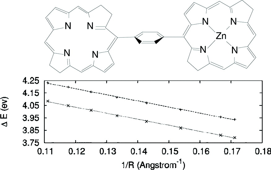
Additionally, CDFT can generate states with partial charges128, this is of particular importance, for example in metal–ligand CT processes, where the diabatic states can be generated by constraining the charge on the ligand and metal center.
Recently, the CDFT implementation of CPMD was tested against high-level wave function methods in the computation of electronic couplings for hole and excess electron transfer 37, 135. CDFT was shown to be on average within 5.3% of the benchmark calculations if 50% HF exchange was introduced (the average deviation goes up to 38.7% if HF exchange is not used).
2.3 Fragment Orbital DFT
The fragment orbital DFT or FODFT is a computationally low-cost method to calculate electronic couplings. This is because the wavefunctions of each diabatic state are approximated by the fronteer orbitals of the isolated donor/acceptor fragments 136, 137, 138. The underlying approximations in FODFT are that (1) the interactions between donor and acceptor have not effect on the orbital shape, (2) the coupling component related to orbitals below the fronteer is neglected (e.g. frozen core). In FODFT, the wavefunctions can be described by a single determinant of spin-orbitals , where i.e. the sum of the number of electrons of the neutral donor and acceptor. These determinants are built from the KS orbitals of the noninteracting isolated donor and acceptor fragments.
| (14) |
The Hamiltonian used to calculate the CT matrix elements is the KS-Hamiltonian. Namely,
| (15) |
where are the one–particle KS–Hamiltonians for either the ”a” diabat or the ”b” diabat. One feature of these Hamiltonians is that they are state dependent, thus, they are made of the combination of orbitals of donor and acceptor species at the given state. The transfer integral, or coupling between states, is calculated as:
| (16) |
Where above is the fronteer orbital for or . Recently, Kubas et al.37 have shown the differences of two FODFT flavors in the calculation of the hole transfer coupling for the HAB11 database. As we can see in Table 3, the implementation including orbitals in the KS Hamiltonian (indicated by in the table) as done in ADF 136 is more accurate than the implementation using one of the Hamiltonians in Eq. (2.3) (which is indicated by in the table).
| FODFT(2N-1) | FODFT(2N) | ADF(2N) | FODFTB | REF | |
|---|---|---|---|---|---|
| Ethylene | 367.7 | 389.2 | 388.4 | 343.7 | 519.2 |
| Acetylene | 316.9 | 345.8 | 345.3 | 212.0 | 460.7 |
| Cyclopropene | 418.8 | 443.7 | 439.4 | 367.4 | 536.6 |
| Cyclobutadiene | 323.3 | 346.9 | 345.6 | 261.6 | 462.7 |
| Cyclopentadiene | 343.3 | 360.6 | 358.7 | 283.2 | 465.8 |
| Furane | 315.6 | 334.0 | 333.7 | 280.3 | 440.3 |
| Pyrrole | 328.7 | 347.8 | 347.7 | 286.2 | 456.3 |
| Thiophene | 341.2 | 357.8 | 356.1 | 264.8 | 449.0 |
| Imidazole | 310.7 | 328.9 | 328.2 | 277.5 | 411.6 |
| Benzene | 342.4 | 353.5 | 354.1 | 299.9 | 435.2 |
| Phenol | 190.5 | 211.3 | 279.5 | 231.4 | 375.0 |
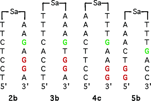
FODFT has been successfully applied to models of CT in molecular semiconductors 139, 140 and also for modeling CT in biosystems. In the following, we provide applications of FODFT to biological CT: such as the determination of the hole rates on DNA hairpins linked by stilbenedicarboxamide (Figure 6), and the elucidation of the electron transfer between two cofactors in the SO enzyme.
2.3.1 Hole transfer rates on DNA hairpins
The absolute rates were determined by using Marcus theory, in Eq. (1), where the electronic coupling was calculated according to FODFT and the superexchange regime (see section 2.1.1). Knowledge of the rates forward and backward (see table 4) enables one to determine the equilibrium constant and the free energy change . Comparable results with experimental results 141 were obtained.
| Sequence | ||
|---|---|---|
| 2b | 3.5 | 0.032 |
| (7.5) | (0.052) | |
| 3b | 39.8 | 0.093 |
| (6.7) | (0.049) | |
| 4b | 20.0 | 0.076 |
| () | (-0.02) | |
| 5b | 10.0 | 0.058 |
| () | () |
2.3.2 The Curious Case of Sulfite Oxidase
An interesting and elusive candidate for electron transfer studies is the Sulfite Oxidase protein143. For this protein, theory predicts an electron transfer rate between the cofactors (a heme and a molybdenum complex) about two orders of magnitude lower than what is measured experimentally2. To address this issue, Beratan et. al., using the Pathways model, suggested that the donor and the acceptor are joined together by a flexible tether144. As the tether allows the two cofactors to come sufficiently close to each other, electron transfer occurs at the rate shown by experiment. A recent simulation of this mechanism was carried out so that the protein was taken out of equilibrium and positioned in a new folded state featuring a much decreased cofactor distance (about 10 Å). However, recent pulsed electron paramagnetic resonance measurements143 indicated that the distance between the cofactors is unchanged on average from the one available in the crystal structure (32 Å). To approach this problem using FODFT, the crystal structure of the protein is obtained and only the chains of the protein between the two cofactors are considered. The chains of interest are broken into individual molecules and treated as separate bridges.
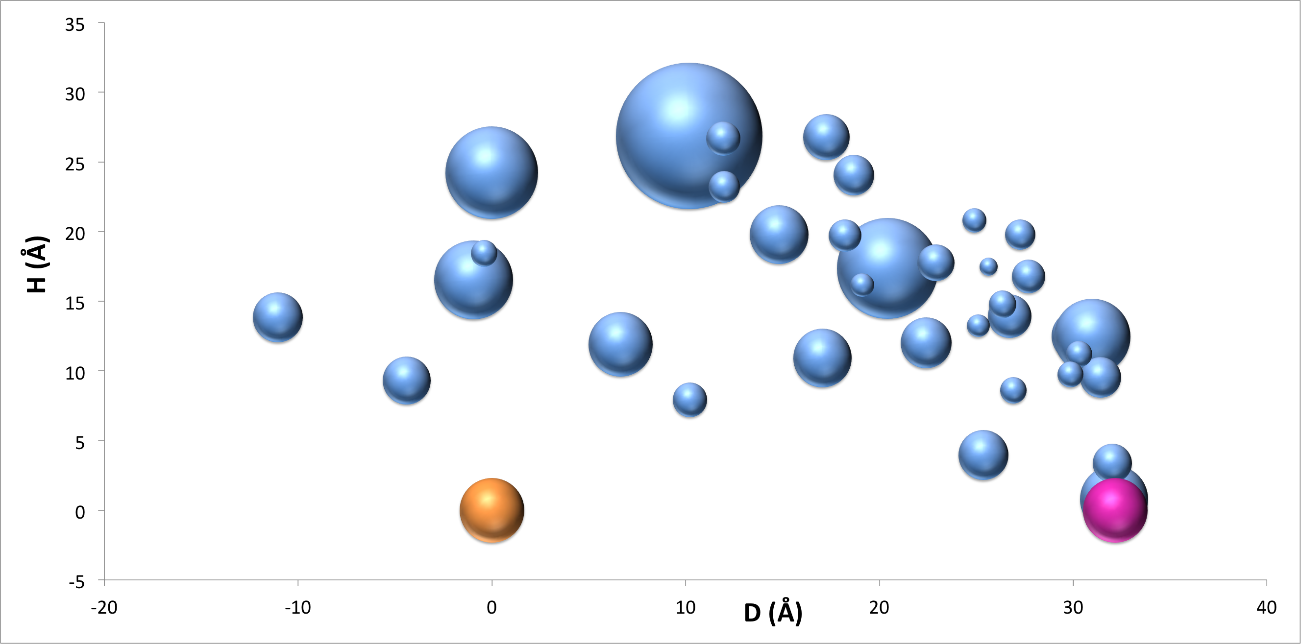
The FODFT computations that we present here will be part of a more in–depth study in a future publication145. Two ingredients are available from the simulations, the site energies, and the couplings between the sites. The energies of the hole transfer pathway for the electron transfer between the iron and the molybdenum is presented in Figure 7. With the aid of Koopman’s theorem, the HOMO energies computed with FODFT are taken here as a measure of the ionization potential of each site. The simulation was able to shed light on some very interesting aspects of the couplings and the energy landscapes. Looking at the energy landscape in Figure 7, it is clear that a path with highest couplings can be directly drawn between the donor and the acceptor. The landscape also shows the possibility of hopping stations–molecules that exist between the donor and acceptor and are close to them in energy. These two aspects of the landscape alone hint the possibility of a hole transfer occurring over the 32 Å. However, proteins are very complex structures with many variables such as size, dynamics and environment. Therefore, providing a quantitative analysis of the kinetic constant would require incorporating unbiased molecular dynamics and a more comprehensive structure to further characterize the role of these hopping stations.
2.4 Ultrafast computations of the electronic couplings: The AOM method
Recently, an ultrafast method to calculate electronic couplings was developed by Blumberger and coworkers135. The analytic overlap method or AOM is a useful method if CT simulations need to be coupled with molecular dynamics, like in proteins146 or in organic semiconductors147. This quest requires hundreds or maybe thousands of and site energy calculations. AOM offers an interesting alternative for such simulations. As in FODFT, AOM assumes that CT is only mediated by two SOMO orbitals (fronteer orbitals, similarly to FODFT), which correspond to each fragment. Then, small Slater type orbital basis for the valence states is generated. Thus the overlap integral is evaluated as follows:
AOM further assumes contributions only from -orbitals, particularly in organic compounds with conjugation, the p–orbital considered is that one perpendicular to the plane of conjugation.
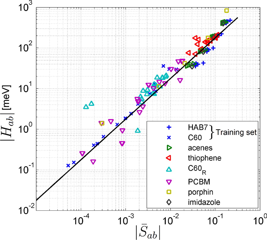
Correlation of the overlap and the electronic coupling given by FODFT is shown in Figure 8 for a set of dimers and their geometries. In this picture, a satisfactory linearity between these two parameters is witnessed, therefore reliable proportionality constants can be achieved. is used in order to obtain the constant C that can be used to get couplings of similar compounds. This first version of this method shows transferability for homo-dimers. However, transferability when non equivalent donor and acceptor systems are considered needs to be explored. Nevertheless, AOM’s speed makes it a very valuable option.
2.5 Note on orthogonality
When carrying out a large number of coupling calculations, one encounters all those low probability situations in which a method fails. In the case of FDE-ET, we probed a large number of so-called “difficult cases”. Specifically, we faced two limitations of the FDE-ET method. If the diabats are orthogonal, or quasi orthogonal, numerical inaccuracies arise in the inversion of the transition overlap matrix in equation 7. This is not specific to FDE-ET, but is a problem shared by all those methods that assume the diabatic states to be nonorthogonal 30, 148, 149, 150, 115. When they are orthogonal, some of the equations previously developed simply do not hold anymore. Yu et al. have applied equations similar to Eq. (5–8) and obtained a picture of the behavior of the electronic couplings in the photosynthetic reaction center, see Figure 9.
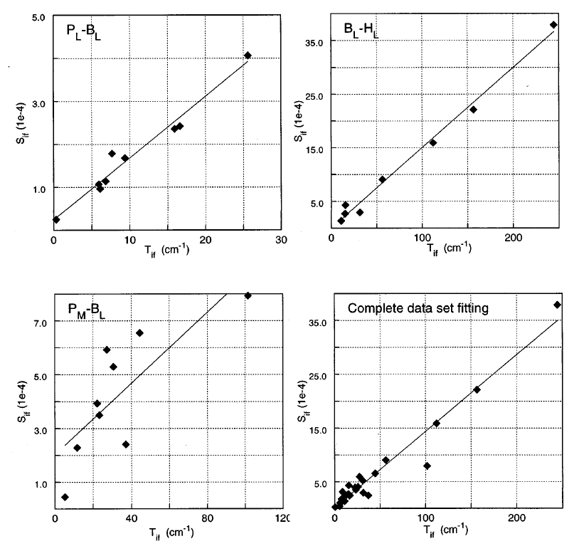
If we concentrate on the left-side panels, we notice that in some cases the electronic coupling is proportional to the coupling, but in other cases (see lower left panel) the coupling seems to behave somewhat erratically as a function of the diabatic overlap. To understand this, let us consider two distinct limiting cases: (1) orthogonality by symmetry considerations, and (2) spatial separation of the orbitals. We found that the second case is the predominant, as the distance between donor–acceptor increases the diabatic overlap becomes increasingly small. In the asymptotic limit 44, there is a linear relationship between the coupling, the diabatic energy difference, and the diabatic overlap. If the overlap is small due to case (2), the asymptotic formula is not expected to hold. This explains the apparently contraddictory results presented in Figure 9.
Regarding FDE-ET, both cases can be circumvented computationally by performing a singular value decomposition of the overlap matrix and then invert only those values which are larger than a threshold (i.e. Penrose inversion). For DNA presented in section 2.1.3, the default inversion threshold of was appropriate in most cases67. However, three systems stood out: AG, GA and TT nucleobase pairs. All the systems above showed erratic behavior of the computed couplings for some specific donor-acceptor distances, specifically 4.0 Å for AG, 3.5 Å and 8.0 Å for GA and 9.0 Å for TT. We found that at those distances, the near singularity of the overlap matrix due to symmetry considerations (case 1 above) was the source of the erratic behavior. To circumvent these numerical issues, a threshold of was adopted in these cases.
We thus conclude that although there is a formal relationship linking the diabatic overlap with the value of the coupling at large donor–acceptor distances 44, generally assuming linearity in the coupling vs. overlap (as mentioned briefly in the previous section regarding the AOM method) can lead to large errors in the magnitude of the computed couplings. In the future, inspired by a recent work by Evangelista et al.131, a more stable algorithm that invokes an orthogonalization first, and then the computation of the couplings will be developed in our group.
2.6 A fully semiempirical method: Pathways
Pathways 50, 2 is a semiempirical model which it is designed to reproduce electron transfer rates between cofactors in proteins.50, 151. In essence, Pathways includes the contributions to the electronic tunneling from a stepwise path covering all nonbonded interactions, as well as the bonded ones at the nearest neighbor level. Namely:
| (17) |
where are the steps the charge need to make from donor to acceptor. For example, a hydrogen bond is one of such steps. The above product is maximized by searching all possible steps that contribute to the tunneling. The coupling is further split into three kinds of interactions:
| (18) |
Pathways can yield reliable predictions of the electronic couplings, where the CT process in proteins are mediated by the interactions of a single or multiple configurations that the protein can adopt50. Pathways has been successfully applied to a number of CT processes in protein environment. For instance, the electron transfer between the proteins cytochrome c2 (cytc2) and the photosynthetic reaction center (RC)152 in order to determine the protein structural dependence of this CT reaction, also, to look at the impact of structural and conformational variations on the electronic coupling between the proteins methylamine dehydrogenase and amicyanin from Paracoccus denitrificans153.
3 High-accuracy electronic couplings
This section is devoted to describing those methods which are able to predict the electronic couplings accurately given a certain definition of the corresponding diabatic states. These methods start with a mathematical definition of diabatic states (usually a definition that involves localization of the electronic structure) such that the resulting states resemble the donor and acceptor states in the electron transfer reaction. Once this is achieved, an adiabatic-to-diabatic transformation matrix is generated which can be applied to the adiabatic Hamiltonian to result in the diabatic Hamiltonian featuring the sought electronic couplings in the off-diagonal elements. Usually, an accurate, wave function based level of theory is used for computing the adiabatic states and Hamiltonian 29. Examples of such techniques are, the Generalized Mulliken–Hush method developed by Newton and Cave30, 29, 154, Boys and Edmiston-Ruedenberg localizations of Subotnik et al.154, 155, 38, and fragment charge difference proposed by Voityuk and Rösch 42, 156. Their utility lies on the possibility of a very accurate computation of the corresponding adiabatic states, as was done for the hole transfer on –stack DNA nucleobases at a CASPT2 and CASSCF level of theory accomplished by Voityuk et. al35. That computation has served as the benchmark reference for many recently developed methodologies137, 99, 49, 157.
Taking this as a motivation, let us briefly introduce each of the above methods, followed by some examples in which these methods where employed.
3.1 GMH method
In the two-state model, the charge localized diabatic states are related with the adiabatic states through the formula:
| (19) |
where are the energies of the donor and acceptor states respectively, and are the energies of the adiabatic states, this means the energy of the ground state () and the first excited state (). and can be obtained with any quantum chemistry method, however when highly accurate wavefunctions methods are employed also the resulting couplings will be of high quality. We now distinguish two cases: a symmetric case, for instance homo–dimers, and the general asymmetric case. In the symmetric case, we have that , and thus the electronic coupling does not depend on the diabatization procedure. Namely,
| (20) |
is the difference on energy between the adiabatic ground state and the first adiabatic excited state .
For asymmetric cases the GMH method prescribes that the proper diabatic states are those that diagonalize the adiabatic dipole moment matrix. In the two–state problem this is calculated as follows:
| (21) |
where , with being the dipole moment in the direction of the electron transfer. The power of GMH lies on the way one calculates the adibatic states. As we have seen through all sections is that the authors benchmark their own method by calculating proper diabatic couplings by using ab initio methods as multireference CI37 and CASPT2 35.
As an example of this method, let us discuss the very first example given by Cave and Newton on their paper34. The system , the transfer of a hole is done over the Zn atoms. However, the water molecule, which is located at a fixed distance opposite to the Zn distance, causes an energy splitting of the Zn orbitals. Thus the electronic coupling is determined for the following diabatic states:
| (22) | ||||
| (23) | ||||
| (24) | ||||
| (25) |
where the transition are between the orbitals of the diabatic states (prime correspond to the acceptor state) (s-s’), (p-p’), (s-p’) and (p-s’). CASSCF wavefunction method was used in all calculations. In Table 5 we collect the values for the different couplings and . There the analysis of the distance dependence of the coupling is carried out for several distances. Note that for an infinite distance, the couplings for (s-p’) and (p-s’) are equal.
| (Å) | ||||
|---|---|---|---|---|
| (a) | ||||
| 4.0 | 28.3 | 23.6 | 50.4 | 42.7 |
| 5.0 | 10.5 | 13.0 | 51.7 | 22.3 |
| 6.0 | 3.73 | 7.55 | 41.1 | 10.1 |
| 7.0 | 1.09 | 4.23 | 21.8 | 4.08 |
| 8.0 | 0.340 | 2.57 | 13.8 | 1.62 |
| 9.0 | 0.0958 | 1.44 | 7.36 | 0.611 |
| 2.28 | 1.11 | 0.81 | 1.71 | |
| (b) | ||||
| 4.0 | 29.7 | 34.4 | 59.3 | 41.7 |
| 5.0 | 7.95 | 14.7 | 38.5 | 22.1 |
| 6.0 | 2.34 | 7.83 | 19.1 | 9.44 |
| 7.0 | 0.698 | 4.25 | 9.56 | 3.84 |
| 8.0 | 0.203 | 2.27 | 4.78 | 1.51 |
| 9.0 | 0.0558 | 1.16 | 2.32 | 0.574 |
| 2.49 | 1.32 | 1.32 | 1.74 |
3.2 Other adiabatic-to-diabatic transformation methods
Inspired by GMH, the electronic coupling can generally be obtained by rotating the corresponding adiabtic states into a set of diabatic states. Thus, each diabatic state can be expressed as a linear combination of rotated adiabatic states as154:
| (26) |
Under specific assumptions of the nature of the system–bath interaction (the following is valid for the condensed phase), the coupling can be estimated by constructing diabatic states based on Boys, Edminton–Ruedenberg (ER) or von Niessen–Edminton–Ruedenberg (VNER) localizations. In Boys diabatization, the bath exerts a linear electrostatic potential on the system, thus the rotation matrix can be found by minimizing the following localizing function39, 24:
| (27) |
Boys localization was shown to be equivalent to GMH for CT reactions 39.
ER diabatization, dictates that the bath exerts an electrostatic potential that responds linearly to the field generated by the molecular system (sum of donor and acceptor) system:
| (28) |
In VNER diabatization, the bath exerts an electrostatic potential that responds linearly to the field of the total system, but the interaction potential is a Dirac delta function:
| (29) |
Just like GMH, once diabats are generated, the electronic coupling readily arises from the off-diagonal element of the electronic Hamiltonian and is equal to:
| (30) |
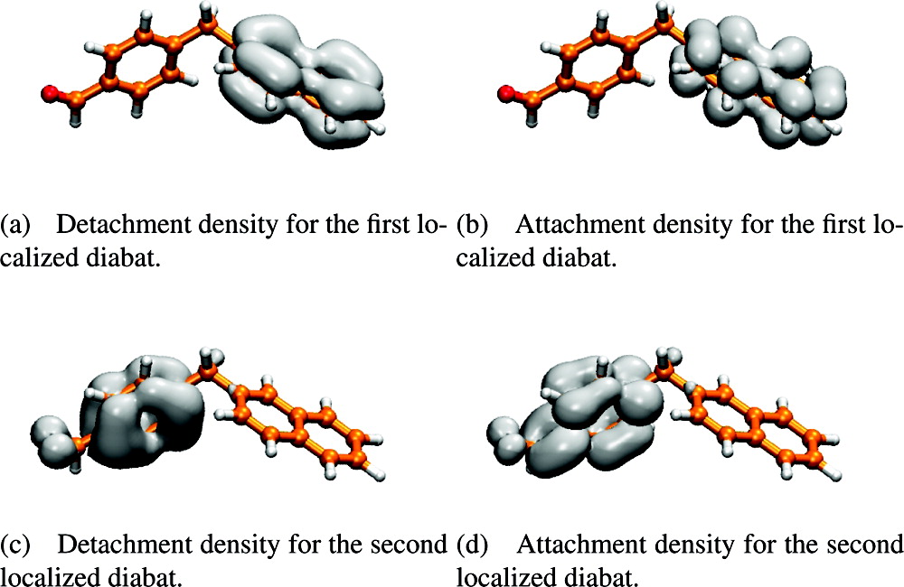
For a series of bridge mediated excitation energy transfer experiments, where the donor is benzaldehyde and the acceptor naphthalene159, 160, the transfer rates and couplings were calculated using Boys diabatization. In Figure 10 it is shown how well the orbitals are localized on either donor or acceptor edges in the various diabatic states. Although this computation does not concern a CT process, we want to stress the ability of this localization procedures in generating true diabatic states.
3.3 Fragment charge difference
Similarly to the GMH method, the fragment charge difference (FCD) method yields a donor to acceptor coupling 42:
| (31) |
where and are the donor–acceptor charges differences in the respective adiabatic states and . is the off diagonal term and is defined in a general form as , i.e. the difference of the populations of the transition charges.
Finally, when donor and acceptor are in resonance, i.e when or , .
FCD method and its simplfied from (SFCD) are compared against GMH in the calculation of for two Watson-Crick pairs and . In Table 6, the couplings calculated from FCD are in good agreement with GMH, the SCFD is also quite reasonable. Because the energy gap between donor and acceptor are large, the charge is completely localized on purines (lowest IP). However, if an electric field F (water molecule for instance) is tuned on near the pairs, the energy gap is then reduced and the coupling strength is enhanced. Overall, FCD is another good alternative to compute accurate couplings. However, the computation of the charges and the transition charges is dependent on the specific population analysis chosen. To our knowledge, only the Mulliken population analysis was used so far (i.e. the transition charges are evaluated on the basis of the MO coefficients over the atomic orbital basis set).
| GMH | SFCD | FCD | ||||
|---|---|---|---|---|---|---|
| Basis set | ||||||
| GC | 6-31G* | 2.163 | 2.159 | 0.0569 | 0.0663 | 0.0547 |
| 6-311++G** | 2.092 | 2.086 | 0.0679 | 0.0760 | 0.0705 | |
| AT | 6-31G* | 1.505 | 1.502 | 0.0421 | 0.0524 | 0.0363 |
| 6-311++G** | 1.462 | 1.459 | 0.0474 | 0.0528 | 0.0425 |
4 Practical Aspects: A protocol for running FDE-ET calculations
In order to obtain the electronic coupling for a CT reaction using FDE-ET, three different single point (SP) calculations have to be performed. FDE-ET is available in ADF51. In Figures 11, 12 and 13, the input files corresponding to the FDE-ET methodology are described. First, a single point calculation for each isolated fragment present in the system is carried out. This gives the initial density and energy of each subsystem without any interaction between them. It is important to save the check point files (TAPE files in ADF), because they contain all fragment information needed in the subsequent calculations. Following all SP jobs for each isolated fragment, an FDE calculation is performed by taking into the account the whole supramolecular structure. So that, we create a diabatic state for each of the present subsystems. This is done by placing a charge different from neutrality in each subsystem, see Figure 12. In this manner, two different directories are made: one in which an FDE calculation is carried out with subsystem 1 positively charged and one where subsystem 2 has the positive charge. In both cases, the SCF converges on the basis of subsystem DFT, thus, a series of three freeze–and–thaw procedure are done for each subsystem in each diabat.


Once both isolated and embedded densities are obtained from the FDE calculations, a electrontransfer job is run whose purpose is to compute Eq. (5–8). As in the FDE calculation, the information about the fragment is of paramount importance in this electrontransfer job. In figure 13, the input file that calculates the diabatic energies and the electronic coupling between them is showed, in pink we can see that the check point files (t21.emb.rho* in the figure) corresponding to the embedded fragments in each diabat is copied directly to the ET directory, where the electrontransfer calculation is done. These files are renamed as fragA*.t21 for those ones from the diabat A (positive charge on the donor fragment) and fragB*.t21 for those that come from diabat B (positive charge on the acceptor fragment). It is worth mentioning that special care has to be taken in the management of the file names. As it is illustrated in Figure 13, the fragments are numerated as 1 and 2, that means that the charge, departs from fragA1 while fragA2 is neutral and arrive to fragB2 while fragB1 becomes neutral. This is very important when the system is comprised of more than two fragments, and the charge is moving throughout all of them.
5 Conclusions and future directions
To conclude, we have presented our (fairly subjective) view of what tools are available nowadays to compute electronic couplings for charge transfer processes. We have surveyed in detail the FDE-ET method simply because we are among the developers of this method. Other methods based on DFT, and those that are best suited for being coupled with wavefunction based methods have also been discussed. The discussion also touches on the strengths and limitations of the various methods.
When discussing the practical aspect of a coupling calculation, one must expose completely the methodology. We have done so for the FDE-ET method, and provided the reader with a step-by-step protocol on how to run such computations. This is important also for outsiders (such as experimentalists) as they can appreciate the kind of effort the theoretitians have to put in computing quantities relevant for the interpretation of the experiments.
We apologize in advance to those authors who have developed all those methods that we have omitted from this presentation. Admittedly, we provide here a subjective view of the field.
6 Acknowledgements
This work was funded by a grant from the National Science Foundation, Grant CBET-1438493. We also acknowledge Dr. Eric Klein for bringing to our attention the problematics related to the electron transfer dynamics in the Sulfite Oxidase.
References
- Gray and Winkler 1996 Gray, H. B.; Winkler, J. R. Annu. Rev. Biochem. 1996, 65, 537–561
- Kawatsu and Beratan 2006 Kawatsu, T.; Beratan, D. N. Chem. Phys. 2006, 326, 259 – 269
- Larsson 1983 Larsson, S. J. Chem. Soc., Faraday Trans. 2 1983, 79, 1375–1388
- Farid et al. 1993 Farid, R. S.; Moser, C. C.; Dutton, P. L. Current Opinion in Structural Biology 1993, 3, 225 – 233
- Winkler et al. 2005 Winkler, J. R.; Gray, H. B.; Prytkova, T. R.; Kurnikov, I. V.; Beratan, D. N. Bioelectronics; Wiley-VCH Verlag GmbH & Co. KGaA, 2005; pp 15–33
- Trumpower and Gennis 1994 Trumpower, B. L.; Gennis, R. B. Annu. Rev. Biochem. 1994, 63, 675–716, PMID: 7979252
- Bixon et al. 1999 Bixon, M.; Giese, B.; Wessely, S.; Langenbacher, T.; Michel-Beyerle, M. E.; Jortner, J. Proc. Natl. Acad. Sci. USA 1999, 96, 11713–11716
- Giese 2004 Giese, B. In Longe-Range Charge Transfer in DNA I; Schuster, G., Ed.; Topics in Current Chemistry; Springer Berlin, Heidelberg, 2004; Vol. 236; pp 27–44
- Giese 2002 Giese, B. Annu. Rev. Biochem. 2002, 71, 51–70
- Stowell et al. 1997 Stowell, M. H. B.; McPhillips, T. M.; Rees, D. C.; Soltis, S. M.; Abresch, E.; Feher, G. Science 1997, 276, 812–816
- Balabin and Onuchic 2000 Balabin, I. A.; Onuchic, J. N. Science 2000, 290, 114–117
- Troisi 2011 Troisi, A. Chem. Soc. Rev. 2011, 40, 2347–2358
- McMahon and Troisi 2011 McMahon, D. P.; Troisi, A. Phys. Chem. Chem. Phys. 2011, 13, 10241–10248
- Marcus 1956 Marcus, R. A. J. Chem. Phys. 1956, 24, 966–978
- Marcus and Sutin 1985 Marcus, R. A.; Sutin, N. Biochim. Biophys. Acta 1985, 811, 265–322
- Landau 1932 Landau, L. D. Physics of the Soviet Union 1932, 2, 46–51
- Zener 1932 Zener, C. Proc. R. Soc. A 1932, 137, 696–702
- Condon 1928 Condon, E. Phys. Rev. 1928, 32, 858–872
- Franck and Dymond 1926 Franck, J.; Dymond, E. G. Trans. Faraday Soc. 1926, 21, 536–542
- Ladanyi and Skaf 1993 Ladanyi, B. M.; Skaf, M. S. Annu. Rev. Phys. Chem. 1993, 44, 335–368
- Ingram et al. 2003 Ingram, J. A.; Moog, R. S.; Ito, N.; Biswas, R.; Maroncelli, M. J. Phys. Chem. B 2003, 107, 5926–5932
- Maroncelli 1993 Maroncelli, M. J. Mol. Liq. 1993, 57, 1
- Nitzan 2006 Nitzan, A. Chemical Dynamics in Condensed Phases; Oxford University Press: Oxford, 2006
- Subotnik et al. 2009 Subotnik, J. E.; Cave, R. J.; Steele, R. P.; Shenvi, N. J. Chem. Phys. 2009, 130, 234102
- Van Voorhis et al. 2010 Van Voorhis, T.; Kowalczyk, T.; Kaduk, B.; Wang, L.-P.; Cheng, C.-L.; Wu, Q. Annu. Rev. Phys. Chem. 2010, 61, 149–170
- London 1932 London, F. Z. Phys. 1932, 74, 143
- Mead and Truhlar 1982 Mead, C. A.; Truhlar, D. G. J. Chem. Phys. 1982, 77, 6090–6098
- Pavanello and Neugebauer 2011 Pavanello, M.; Neugebauer, J. J. Chem. Phys. 2011, 135, 134113
- Cave and Newton 1997 Cave, R. J.; Newton, M. D. J. Chem. Phys. 1997, 106, 9213–9226
- Newton 1991 Newton, M. D. Chem. Rev. 1991, 91, 767–792
- Warshel and Weiss 1980 Warshel, A.; Weiss, R. M. J. Am. Chem. Soc. 1980, 102, 6218–6226
- Mulliken 1952 Mulliken, R. S. J. Am. Chem. Soc. 1952, 64, 811
- Hush 1968 Hush, N. Electrochimica Acta 1968, 13, 1005 – 1023
- Cave and Newton 1996 Cave, R. J.; Newton, M. D. Chem. Phys. Lett. 1996, 249, 15 – 19
- Blancafort and Voityuk 2006 Blancafort, L.; Voityuk, A. A. J. Phys. Chem. A 2006, 110, 6426–6432
- Voityuk 2007 Voityuk, A. A. J. Chem. Phys. 2007, 128, 115101
- Adam et al. 2014 Adam, K.; Hoffmann, F.; Heck, A.; Oberhofer, H.; Elstner, M.; Blumberger, J. J. Chem. Phys. 2014, 140, 104105
- Fatehi et al. 2013 Fatehi, S.; Alguire, E.; Subotnik, J. E. J. Chem. Phys. 2013, 139, –
- Subotnik et al. 2008 Subotnik, J. E.; Yeganeh, S.; Cave, R. J.; Ratner, M. A. J. Chem. Phys. 2008, 129, –
- Kaduk et al. 2012 Kaduk, B.; Kowalczyk, T.; Van Voorhis, T. Chem. Rev. 2012, 112, 321–370
- Wu and Van Voorhis 2006 Wu, Q.; Van Voorhis, T. J. Chem. Phys. 2006, 125, 164105
- Voityuk and Rösch 2002 Voityuk, A. A.; Rösch, N. J. Chem. Phys. 2002, 117, 5607
- Hsu et al. 2008 Hsu, C.-P.; You, Z.-Q.; Chen, H.-C. J. Phys. Chem. C 2008, 112, 1204–1212
- Migliore 2011 Migliore, A. J. Chem. Theory Comput. 2011, 7, 1712–1725
- Migliore et al. 2006 Migliore, A.; Corni, S.; Di Felice, R.; Molinari, E. J. Chem. Phys. 2006, 124, –
- Migliore et al. 2009 Migliore, A.; Sit, P. H.-L.; Klein, M. L. J. Chem. Theory Comput. 2009, 5, 307–323
- Cembran et al. 2009 Cembran, A.; Song, L.; Mo, Y.; Gao, J. J. Chem. Theory Comput. 2009, 5, 2702–2716
- Pavanello and Neugebauer 2011 Pavanello, M.; Neugebauer, J. J. Chem. Phys. 2011, 135, 234103
- Pavanello et al. 2013 Pavanello, M.; Van Voorhis, T.; Visscher, L.; Neugebauer, J. J. Chem. Phys. 2013, 138, 054101
- Beratan et al. 1991 Beratan, D.; Betts, J.; Onuchic, J. Science 1991, 252, 1285–1288
- 51 Amsterdam Density Functional program. Theoretical Chemistry, Vrije Universiteit, Amsterdam, URL: http://www.scm.com
- Jacob and Neugebauer 2014 Jacob, C. R.; Neugebauer, J. WIREs: Comput. Mol. Sci. 2014, 4, 325–362
- Wesolowski and Warshel 1993 Wesolowski, T. A.; Warshel, A. J. Chem. Phys. 1993, 97, 8050
- Wesolowski 2006 Wesolowski, T. A. In Computational Chemistry: Reviews of Current Trends; Leszczynski, J., Ed.; World Scientific: Singapore, 2006; Vol. 10; pp 1–82
- Neugebauer et al. 2005 Neugebauer, J.; Louwerse, M. J.; Baerends, E. J.; Wesolowski, T. A. J. Chem. Phys. 2005, 122, 094115
- Neugebauer 2010 Neugebauer, J. Phys. Rep. 2010, 489, 1–87
- Humbert-Droz et al. 2013 Humbert-Droz, M.; Zhou, X.; Shedge, S.; Wesolowski, T. Theor. Chem. Acc. 2013, 133
- Jacob and Visscher 2006 Jacob, C. R.; Visscher, L. J. Chem. Phys. 2006, 125, 194104
- Bulo et al. 2008 Bulo, R. E.; Jacob, C. R.; Visscher, L. J. Phys. Chem. A 2008, 112, 2640–2647
- Neugebauer et al. 2005 Neugebauer, J.; Louwerse, M. J.; Belanzoni, P.; Wesolowski, T. A.; Baerends, E. J. J. Chem. Phys. 2005, 123, 114101
- Kevorkyants et al. 2013 Kevorkyants, R.; Wang, X.; Close, D. M.; Pavanello, M. J. Phys. Chem. B 2013, 117, 13967–13974
- Wesolowski 1999 Wesolowski, T. A. Chem. Phys. Lett. 1999, 311, 87–92
- Neugebauer et al. 2010 Neugebauer, J.; Curutchet, C.; Munioz-Losa, A.; Mennucci, B. J. Chem. Theory Comput. 2010, 6, 1843–1851
- Casida and Wesolowski 2004 Casida, M. E.; Wesolowski, T. A. Int. J. Quantum Chem. 2004, 96, 577–588
- Pavanello 2013 Pavanello, M. J. Chem. Phys. 2013, 138, 204118
- García-Lastra et al. 2006 García-Lastra, J. M.; Wesolowski, T. A.; Barriuso, M. T.; Aramburu, J. A.; Moreno, M. J. Phys.: Condens. Matter 2006, 18, 1519–1534
- Ramos and Pavanello 2014 Ramos, P.; Pavanello, M. J. Chem. Theory Comput. 2014, 10, 2546–2556
- Solovyeva et al. 2014 Solovyeva, A.; Pavanello, M.; Neugebauer, J. J. Chem. Phys. 2014, 140, 164103
- Jacob et al. 2008 Jacob, C. R.; Neugebauer, J.; Visscher, L. J. Comput. Chem. 2008, 29, 1011–1018
- Helgaker et al. 2001 Helgaker, T.; Jensen, H. J. A.; Jørgensen, P.; Olsen, J.; Ruud, K.; Ågren, H.; Auer, A. A.; Bak, K. L.; Bakken, V.; Christiansen, O.; Coriani, S.; Dahle, P.; Dalskov, E. K.; Enevoldsen, T.; Fernandez, B.; Hättig, C.; Hald, K.; Halkier, A.; Heiberg, H.; Hettema, H.; Jonsson, D.; Kirpekar, S.; Kobayashi, R.; Koch, H.; Mikkelsen, K. V.; Norman, P.; Packer, M. J.; Pedersen, T. B.; Ruden, T. A.; Sanchez, A.; Saue, T.; Sauer, S. P. A.; Schimmelpfennig, B.; Sylvester-Hvid, K. O.; Taylor, P. R.; Vahtras, O. DALTON, a molecular electronic structure program, Release 1.2. 2001
- Aidas et al. 2014 Aidas, K.; Angeli, C.; Bak, K. L.; Bakken, V.; Bast, R.; Boman, L.; Christiansen, O.; Cimiraglia, R.; Coriani, S.; Dahle, P.; Dalskov, E. K.; Ekström, U.; Enevoldsen, T.; Eriksen, J. J.; Ettenhuber, P.; Fernández, B.; Ferrighi, L.; Fliegl, H.; Frediani, L.; Hald, K.; Halkier, A.; Hättig, C.; Heiberg, H.; Helgaker, T.; Hennum, A. C.; Hettema, H.; Hjertenæs, E.; Høst, S.; Høyvik, I.-M.; Iozzi, M. F.; Jansík, B.; Jensen, H. J. A.; Jonsson, D.; Jørgensen, P.; Kauczor, J.; Kirpekar, S.; Kjærgaard, T.; Klopper, W.; Knecht, S.; Kobayashi, R.; Koch, H.; Kongsted, J.; Krapp, A.; Kristensen, K.; Ligabue, A.; Lutnæs, O. B.; Melo, J. I.; Mikkelsen, K. V.; Myhre, R. H.; Neiss, C.; Nielsen, C. B.; Norman, P.; Olsen, J.; Olsen, J. M. H.; Osted, A.; Packer, M. J.; Pawlowski, F.; Pedersen, T. B.; Provasi, P. F.; Reine, S.; Rinkevicius, Z.; Ruden, T. A.; Ruud, K.; Rybkin, V. V.; Sałek, P.; Samson, C. C. M.; de Merás, A. S.; Saue, T.; Sauer, S. P. A.; Schimmelpfennig, B.; Sneskov, K.; Steindal, A. H.; Sylvester-Hvid, K. O.; Taylor, P. R.; Teale, A. M.; Tellgren, E. I.; Tew, D. P.; Thorvaldsen, A. J.; Thøgersen, L.; Vahtras, O.; Watson, M. A.; Wilson, D. J. D.; Ziolkowski, M.; Ågren, H. WIREs: Computational Molecular Science 2014, 4, 269–284
- Goodpaster et al. 2010 Goodpaster, J. D.; Ananth, N.; ; Manby, F. R.; Miller, III, T. F. J. Chem. Phys. 2010, 133, 084103
- Shao et al. 2006 Shao, Y.; Fusti-Molnar, L.; Jung, Y.; Kussmann, J.; Ochsenfeld, C.; Brown, S. T.; Gilbert, A. T. B.; Slipchenko, L. V.; Levchenko, S. V.; O’Neill, D. P.; DiStasio Jr., R. A.; Lochan, R. C.; Wang, T.; Beran, G. J. O.; Besley, N. A.; M., J.; Herbert,; Lin, C. Y.; Voorhis, T. V.; Chien, S. H.; Sodt, A.; Steele, R. P.; Rassolov, V. A.; Maslen, P. E.; Korambath, P. P.; Adamson, R. D.; Austin, B.; Baker, J.; Byrd, E. F. C.; Dachsel, H.; Doerksen, R. J.; Dreuw, A.; Dunietz, B. D.; Dutoi, A. D.; Furlani, T. R.; Gwaltney, S. R.; Heyden, A.; Hirata, S.; Hsu, C.-P.; Kedziora, G.; Khalliulin, R. Z.; Klunzinger, P.; Lee, A. M.; Lee, M. S.; Liang, W.; Lotan, I.; Nair, N.; Peters, B.; Proynov, E. I.; Pieniazek, P. A.; Rhee, Y. M.; Ritchie, J.; Rosta, E.; Sherrill, C. D.; Simmonett, A. C.; Subotnik, J. E.; Woodcock III, H. L.; Zhang, W.; Bell, A. T.; Chakraborty, A. K.; Chipman, D. M.; Keil, F. J.; Warshel, A.; Hehre, W. J.; Schaefer III, H. F.; Kong, J.; Krylov, A. I.; Gill, P. M. W.; Head-Gordon, M. Phys. Chem. Chem. Phys. 2006, 8, 3172 – 3191, URL: http://www.q-chem.com
- 74 R. Ahlrichs et al., http://www.cosmologic.de/turbomole.html
- Ahlrichs et al. 1989 Ahlrichs, R.; Bär, M.; Häser, M.; Horn, H.; Kölmel, C. Chem. Phys. Lett. 1989, 162, 165–169
- Laricchia et al. 2010 Laricchia, S.; Fabiano, E.; Sala, F. D. J. Chem. Phys. 2010, 133, 164111
- cp 2 CP2k: A General Program to Perform Molecular Dynamics Simulations. URL: http://www.cp2k.org
- Iannuzzi et al. 2006 Iannuzzi, M.; Kirchner, B.; Hutter, J. Chem. Phys. Lett. 2006, 421, 16–20
- Clark et al. 2005 Clark, S. J.; Segall, M. D.; Pickard, C. J.; Hasnip, P. J.; Probert, M. I. J.; Refson, K.; Payne, M. C. Z. Kristallogr. 2005, 220, 567–570
- Lahav and Klüner 2007 Lahav, D.; Klüner, T. J.Phys: Cond. Mat 2007, 19, 226001
- Genova et al. 2013 Genova, A.; Krishtal, A.; Ceresoli, D.; Pavanello, M. Frozen Density Embedding Project of Quantum Espresso: http://qe-forge.org/gf/project/fde. 2013; \urlhttp://qe-forge.org/gf/project/fde
- Giannozzi et al. 2009 Giannozzi, P.; Baroni, S.; Bonini, N.; Calandra, M.; Car, R.; Cavazzoni, C.; Ceresoli, D.; Chiarotti, G. L.; Cococcioni, M.; Dabo, I.; Dal Corso, A.; de Gironcoli, S.; Fabris, S.; Fratesi, G.; Gebauer, R.; Gerstmann, U.; Gougoussis, C.; Kokalj, A.; Lazzeri, M.; Martin-Samos, L.; Marzari, N.; Mauri, F.; Mazzarello, R.; Paolini, S.; Pasquarello, A.; Paulatto, L.; Sbraccia, C.; Scandolo, S.; Sclauzero, G.; Seitsonen, A. P.; Smogunov, A.; Umari, P.; Wentzcovitch, R. M. J. Phys.: Cond. Mat. 2009, 21, 395502
- Genova et al. 2014 Genova, A.; Ceresoli, D.; Pavanello, M. J. Chem. Phys. 2014, 141, 174101
- Gonze et al. 2009 Gonze, X.; Amadon, B.; Anglade, P.-M.; Beuken, J.-M.; Bottin, F.; Boulanger, P.; Bruneval, F.; Caliste, D.; Caracas, R.; Côté, M.; Deutsch, T.; Genovese, L.; Ghosez, P.; Giantomassi, M.; Goedecker, S.; Hamann, D.; Hermet, P.; Jollet, F.; Jomard, G.; Leroux, S.; Mancini, M.; Mazevet, S.; Oliveira, M.; Onida, G.; Pouillon, Y.; Rangel, T.; Rignanese, G.-M.; Sangalli, D.; Shaltaf, R.; Torrent, M.; Verstraete, M.; Zerah, G.; Zwanziger, J. Computer Physics Communications 2009, 180, 2582 – 2615
- Govind et al. 1998 Govind, N.; Wang, Y. A.; da Silva, A. J. R.; Carter, E. A. Chem. Phys. Lett. 1998, 295, 129–134
- Senatore and Subbaswamy 1986 Senatore, G.; Subbaswamy, K. R. Phys. Rev. B 1986, 34, 5754–5757
- Cortona 1991 Cortona, P. Phys. Rev. B 1991, 44, 8454
- Kolos and Radzio 1978 Kolos, W.; Radzio, E. Int. J. Quantum Chem. 1978, 13, 627–634
- Gordon and Kim 1972 Gordon, R. G.; Kim, Y. S. J. Chem. Phys. 1972, 56, 3122
- Götz et al. 2009 Götz, A.; Beyhan, S.; Visscher, L. J. Chem. Theory Comput. 2009, 5, 3161–3174
- Wesolowski et al. 1996 Wesolowski, T. A.; Chermette, H.; Weber, J. J. Chem. Phys. 1996, 105, 9182
- Ludeña et al. 2003 Ludeña, E. V.; Karasiev, V. V.; Nieto, P. Theor. Chem. Acc. 2003, 110, 395–402
- Ludeña and Karasiev 2002 Ludeña, E. V.; Karasiev, V. V. Reviews of Modern Quantum Chemistry; 2002; Chapter 22, pp 612–665
- Fux et al. 2008 Fux, S.; Kiewisch, K.; Jacob, C. R.; Neugebauer, J.; Reiher, M. Chem. Phys. Lett. 2008, 461, 353–359
- Kiewisch et al. 2008 Kiewisch, K.; Eickerling, G.; Reiher, M.; Neugebauer, J. J. Chem. Phys. 2008, 128, 044114
- Wesolowski and Weber 1996 Wesolowski, T. A.; Weber, J. Chem. Phys. Lett. 1996, 248, 71–76
- Luber 2014 Luber, S. J. Chem. Phys. 2014, 141, 234110
- Krishtal et al. 2015 Krishtal, A.; Sinha, D.; Genova, A.; Pavanello, M. J. Phys.: Condens. Matter 2015, accepted
- Kumar and Sevilla 2011 Kumar, A.; Sevilla, M. D. J. Phys. Chem. B 2011, 115, 4990–5000
- Mantz et al. 2007 Mantz, Y. A.; Gervasio, F. L.; Laino, T.; Parrinello, M. Phys. Rev. Lett. 2007, 99, 058104
- Burin and Uskov 2008 Burin, A. L.; Uskov, D. B. J. Chem. Phys. 2008, 129, 025101
- Solovyeva et al. 2012 Solovyeva, A.; Pavanello, M.; Neugebauer, J. J. Chem. Phys. 2012, 136, 194104
- Ramos et al. 0 Ramos, P.; Papadakis, M.; Pavanello, M. J. Phys. Chem. B 0, 0, null, PMID: 25845645
- Jacob et al. 2007 Jacob, C. R.; Beyhan, S. M.; Visscher, L. J. Chem. Phys. 2007, 126, 234116
- Dulak and Wesolowski 2006 Dulak, M.; Wesolowski, T. A. J. Chem. Phys. 2006, 124, 164101
- Wang and Carter 2000 Wang, Y. A.; Carter, E. A. In Theoretical Methods in Condensed Phase Chemistry; Schwartz, S. D., Ed.; Kluwer: Dordrecht, 2000; pp 117–184
- Thakkar 1992 Thakkar, A. J. Phys. Rev. A 1992, 46, 6920–6924
- Perdew 1992 Perdew, J. P. Phys. Lett. A 1992, 165, 79–82
- Fux et al. 2010 Fux, S.; Jacob, C. R.; Neugebauer, J.; Visscher, L.; Reiher, M. J. Chem. Phys. 2010, 132, 164101
- Lewis et al. 1997 Lewis, F. D.; Wu, T.; Zhang, Y.; Letsinger, R. L.; Greenfield, S. R.; Wasielewski, M. R. Science 1997, 277, 673–676
- Jortner et al. 1998 Jortner, J.; Bixon, M.; Langenbacher, T.; Michel-Beyerle, M. E. Proc. Natl. Acad. Sci. USA 1998, 95, 12759–12765
- Renaud et al. 2013 Renaud, N.; Berlin, Y. A.; Lewis, F. D.; Ratner, M. A. J. Am. Chem. Soc. 2013, 135, 3953–3963
- Giese 2000 Giese, B. Acc. Chem. Res. 2000, 33, 631–636
- Thom and Head-Gordon 2009 Thom, A. J. W.; Head-Gordon, M. J. Chem. Phys. 2009, 131, 124113
- Farazdel et al. 1990 Farazdel, A.; Dupuis, M.; Clementi, E.; Aviram, A. J. Am. Chem. Soc. 1990, 112, 4206–4214
- Mayer 2002 Mayer, I. Int. J. Quantum Chem. 2002, 90, 63–65
- Hatcher et al. 2008 Hatcher, E.; Balaeff, A.; Keinan, S.; Venkatramani, R.; Beratan, D. N. J. Am. Chem. Soc. 2008, 130, 11752–11761
- Marcus 1987 Marcus, R. Chem. Phys. Lett. 1987, 133, 471 – 477
- Voityuk 2012 Voityuk, A. A. Phys. Chem. Chem. Phys. 2012, 14, 13789–13793
- Evenson and Karplus 1992 Evenson, J. W.; Karplus, M. J. Chem. Phys. 1992, 96, 5272–5278
- Löwdin 1963 Löwdin, P.-O. J. Mol. Spectrosc. 1963, 10, 12 – 33
- Larsson 1981 Larsson, S. J. Am. Chem. Soc. 1981, 103, 4034–4040
- Priyadarshy et al. 1996 Priyadarshy, S.; Skourtis, S. S.; Risser, S. M.; Beratan, D. N. J. Chem. Phys. 1996, 104, 9473–9481
- Fradelos and Wesołowski 2011 Fradelos, G.; Wesołowski, T. A. J. Phys. Chem. A 2011, 115, 10018–10026
- Fradelos and Wesolowski 2011 Fradelos, G.; Wesolowski, T. A. J. Chem. Theory Comput. 2011, 7, 213–222
- Miller 1990 Miller, K. J. J. Am. Chem. Soc. 1990, 112, 8533–8542
- Dederichs et al. 1984 Dederichs, P. H.; Blügel, S.; Zeller, R.; Akai, H. Phys. Rev. Lett. 1984, 53, 2512–2515
- Wu and Van Voorhis 2005 Wu, Q.; Van Voorhis, T. Phys. Rev. A 2005, 72, 024502
- Valiev et al. 2010 Valiev, M.; Bylaska, E.; Govind, N.; Kowalski, K.; Straatsma, T.; Dam, H. V.; Wang, D.; Nieplocha, J.; Apra, E.; Windus, T.; de Jong, W. Computer Physics Communications 2010, 181, 1477 – 1489
- Oberhofer and Blumberger 2010 Oberhofer, H.; Blumberger, J. J. Chem. Phys. 2010, 133, 244105
- Evangelista et al. 2013 Evangelista, F. A.; Shushkov, P.; Tully, J. C. J. Phys. Chem. A 2013, 117, 7378–7392, PMID: 23590595
- Souza et al. 2013 Souza, A. M.; Rungger, I.; Pemmaraju, C. D.; Schwingenschloegl, U.; Sanvito, S. Phys. Rev. B 2013, 88, 165112
- Ramos and Pavanello 2015 Ramos, P.; Pavanello, M. J. Chem. Phys. 2015, submitted
- Dreuw and Head-Gordon 2004 Dreuw, A.; Head-Gordon, M. J. Am. Chem. Soc. 2004, 126, 4007–4016
- Gajdos et al. 2014 Gajdos, F.; Valner, S.; Hoffmann, F.; Spencer, J.; Breuer, M.; Kubas, A.; Dupuis, M.; Blumberger, J. Journal of Chemical Theory and Computation 2014, 10, 4653–4660
- Senthilkumar et al. 2003 Senthilkumar, K.; Grozema, F. C.; Bickelhaupt, F. M.; Siebbeles, L. D. A. J. Chem. Phys. 2003, 119, 9809–9817
- Kubar and Elstner 2013 Kubar, T.; Elstner, M. Phys. Chem. Chem. Phys. 2013, 15, 5794–5813
- Oberhofer and Blumberger 2012 Oberhofer, H.; Blumberger, J. Phys. Chem. Chem. Phys. 2012, 14, 13846–13852
- Coropceanu et al. 2007 Coropceanu, V.; Cornil, J.; da Silva Filho, D. A.; Olivier, Y.; Silbey, R.; Brédas, J.-L. Chem. Rev. 2007, 107, 926–952
- Gajdos et al. 2013 Gajdos, F.; Oberhofer, H.; Dupuis, M.; Blumberger, J. J. Phys. Chem. Lett. 2013, 4, 1012–1017
- Lewis et al. 2003 Lewis, F. D.; Liu, J.; Zuo, X.; Hayes, R. T.; Wasielewski, M. R. J. Am. Chem. Soc. 2003, 125, 4850–4861, PMID: 12696904
- Senthilkumar et al. 2005 Senthilkumar, K.; Grozema, F.; Guerra, C.; Bickelhaupt, F.; Lewis, F.; Berlin, Y.; Ratner, M.; Siebbeles, L. J. Am. Chem. Soc. 2005, 127, 14894–14903
- Astashkin et al. 2012 Astashkin, A. V.; Rajapakshe, A.; Cornelison, M. J.; Johnson-Winters, K.; Enemark, J. H. J. Phys. Chem. B 2012, 116, 1942–1950
- Utesch and Mroginski 2010 Utesch, T.; Mroginski, M. A. J. Phys. Chem. Lett. 2010, 2159–2164
- Ramos et al. 2015 Ramos, P.; Mankarious, M.; Klein, E.; Pavanello, M. J. Phys. Chem. B 2015, submitted
- Balabin et al. 2008 Balabin, I. A.; Beratan, D. N.; Skourtis, S. S. Phys. Rev. Lett. 2008, 101, 158102
- Troisi and Orlandi 2006 Troisi, A.; Orlandi, G. Phys. Rev. Lett. 2006, 96, 086601
- Zhang et al. 1997 Zhang, L. Y.; Friesner, R. A.; Murphy, R. B. J. Chem. Phys. 1997, 107, 450–459
- Cave et al. 1987 Cave, R. J.; Baxter, D. V.; Goddard, W. A.; Baldeschwieler, J. D. J. Chem. Phys. 1987, 87, 926–935
- King et al. 1967 King, H. F.; Stanton, R. E.; Kim, H.; Wyatt, R. E.; Parr, R. G. J. Chem. Phys. 1967, 47, 1936–1941
- Jones et al. 2002 Jones, M. L.; Kurnikov, I. V.; Beratan, D. N. J. Phys. Chem. A 2002, 106, 2002–2006
- Aquino et al. 1995 Aquino, A.; Beroza, P.; Beratan, D.; Onuchic, J. Chem. Phys. 1995, 197, 277 – 288
- de la Lande et al. 2010 de la Lande, A.; Babcock, N. S.; Řezáč, J.; Sanders, B. C.; Salahub, D. R. Proc. Natl. Acad. Sci. USA 2010, 107, 11799–11804
- Subotnik et al. 2010 Subotnik, J. E.; Vura-Weis, J.; Sodt, A. J.; Ratner, M. A. J. Phys. Chem. A 2010, 114, 8665–8675
- Vura-Weis et al. 2010 Vura-Weis, J.; Newton, M. D.; Wasielewski, M. R.; Subotnik, J. E. J. Phys. Chem. C 2010, 114, 20449–20460
- Hsu 2009 Hsu, C.-P. Acc. Chem. Res. 2009, 42, 509–518
- Migliore 2009 Migliore, A. J. Chem. Phys. 2009, 131, 114113
- Yang and Hsu 2013 Yang, C.-H.; Hsu, C.-P. J. Chem. Phys. 2013, 139, 154104
- Closs et al. 1988 Closs, G. L.; Piotrowiak, P.; MacInnis, J. M.; Fleming, G. R. J. Am. Chem. Soc. 1988, 110, 2652–2653
- Closs et al. 1989 Closs, G. L.; Johnson, M. D.; Miller, J. R.; Piotrowiak, P. J. Am. Chem. Soc. 1989, 111, 3751–3753