Determining physical properties of the cell cortex
Keywords– Actomyosin Cortex, Active gel theory, Viscoelastic fluid, Laser ablation
1 Abstract
Actin and myosin assemble into a thin layer of a highly dynamic network underneath the membrane of eukaryotic cells. This network generates the forces that drive cell- and tissue-scale morphogenetic processes. The effective material properties of this active network determine large-scale deformations and other morphogenetic events. For example, the characteristic time of stress relaxation (the Maxwell time ) in the actomyosin sets the time scale of large-scale deformation of the cortex. Similarly, the characteristic length of stress propagation (the hydrodynamic length ) sets the length scale of slow deformations, and a large hydrodynamic length is a prerequisite for long-ranged cortical flows. Here we introduce a method to determine physical parameters of the actomyosin cortical layer in vivo. For this we investigate the relaxation dynamics of the cortex in response to laser ablation in the one-cell-stage C. elegans embryo and in the gastrulating zebrafish embryo. These responses can be interpreted using a coarse grained physical description of the cortex in terms of a two dimensional thin film of an active viscoelastic gel. To determine the Maxwell time , the hydrodynamic length and the ratio of active stress and per-area friction , we evaluated the response to laser ablation in two different ways: by quantifying flow and density fields as a function of space and time, and by determining the time evolution of the shape of the ablated region. Importantly, both methods provide best fit physical parameters that are in close agreement with each other and that are similar to previous estimates in the two systems. Our method provides an accurate and robust means for measuring physical parameters of the actomyosin cortical layer. It can be useful for investigations of actomyosin mechanics at the cellular-scale, but also for providing insights in the active mechanics processes that govern tissue-scale morphogenesis.
2 Introduction
Cells need to adopt their shape in order to drive tissue scale morphogenetic processes [1, 2, 3, 4]. A classical example is convergent extension, where cells reshape and intercalate in order to allow an epithelium to shrink in one direction, while expanding in the other [5]. The actomyosin cortex also endows cells with the ability to reshape themselves [6]. This thin layer beneath the membrane largely consists of cross-linked actin filaments and non-muscle myosin motor proteins. Importantly, this thin structure can generate active stresses and contract [7, 8, 9]. Active stresses emerge from the force-generation of myosin motors interacting with actin filaments, fuelled by ATP hydrolysis [10]. Such active molecular processes lead to the buildup of mechanical stress () on larger scales [11]. To gain a physical insight into the stresses and the force balances that govern large-scale deformation and flows of the cell cortex, coarse grained continuum mechanical descriptions have played an important role [8, 9, 12]. Furthermore, cortical laser ablation (COLA) has emerged as a useful tool for investigating forces and stresses in the cortical layer [3, 8, 9]. The aim of this manuscript is to extend the analysis of the response of the actomyosin cortex to COLA by use of thin film active viscoelastic gel theory [13, 14, 15], in order to determine physical parameters that characterize the actomyosin cortical layer.
One biological example where a continuum mechanics description in the framework of active gels has been useful is epiboly in zebrafish gastrulation [8]. Here, a ring of actomyosin cortex forms on the surface of the yolk cell [16]. This ring contracts to generate circumferential stresses, but also to drive flow of actomyosin into the ring. The mechanical stresses that are generated in this process play an important role to pull the connected enveloping layer of cells (EVL) of the blastomere from the animal pole of the embryo towards the vegetal one. The stress distribution and the emerging large-scale actomyosin flow velocity field can be understood using thin active gel theory. Cortical tension can also be investigated in experimental terms by cortical laser ablation (COLA). Anisotropies of recoil velocities can be related to tension anisotropies resulting from flow, and viscous tension and the emerging flow fields depend on the large-scale physical parameters of the actomyosin cortical layer. Therefore, in order to understand the mechanics that underlie flow and deformation of the actomysoin cortical layer it is key to determine its large scale physical properties, which has been difficult to achieve.
A second example that highlights the role of actomyosin in morphogenesis is the polarizing one-cell stage C. elegans embryo [17, 18]. Here, gradients of active tension generate cortical flows that lead to the establishment of anterior-posterior cell polarity, which in turn is key for the subsequent asymmetric cell division [19, 20]. Notably, cortical flow in this system can be well described by a thin film active gel theory [9]. Also here, COLA has permitted the characterization of tension profiles [9]. However, an estimation of other physical parameters that characterize the actomyosin cortex, such as the effective 2D viscosity , the friction coefficient with respect to the membrane and the cytosol have remained elusive. Furthermore, the cortex behaves as an elastic solid on short times, while it is essentially viscous on long times [21]. Thus, a dynamic description needs to take into account that the actomyosin cortical layer is viscoelastic. A simple model for viscoelastic behaviors which is limited to the slowest relaxation processes is the Maxwell model which can be incorporated in the theory of active gels [13, 14, 15]. Viscoelastic behavior of the cortex stems from the continuous remodeling of the cortical network by the turnover of actin filaments and other cortical constituents [22, 23, 24]. This turnover occurs under the influence of regulatory and signaling molecules [10] . Because of turnover, the strain energy stored in the cortex elastic stress relaxes and the corresponding strain energy is dissipated [25, 26]. The corresponding time scale of stress relaxation (the Maxwell time ) is an important physical quantity that determines cortex behavior. Mechanical perturbations that persist on times large compared to will lead to viscous deformations and flows [9]. If, however a mechanical perturbation persist only for times that are shorter than , strain energy will be stored and the the cortical layer will respond elastically. To conclude, the time scale of cortical remodelling determines the characteristic timescale of stress relaxation , which in turn governs the cortical response to mechanical perturbations.
An second important parameter characterizing the coarse-grained spatiotemporal dynamics of the actomyosin cortex is its hydrodynamic length, . This length determines the range of stress propagation and sets the correlation length of the cortical flow field. Hence, a large hydrodynamic length leads to long ranged cortical flows, which, for the case of C. elegans, is important for polarization [20]. In this manuscript we sought to determine key coarse grained physical parameters of the cell cortex , by use of COLA experiments and by use of a theoretical description of the cortical layer in terms of a two dimensional active viscoelastic gel. We apply our method to two model systems: the actomyosin ring that drives zebrafish epiboly, and the actomyosin cortical layer in the single cell embryo of C. elegans that drives cell polarization. We measure distinct physical properties of the cortex in the two systems, thus demonstrating a broad applicability of our method.
3 Materials and Methods
3.1 C. elegans strain and sample preparation
To image NMY-2 in single cell embryos, we used transgenic line LP133 (nmy-2(cp8[NMY-2::GFP + unc-119(+)])I; unc-119 (ed3) III). C. elegans maintenance and handling was as previously described [27]. We cultured the line at 20 C∘ and shifted temperature up to 24 C∘ 24 hours prior to microscope imaging and COLA. Embryos were dissected in M9 buffer (0.1 M NaCl and 4 % sucrose) and mounted onto the agar pads (2 % agarose in water) to squish the embryos gently. All experiments were performed at 23 - 24 C∘.
3.2 Zebrafish transgenic lines and sample preparation
To visualize NMY-2 in the yolk syntial layer (YSL) of zebrafish embryos throughout epiboly, we used transgenic line Tg(actb2:myl12.1-EGFP) [28]. Maintenance of the fish line as well as embryo collection were conducted as previously described [29]. Zebrafish embryos were incubated at 25-31∘C in E3 medium and staged according to morphological criteria [30]. For imaging and the performance of COLA embryos were mounted in 1 % low melting point agarose (Invitrogen) inside E3 medium on a glass bottom petri dish (MatTek).
3.3 Imaging and COLA
Zebrafish embryos and C. elegans embryos were imaged and ablated using modified versions of previously described spinning disk laser ablation systems [8, 9]. In brief, the spinning disc system (Andor Revolution Imaging System, Yokogawa CSU-X1) was assembled onto a the Axio Observer Z1 inverted microscope (Zeiss) equipped with an 63x water immersion objective. Fluorescent images were acquired by an EMCCD (Andor iXon) at the specified time intervals (for C. elegans: 1 sec, for zebrafish 0.5 sec). The pulsed 355nm-UV laser (Powerchip, Teem Photonics) with a repetition rate of 1kHz was coupled into the Axio Observer and steered for point-wise ablation by galvanometric mirrors (Lightning DS, Cambridge Technology). A custom-built LabView program integrated all devices for simultaneous COLA and imaging. To see the cortical response in C. elegans embryos, we applied 20 pulses for each points spaced every 0.5 along a 10 line. The cut lines were chosen to be parallel with the long axis of the embryos, the future AP axis. For zebrafish embryos we applied 25 pulses per point spaced at 0.5 along a 20 line. The cut line was placed within the YSL actomyosin ring at a distance of 20 from the EVL margin and parallel to that margin. The intensity of the UV laser was adjusted to achieve successful COLA in zebrafish embryos. Successful COLA was characterized by the visible opening of the cortex in response to the cut, subsequent recovery of the actomyosin cortex within the cut opening and no ‘wound healing’ response, as previously described [8, 9].
3.4 Comparison to theory
We convert the NMY-2 fluorescence intensity to a scalar height field according to
| (1) |
Here, denotes the ‘background’ signal obtained from the average intensity within the box located at the center of the cut opening in the first post-cut frame, is the maximum intensity in each recorded image, and and denoting spatial cartesian coordinates within the plane of the cortical layer and denoting time. Note that this conversion assumes that myosin intensity is proportional to cortex height.
To determine the best fit non-dimensionalized model parameters and the characteristic time (see Appendix for the detail), we performed the least square fitting for the COLA responses in experimental observation by theoretical one. We evaluated the spatial velocity profile at a time just following the COLA, temporal evolution of the width of the cut opening boundary, and the recovery timecourse (see Fig. 2). Fitting was performed iteratively by using the Nelder-Mead method [31]. First we obtained the best fit value of with the arbitrary values of by fitting the recovery timecourse. We then nondimensionalized the experimental time course with determined value and obtained the best fit values of through fitting the spatial velocity profile and the time evolution of the cut opening boundary. In next iteration step, we determined the best fit value of with values obtained in the previous iteration step. We stopped the iteration after the convergence of the fitting parameters.
In parallel to this, we tested to find the best fit parameter values by comparing the temporal development of the cut boundary between the experimental data and theoretical prediction. We determined the cut opening boundary by automatically detecting the cut opening region. The edge points were detected by using active contour method, a built-in function in Matlab (Mathworks). Then we obtained the best fit parameter values iteratively by using the Nelder-Mead method [31]. In each iterations, we first determined the best fit value of by fitting the recovery curve with the arbitrary values of , to set the time scale for the non-dimensionalization. Then we computed the cut response numerically with the given parameter values. We next compared the edge points of the cut opening region between the experimental observation, , and theoretical prediction, , at the given time point, n. We computed the pairwise distances between and , , and then calculated the minimum distances, with respect to each j, . Then we took sum with respect to , and average for the frames in analysis, to get the distance measure,
| (2) |
where N represents the number of frames in analysis. We minimized to obtain the best fit parameter values of ’s with the given value of . The iteration is stopped after the convergence of the parameter values.
4 Results
4.1 Cortical response to COLA
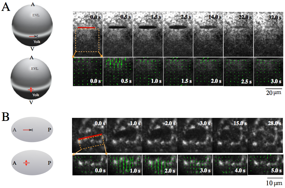
We first sought to quantify precisely the spatiotemporal dynamics of cortical non-muscle myosin II (NMY-2) that arises in response to ablating the cortex along a line. For this we used spinning disc microscopy to image NMY-2 fluorescence in combination with a UV laser ablation setup to sever the cortical layer along a line [8, 9, 32]. We recorded the spatiotemporal evolution of the surrounding NMY-2 in the cortex to following the resealing process.
In case of the zebrafish embryo, at the stage of - epiboly, COLA was performed within the YSL actomyosin ring and along a line parallel to EVL. In case of the C. elegans one cell embryo, the cortex was ablated just before the onset of cortical flow in the anterior half of the embryo and in a direction along AP axis. In both systems the area surrounding the cut was imaged until the hole was no longer visible due to turnover and regrowth (Fig. 1 A and B right upper).
COLA severs all connections within the cortex along the cut line and sets tension in the direction orthogonal to the cut line to zero. This results in tension gradients that drive an outward movement of the adjacent cortex away from the cut line (Fig. 1 A and B right). Notably, despite significant differences in cortical structure and dynamics, both zebrafish and C. elegans share large similarities in the overall response to COLA and the spatiotemporal dynamics of cut opening and resealing. In both systems the outward movement of the cut boundary lasts for several seconds and turns the cut line into an approximately elliptically shaped opening. Furthermore, the adjacent cortex moves outward with a velocity that decays over time, followed by cortical regrowth within the cleared region until no visible mark of the COLA procedure remains.
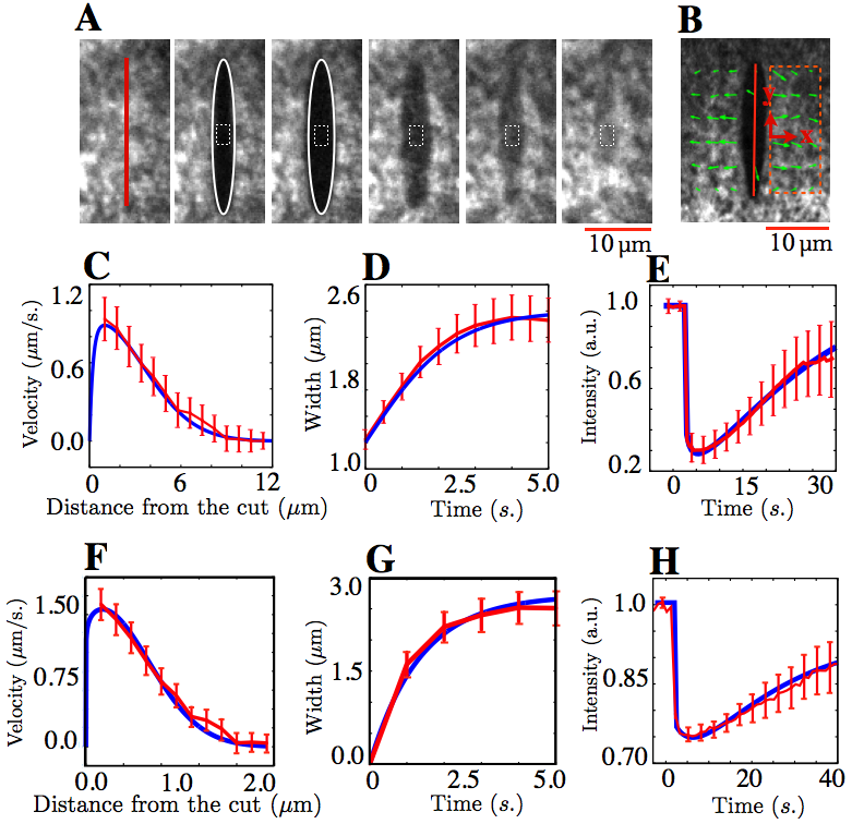
To extract the characteristics of the response of the cortex to COLA and the characteristics of the resealing process, we analyzed the spatiotemporal dynamics of cortical NMY-2 after COLA in several ways. We first determined the outward velocity of cortical NMY-2 adjacent to the cut line by Particle Image Velocimetry (PIV) at a time just following the cut, as indicated by the arrows in Fig. 1 A and B right lower panels. Remarkably, the velocity profile along the direction perpendicular to the cut line was not uniform, but rather decayed over a characteristic distance away from the cutline as shown in Fig. 2 C and F. This spatial decay entails information about the characteristic distance over which mechanical stresses communicate in the 2D cortical layer. Second, we quantified the temporal evolution of the cut opening. For this, we determined the extent of the opening generated by COLA, by fitting an ellipse to this opening and determining the time evolution the minor radius of this fitted ellipse as a measure of the width of the cut opening (Fig. 2 A). Notably, the width of the cut opening increased with time and reached maximum after approximately 3-5 seconds (Fig. 2 D and G). The minor radius of the opening grows with a characteristic time governed by processes of stress relaxation in the actomyosin cortical layer. Third, we analyzed NMY-2 regrowth within the opening, by quantifying the average fluorescence intensity in a box that is placed at the center of the COLA opening (white broken line in Fig. 2 A). Following an initial drop in intensity due to ablation and outward movement, NMY-2 intensity gradually recovered over approximately 30 seconds (Fig. 2 E and H). Similar time scales have been observed in FRAP measurements that characterize NMY-2 turnover [9].
These observations lend credence to the assumption that the cortex behaves as an active viscoelastic material. The shape evolution of the cut opening is determined largely by elastic properties of the cortex as well as active tension provided by NMY-2. The velocity decay away from the cut line is largely determined by the decay length of tension in the layer, and thus by and . Finally, re-association of myosin at the cut site is determined by actomyosin turnover. In what follows, we will compare the dynamics of opening and regrowth, as determined in our experiments by characterizing the spatial decay of the velocity field and the time evolution of the cut opening width and the myosin levels at the center of the hole, with theory.


4.2 Physical description of the actomyosin cortex
We next sought to calculate the cut response in a coarse grained physical description of the cortical layer. Considering that the thickness of the cortex is small compared to the size of the cell we describe the actomyosin cortex as an active 2D viscoelastic compressible fluid. We introduce a scalar field which denotes the local height of the cortical layer in the direction, with and denoting spatial cartesian coordinates within the plane of the cortical layer, and denoting time. Considering a viscoelastic isotropic active fluid in the plane and integrating over the height of the cortex we write the following constitutive equation:
where indices . The dynamic variables are the 2D stress tensor , the 2D active stress tensor and the 2D velocity field . The material properties are characterized by the 2D shear viscocity , 2D bulk viscocity and a characteristic Maxwell time of stress relaxation . Here, denotes the material time derivative. Turnover of the film is captured by the dynamics of the height given by
| (4) |
where the first term on the r.h.s. accounts for advection of the gel by cortical flow, and the second term describes turnover which relaxes to the steady state hight . Note that both the turnover time and the relaxed height depend on actin and myosin turnover. The force balance equation reads [7, 8, 9, 33, 34]
| (5) |
where inertial forces have been neglected and is a friction coefficient that describes frictional interactions between the cortex and it its surrounding cytosol and membrane.
We consider an active gel that is incompressible in 3D, with constant density through the height of the cortical layer. In this case, both the active stress and the 2D viscosities and of the cortex are proportional to cortex height and are written as
| (6) | |||||
| (7) | |||||
| (8) |
Here, denotes the isotropic active stress generated through ATP consumption of myosin, positive for contraction and dependent on the change in chemical potential associated with ATP hydrolyosis . Furthermore, and denote the shear and bulk viscosities of the layer when . 3D incompressibility condition couples divergences in the 2D flow velocity field to change the cortex height according to Eq. 4, and we set . Eqs. 4.2-5 complete the model. For nondimensionalization we choose the characteristic time of regrowth and the COLA cut length as the respective time and length scales. Model equations with dimensionless variables can be rewritten with dimensionless parameters, , , (see Appendix).
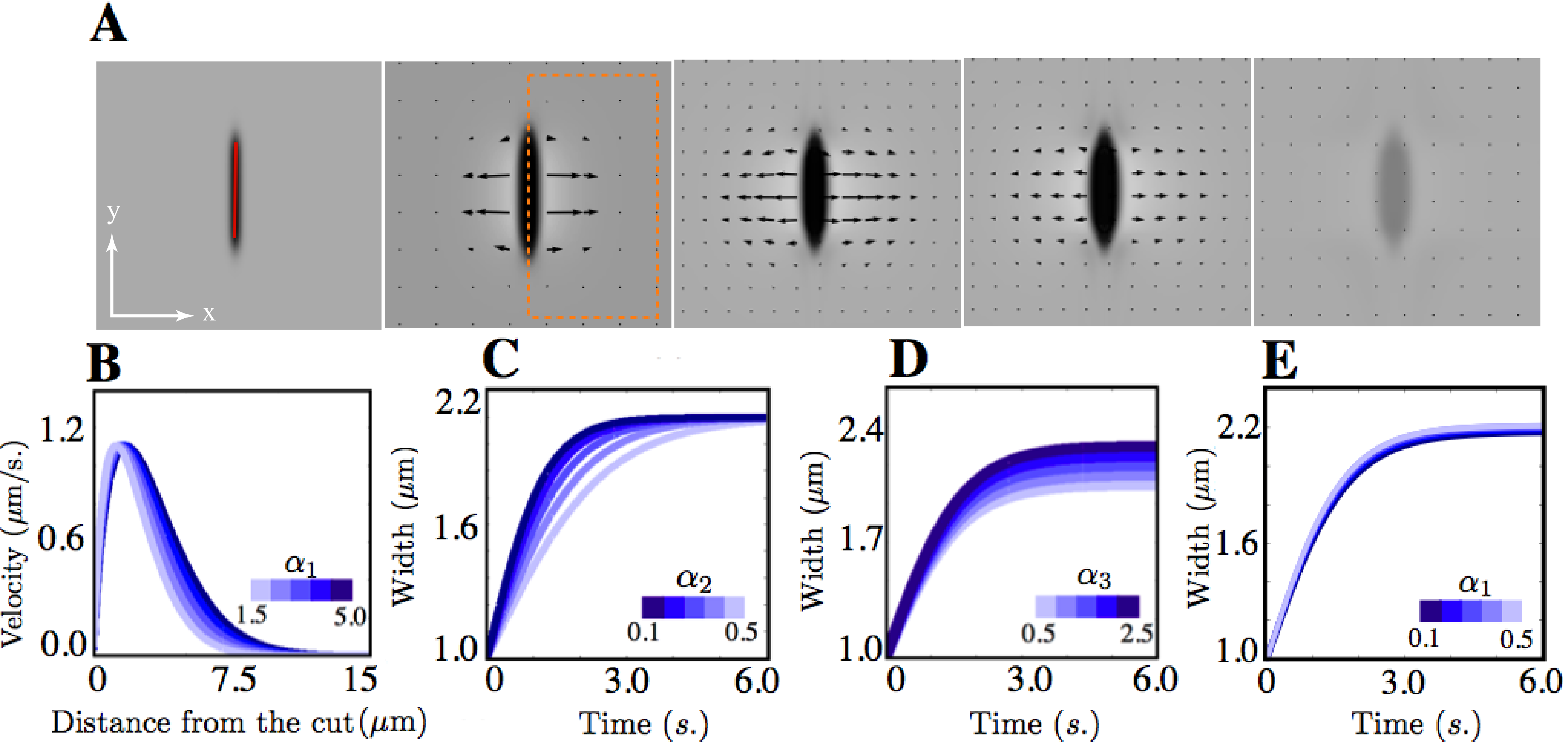
We next asked whether this description can reproduce the relaxation dynamics in response to COLA that we observed in our experiments. To this end we numerically solved non-dimensional versions of Eq. 4.2-5 (see Appendix) in a rectangular box of width ( ) in and , with periodic boundary conditions. As an initial condition we choose uniform height and stress fields that correspond to the unperturbed stationary solution with . To account for COLA, this homogenous initial condition is perturbed at by setting height to zero within a thin rectangular strip of length and width . We then computed the resulting spatiotemporal dynamics. Fig. 3 A displays the time evolution of the resultant height and velocity fields. We observe that 1) the velocity field is not uniform through the cortex but decayed over a distance from the cut line; 2) the width of the boundary initially grows and reaches a maximum; and 3) in the the cut region recovers to steady state values on long times. These observations are consistent with those that we have made in our experiments, which lends credence to our approach.
Next we analyzed our results from theory in terms of the spatial profile of the velocity in the direction perpendicular to the cut line, the growth of the cut boundary, and the recovery of the cut region in a manner that is similar to how we analyzed the experimental data. Fig. 3 B to E demonstrates that the essential features observed in the corresponding graphs from our experiments (Fig. 2 C to H) are reproduced in our theory. We next asked how changes of physical parameters of the cortical layer impact on the spatial profile of the velocity in the direction perpendicular to the cut line, the growth of the cut boundary, and the recovery of cortex height. To this end we performed numerical simulations of COLA with different values of . We find that increasing leads to a corresponding increase of the spatial decay length of the velocity profile in a direction perpendicular to the cut line (Fig. 3 B), but has little impact on the growth timecourse of cut boundary (Fig. 3 E). Furthermore, increasing results in a corresponding increase in the relaxation time to reach the maximal width of the opening (Fig. 3 C), with little effect on the final size of the hole. Taken together, key aspects of the relaxation process after COLA are separately determined by the characteristic length and time, and of the cortical layer.

4.3 Comparison of theory and experiment for determination of physical parameters
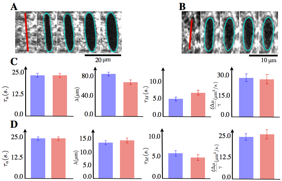
Next we determine coarse grained physical parameters of the cell cortex both in C. elegans single cell embryo and zebrafish actomyosin ring, by comparing the experimentally determined cortical response to COLA in terms of regrowth of the cut region, spatial decay of the outward velocity profile, and the time evolution of the cut opening boundary to theory. We note that the height variable in the theory is proportional to the per-area-concentration of molecules that are uniformly distributed along the direction. Since the thickness of the cortical layer is smaller than the focal depth of the confocal microscope (approximately ), the fluorescence intensity of cortical NMY-2, in our microscope images is proportional to the 2D concentration of NMY-2, integrated over the height of the cortical layer. Hence the local 2D concentration of NMY-2 in our microscope images is proportional to . We determined the best fit non-dimensionalized model parameters within a nonlinear least-square fitting scheme [31] by iteratively calculating the theoretical responses that best fit the experimental profiles (see blue curves in Fig. 3 C to H in comparison to the experimental profile given by red curves). In Fig. 4 C and D, we report the physical parameters of the cell cortex () from the best fit model parameters. We obtained and for zebrafish, and and for C. elegans. Note that these values were obtained by fitting each individual experiment (n=15 for both systems), and we report the respective averages standard error. Note also that the hydrodynamic lengths of for the zebrafish actomyosin ring and for the C. elegans cortex as well as the turnover times of show a close agreement with previous investigations [8, 9]. This supports our method of extracting values of physical parameters by use of fitting the measured COLA response to that expected from theory.
The good agreement between theory and experiment indicates that the shape evolution of the cut opening might entail a sufficient amount of information for accurately determining both and . Since the spatial profile of the velocity field is governed by hydrodynamic length, , the temporal change of the shape of the cut opening boundary is likely to be affected by . On the other hand, the temporal decay of the outward velocity is characterized by the time scale of the stress relaxation, . Thus it is possible that the shape evolution of the cut boundary is to a large extent governed by these two physical parameters. In agreement with this statement, the shapes of the cut opening boundary are distinct between the zebrafish actomyosin ring and C. elegans embryo, which might reflect the respective differences in physical parameters.
Therefore we asked if it is possible to determine the hydrodynamic length and the time scale of stress relaxation from the shape evolution of the cut opening boundary. To this end we determined the ’s by comparing the spatiotemporal development of the cut opening boundary shapes in experiment and theory. We detected the shape of the cut opening boundary observed in experiment to evaluate the difference with the shape from the theoretical prediction at the given parameter values. We computed the minimum distance from th edge point detected in the experiment to the edge points obtained from the theory at the th time frame, , (see Materials and Methods for details). We then evaluated a merit function, , where is the number frames analysed (a useful choice for was between 8 and 12 for a zebrafish, and between 4 and 6 for C. elegans). was minimized iteratively to find the best fit values of ’s and . Fig 4 A and B shows the shape of the cut opening boundary from the theory with the best fit parameter values with respect to the minimization of , successfully reproducing the shape evolution of the cut opening boundary observed in experiment. Consequently we obtained non-dimensionalized model parameters , as well as the physical parameters and for zebrafish and and for C. elegans. Again, these values were obtained by fitting each individual experiment (n=15 for zebrafish and n= 10 for C. elegans, in the latter not all of our experimental datasets converged in the fitting procedure), and we report the respective averages standard error. The values of the parameters of the actomyosin network are in close agreement between this method of determination, and the method used before, see Fig. 4 C and D. We conclude that the shape evolution of the cut opening boundary entails a sufficient amount of information to determine the entire set of non-dimensional physical parameters, and provides a second means of determining physical parameters of the actomyosin cortical layer.

5 Discussion
While COLA is a powerful tool to characterize the tension in the actomyosin cortex, obtaining the physical parameters to describe the cortical mechanics relies on an appropriate analytical method. Here we present a strategy to determine the physical parameters of actomyosin cortex from the cortical response to COLA. The method relies on the fitting of the COLA response to the theoretical prediction from the 2D active viscoelastic fluid model. We show that the best fit value of the hydrodynamic length, ’s are in close agreement with previous estimates both for zebrafish actomyosin ring and C. elegans single cell embryo [9, 8]. Notably we can determine a whole set of parameters in a single experiment, in contrast the previous estimates that require the ensemble averaging for the flow profile, and myosin distribution. As shown in Fig. 4, the standard error of the best fit parameters are small in both methods, signifying the overall robustness of the approach. In addition, our method does not require any assumptions for boundary conditions in the flow and myosin profiles. Taken together, our method allows the precise determination of the physical parameters.
Importantly, our method also allows to determine the characteristic time of stress relaxation, , which governs the time scale between elastic and viscous regime. sets the timescale for the large-scale movement of the actomyosin cortex, and morphogenetic deformations of the cortex that are driven by actives stresses in the layer generally occurs on time scales larger than . Recently active microrheology has been performed to measure the storage and loss moduli by manipulating a bead injected inside the cell [35]. This method allows the precise determination of the characteristic time of stress relaxation of the cytoplasm. However a typical diameter of bead used in the active microrheology measurement, is bigger than the thickness of the cortical layer, one cannot distinguish the cortical and cytoplasmic viscoelastic properties in the measurement. In contrast, our method allows us to directly determine of the cortex.
In summary, we have developed a robust and accurate method with broad applicability to determine the physical parameters of cortical mechanics from COLA experiment in conjunction with the coarse grained mechanical theory. It provides us with the large-scale and biologically relevant parameters in terms of morphogenetic mechanics. Given the simple description of the cortex used in the paper, we suggest that our method can be applied for complex multicellular systems such as epithelial tissues to determine the physical parameters that describes the tissue mechanics.
6 Appendix: Non-dimensional equations
7 Acknowledgments
We are grateful to Daniel Dickinson for providing LP133 C. elegans strain. We thank G. Salbreux, V. K. Krishnamurthy and J. S. Bois for fruitful discussions. SWG acknowledges support by grant no. 281903 from the European Research Council (ERC) and by grant GR 7271/2-1 from the Deutsche Forschungsgemeinschaft (DFG). SWG and CPH acknowledge support through a grant from the Fonds zur Förderung der wissenschaftlichen Forschung (FWF) and the DFG (I930-B20).
8 Author Contributions
AS, FJ and SWG designed the research, MB and MN performed experiments under supervision of CPH and SWG, AS and MN developed the theory and performed numerical simulations as well as the comparison to data under supervision of FJ and SWG, MN and SWG wrote the paper with the help of AS and with support from FJ.
References
- [1] Thomas Lecuit and Pierre-Francois Lenne. Cell surface mechanics and the control of cell shape, tissue patterns and morphogenesis. Nature Reviews Molecular Cell Biology, 8(8):633–644, 2007.
- [2] Suzanne Eaton and Frank Jülicher. Cell flow and tissue polarity patterns. Current opinion in genetics & development, 21(6):747–752, 2011.
- [3] M Shane Hutson, Yoichiro Tokutake, Ming-Shien Chang, James W Bloor, Stephanos Venakides, Daniel P Kiehart, and Glenn S Edwards. Forces for morphogenesis investigated with laser microsurgery and quantitative modeling. Science, 300(5616):145–149, 2003.
- [4] Jerome Solon, Aynur Kaya-Copur, Julien Colombelli, and Damian Brunner. Pulsed forces timed by a ratchet-like mechanism drive directed tissue movement during dorsal closure. Cell, 137(7):1331–1342, 2009.
- [5] Masazumi Tada and Carl-Philipp Heisenberg. Convergent extension: using collective cell migration and cell intercalation to shape embryos. Development, 139(21):3897–3904, 2012.
- [6] Guillaume Salbreux, Guillaume Charras, and Ewa Paluch. Actin cortex mechanics and cellular morphogenesis. Trends in cell biology, 22(10):536–545, 2012.
- [7] Guillaume Salbreux, Jacques Prost, and Jean-Francois Joanny. Hydrodynamics of cellular cortical flows and the formation of contractile rings. Physical review letters, 103(5):058102, 2009.
- [8] Martin Behrndt, Guillaume Salbreux, Pedro Campinho, Robert Hauschild, Felix Oswald, Julia Roensch, Stephan W Grill, and Carl-Philipp Heisenberg. Forces driving epithelial spreading in zebrafish gastrulation. Science, 338(6104):257–260, 2012.
- [9] Mirjam Mayer, Martin Depken, Justin S Bois, Frank Jülicher, and Stephan W Grill. Anisotropies in cortical tension reveal the physical basis of polarizing cortical flows. Nature, 467(7315):617–621, 2010.
- [10] Romain Levayer and Thomas Lecuit. Biomechanical regulation of contractility: spatial control and dynamics. Trends in cell biology, 22(2):61–81, 2012.
- [11] Gijsje H Koenderink, Zvonimir Dogic, Fumihiko Nakamura, Poul M Bendix, Frederick C MacKintosh, John H Hartwig, Thomas P Stossel, and David A Weitz. An active biopolymer network controlled by molecular motors. Proceedings of the National Academy of Sciences, 106(36):15192–15197, 2009.
- [12] Martin Bergert, Anna Erzberger, Ravi A Desai, Irene M Aspalter, Andrew C Oates, Guillaume Charras, Guillaume Salbreux, and Ewa K Paluch. Force transmission during adhesion-independent migration. Nature cell biology, 2015.
- [13] Karsten Kruse, Jean-Francois Joanny, Frank Jülicher, Jacques Prost, and Ken Sekimoto. Asters, vortices, and rotating spirals in active gels of polar filaments. Physical Review Letters, 92(7):078101, 2004.
- [14] Karsten Kruse, Jean-Francois Joanny, Frank Jülicher, Jacques Prost, and Ken Sekimoto. Generic theory of active polar gels: a paradigm for cytoskeletal dynamics. The European Physical Journal E: Soft Matter and Biological Physics, 16(1):5–16, 2005.
- [15] Andrew Callan-Jones and Frank Jülicher. Hydrodynamics of active permeating gels. New Journal of Physics, 13(9):093027, 2011.
- [16] Mathias Köppen, Beatriz García Fernández, Lara Carvalho, Antonio Jacinto, and Carl-Philipp Heisenberg. Coordinated cell-shape changes control epithelial movement in zebrafish and drosophila. Development, 133(14):2671–2681, 2006.
- [17] Adrian A Cuenca, Aaron Schetter, Donato Aceto, Kenneth Kemphues, and Geraldine Seydoux. Polarization of the c. elegans zygote proceeds via distinct establishment and maintenance phases. Development, 130(7):1255–1265, 2003.
- [18] Rebecca J Cheeks, Julie C Canman, Willow N Gabriel, Nicole Meyer, Susan Strome, and Bob Goldstein. C. elegans par proteins function by mobilizing and stabilizing asymmetrically localized protein complexes. Current Biology, 14(10):851–862, 2004.
- [19] Edwin Munro, Jeremy Nance, and James R Priess. Cortical flows powered by asymmetrical contraction transport par proteins to establish and maintain anterior-posterior polarity in the early c. elegans embryo. Developmental cell, 7(3):413–424, 2004.
- [20] Nathan W Goehring, Philipp Khuc Trong, Justin S Bois, Debanjan Chowdhury, Ernesto M Nicola, Anthony A Hyman, and Stephan W Grill. Polarization of par proteins by advective triggering of a pattern-forming system. Science, 334(6059):1137–1141, 2011.
- [21] D Humphrey, C Duggan, D Saha, D Smith, and J Käs. Active fluidization of polymer networks through molecular motors. Nature, 416(6879):413–416, 2002.
- [22] Andreas R Bausch, Florian Ziemann, Alexei A Boulbitch, Ken Jacobson, and Erich Sackmann. Local measurements of viscoelastic parameters of adherent cell surfaces by magnetic bead microrheometry. Biophysical Journal, 75(4):2038–2049, 1998.
- [23] Qi Wen and Paul A Janmey. Polymer physics of the cytoskeleton. Current Opinion in Solid State and Materials Science, 15(5):177–182, 2011.
- [24] Chase P Broedersz, Martin Depken, Norman Y Yao, Martin R Pollak, David A Weitz, and Frederick C MacKintosh. Cross-link-governed dynamics of biopolymer networks. Physical review letters, 105(23):238101, 2010.
- [25] Katsuhisa Tawada and Ken Sekimoto. Protein friction exerted by motor enzymes through a weak-binding interaction. Journal of theoretical biology, 150(2):193–200, 1991.
- [26] Volker Bormuth, Vladimir Varga, Jonathon Howard, and Erik Schäffer. Protein friction limits diffusive and directed movements of kinesin motors on microtubules. Science, 325(5942):870–873, 2009.
- [27] Sydney Brenner. The genetics of caenorhabditis elegans. Genetics, 77(1):71–94, 1974.
- [28] Jean-Léon Maître, Hélène Berthoumieux, Simon Frederik Gabriel Krens, Guillaume Salbreux, Frank Jülicher, Ewa Paluch, and Carl-Philipp Heisenberg. Adhesion functions in cell sorting by mechanically coupling the cortices of adhering cells. Science, 338(6104):253–256, 2012.
- [29] M. Westerfield. The Zebrafish Book: A Guide for the Laboratory Use of Zebrafish (Danio Rerio). Univ. of Oregon Press, Eugene, 2007.
- [30] Charles B Kimmel, William W Ballard, Seth R Kimmel, Bonnie Ullmann, and Thomas F Schilling. Stages of embryonic development of the zebrafish. Developmental dynamics, 203(3):253–310, 1995.
- [31] John A Nelder and Roger Mead. A simplex method for function minimization. The computer journal, 7(4):308–313, 1965.
- [32] Julien Colombelli, Stephan W Grill, and Ernst HK Stelzer. Ultraviolet diffraction limited nanosurgery of live biological tissues. Review of Scientific Instruments, 75(2):472–478, 2004.
- [33] Justin S Bois, Frank Jülicher, and Stephan W Grill. Pattern formation in active fluids. Physical review letters, 106(2):028103, 2011.
- [34] K Vijay Kumar, Justin S Bois, Frank Jülicher, and Stephan W Grill. Pulsatory patterns in active fluids. Physical Review Letters, 112(20):208101, 2014.
- [35] Ming Guo, Allen J Ehrlicher, Mikkel H Jensen, Malte Renz, Jeffrey R Moore, Robert D Goldman, Jennifer Lippincott-Schwartz, Frederick C Mackintosh, and David A Weitz. Probing the stochastic, motor-driven properties of the cytoplasm using force spectrum microscopy. Cell, 158(4):822–832, 2014.