To understanding of the mechanisms of DNA deactivation in ion therapy of cancer cells
Abstract
Changes in the medium of biological cell nucleus under ion beam action is considered as a possible cause of cell functioning disruption in the living body. As the most long-lived molecular product appeared in the cell after the passage of high energy ions, the hydrogen peroxide molecule is picked out. The possibility of the formation of stable complexes of hydrogen peroxide molecules with active sites of DNA nonspecific recognition (phosphate groups of the double helix backbone) is studied, and the formation of stable DNA-peroxide complexes is considered. Due to the negative charge on the oxygen atoms of DNA phosphate group in solution the counterions that under natural conditions neutralize the double helix have been also taken into consideration. The complexes consisting of oxygen atoms of DNA phosphate group, H2O2 and H2O molecules, and Na+ counterion have been considered. Energy of the complexes have been determined based on the electrostatic and van der Waals interactions within the approach of atom-atom potential functions. The stability of various configurations of molecular complexes has been estimated. It has been found that hydrogen peroxide molecules can form the stable complexes with phosphate groups of DNA and counterions which are no less stable than the complexes with water molecules. It is shown that the formation of stable complexes of H2O2–Na+–PO can be detected experimentally by the observation of specific DNA vibrations in the low-frequency Raman spectra. The interaction of H2O2 molecule with phosphate group of the double helix backbone can block the processes of DNA biological functioning and induce the deactivation of the genetic apparatus of the cell. Thus, the new channel of high-energy ions action on living cell has been proposed.
1 Introduction
Invention of new instruments for cancer therapy is known to be one of the most important challenges of modern science. In spite of the increase of funding for the development of new anticancer drugs the illness rate is still very high [1]. In this regard, the elaboration of non pharmacological methods of cancer therapy are stimulated. The last decades, use of high energy ion beams has established itself as an effective treatment of cancer disease. This method is based on well-known Bragg effect [2, 3, 4, 5]. Heavy ions (usually protons or 12C6+), accelerated to the energies of hundreds of MeV, are targeted into tumor. Within organism tissue the ions lose their initial energy mainly at the end of their trajectory of motion, and the energetic peak is formed (the Bragg peak). The initial beam energy is chosen in such a way that the position of the Bragg peak coincide with the location of tumor in human body (it can be 5-10 cm from body surface). The energy which is lost in living tissues destroys cancer cells and consequently the whole tumor. The method of ion beam therapy is much more effective than X-ray therapy, because of local action in the Bragg peak, especially in the case of places of difficult access, such as human brain [3, 4, 5]. In spite of great practical interest and large number of ion therapy facilities already built, there is no clear understanding of the molecular mechanism of action of high energy ions.
Starting with the early study [6] the action of radiation on living organisms is known to be related with the DNA damage. The breaking of DNA double helix affects the mechanism of storage and transfer of genetic information which leads to cell death. Under heavy ion irradiation the breaks of hydrogen bounds in nucleotide pairs occur, which cause DNA melting, single and double strand breaks in double helix [3, 4, 5]. Single strand breaks in DNA and melting can be repaired due to the complementary structure of double helix and cell reparation mechanisms. The double strand breaks in DNA lead to damage of macromolecule, which can not be repaired [7]. Thus the formation of large number of double strand breaks of double helix is necessary for deactivation and destruction of DNA molecule.
The double strand breaks in DNA macromolecule occur due to the large quantity of secondary electrons born in the medium under the action of ion beam [8, 9, 10, 13]. Some molecules decay into atoms and radicals that can break chemical bonds in biological macromolecules. At the same time, the calculations show that the number of the double strand breaks, made by secondary electrons and radicals, is insufficient for cell death [10]. Therefore, additional channels of ion beam action on living tissues should exist.
To describe DNA damage the formation of the shockwave in the medium after passage of high energy ions is suggested [11, 12]. Within the framework of shockwave mechanism the destruction of DNA double helix may occur even in the case of macromolecules situated far from ion hit point in the cell. The molecular dynamics simulations shows that shockwave cause large number of double strand breaks in DNA double helix [13]. However, in spite of high efficiency of proposed mechanism, there are processes which can prevent DNA damage in the living cell, particularly, the histone proteins restraining the macromolecule in frames of chromatin.
The deactivation of DNA macromolecule may be induced by changes of medium that occur after the passage of high energy ions. The goal of the present work is to consider the changes of medium in the Bragg peak region and to pick out new molecular compounds that can influence the structure and dynamics of DNA double helix. The analysis of changes in water medium induced by ion beams is carried out in second section and the most probable molecular compounds formed in the Bragg peak are analyzed. The third section is dedicated to the description of calculation method of interaction of appeared molecules and atoms of DNA double helix. The results of energy calculations are presented in fourth section and the most stable complexes are selected. In the fifth section the low-frequency vibrations of formed complexes are studied, and the specific modes of the complexes are found in the Raman spectra of DNA. The possible action of medium changes on DNA biological functioning is discussed in the sixth section.
2 Medium change under ion beam radiation
The ions of high energy passing through intracellular medium induce the process of decay of water molecules (water radiolysis), different chemical and nuclear reactions [14, 15, 16, 17, 18, 19, 20, 21]. The processes of nuclear fragmentation are showed be not significant for environment [15, 16], whereas the fragmentation of water molecules change essentially the composition and properties of water medium [3, 4, 5, 10]. In the process of water radiolysis the secondary electrons (), radicals (OH∙ and HO), molecules (H2O2 and H2O), and other products (H∙, OH-, H3O+) are produced [17, 18, 19].
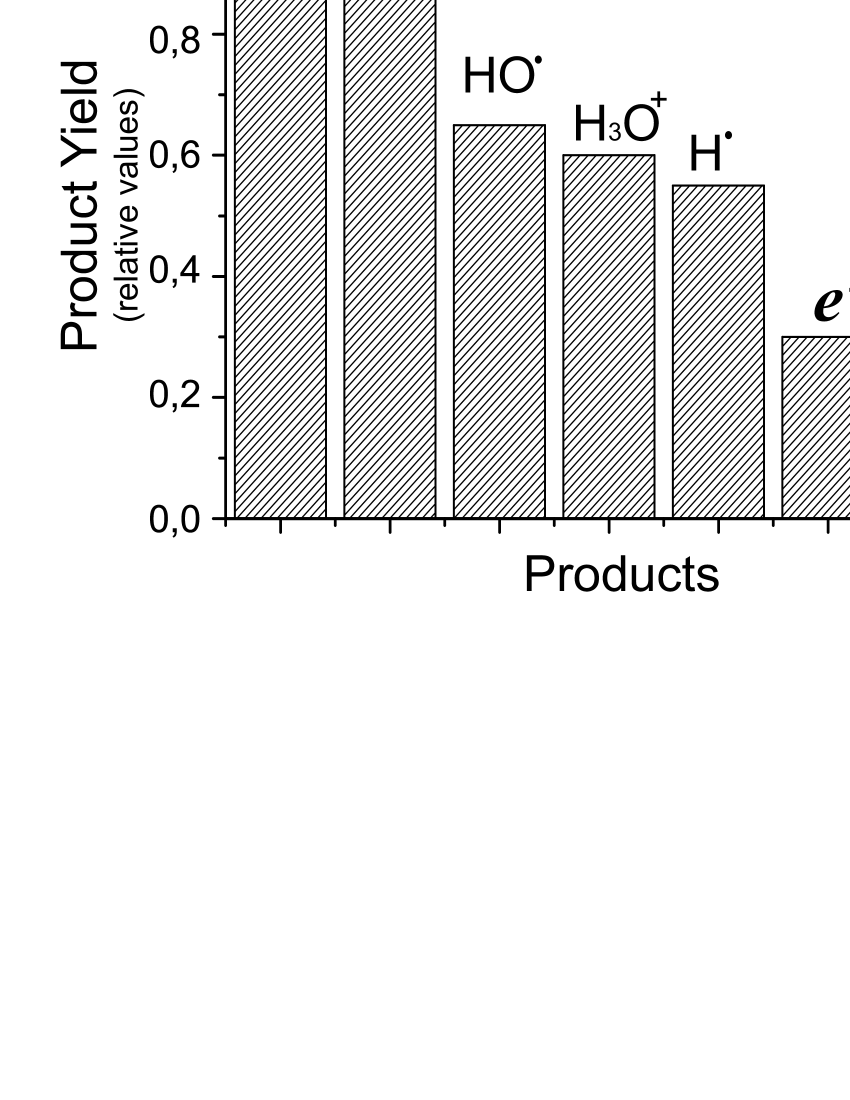
Quantity and composition of radiolysis products depend on absorbed radiation dose. At small doses the number of secondary electrons and radicals is the largest, while the increase of the dose energy leads to the increase of molecular products yields. Due to the high energy transfer the number of molecular products is expected to be the largest in the Bragg peak region. The Monte Carlo simulation data for water radiolysis [20, 21] show that in the Bragg peak the secondary electrons and hydroxyl radicals yields prevail at early stage of radiolysis (time 10-12 s). After the time 10-6 s the reactions OHOHH2O2, HOHOH2OO2 occur, and the yields of molecular products become larger than other yields [20]. Taking into consideration that the microsecond time scale is typical for biological processes, the equilibrium composition of the intracellular medium after the passage of high energy ions must be enriched by the molecular products of water radiolysis.
To analyze the composition of the radiolysis products at the Bragg peak the Monte Carlo simulation data [20] are used. The distribution of radiolysis products at the time 1 s after ion passage are showed in the Figure 1. It is seen that the number of hydrogen peroxide molecules in intracellular medium is larger than other products of water radiolysis. The number of H2 molecules is also considerable, but these molecules don’t have high reactivity and as a result do not threaten biological macromolecules. Radicals which also have substantial yields after some time transform into neutral water molecules or hydrogen peroxide. Thus, the composition of intracellular medium changes after ion beam irradiation and significant amounts of hydrogen peroxide molecules appear.
The concentration of hydrogen peroxide in the living cell is kept constant by special ferments [22], but it may be increased greatly by the action of ion beams. Increasing the amount of hydrogen peroxide molecules raises the probability of their interaction with DNA. The interaction of large number of H2O2 molecules and DNA can deactivate the cell genetical apparatus and induce cell death. Therapeutic effect of the hydrogen peroxide has been found under the treatment of cancer disease (see for example [23]). Ion therapy allows to increase the concentration of hydrogen peroxide in tumor cells with high precision and with minimal damage of healthy tissues.
The hydrogen peroxide molecule can interact with DNA molecule via charged atomic groups of the double helix. It is well known that the phosphate groups of double helix backbone bear the highest charge [7]. Under the natural conditions it is neutralized by metal ions (for example, Na+ or K+). Thus, the interaction of H2O2 molecule with the phosphate groups of the double helix and tethered metal counterions is expected to be the most probable.
3 Model and method of energy calculation
The capability of hydrogen peroxide to interact with DNA is studied by calculating the energy of different complexes. The considered complexes consist of DNA phosphate group, hydrogen peroxide molecule, water molecule and Na+ counterion. For the determination of relative stability of different complexes two oxygen atoms of the phosphate group are sufficient to consider. Other atoms of PO are essential for accurate calculations of energy. Schematic constructions and structural parameters of molecules in complexes are showed at Figure 2.

The method of atom-atom potential functions is used for calculation of energies of complexes [24, 25]. Within the framework of this method the energy is presented as the sum of pair interactions between atoms which belong to different structure elements. The energy of complex can be presented as the sum of two components:
| (1) |
where and are the potential energy of electrostatic and van der Waals interactions between and atoms at the distance .
Electrostatic interaction is defined as follows:
| (2) |
where and are the charges of and atoms, is the dielectric constant of vacuum, is the dielectric constant of medium. Charges of atoms of hydrogen peroxide and water molecules are determined using the values of dipole moments 2,1 D (H2O2) and 1,86 D (H2O) [26]. Charges of atoms in phosphate group are taken only for oxygen atoms in order to equate total charge with - (Table 1). The value of dielectric constant is used in calculations.
In the case of van der Waals interaction the Lennard-Jones potential is used:
| (3) |
where and are the constants (Table 1). The first and the second terms in (3) describe the van der Waals repulsion and short range repulsion between atoms, respectively. To take into consideration the hydrogen bonds (O–HO) the interaction between respective oxygen atoms are calculated using the modified potential [25], where exponent in the first term of equation (3) is changed to -10. Such exponent describes the short-range character of hydrogen bonds.
For the description of interaction between ion and charged oxygen atoms the Born-Mayer potential is used:
| (4) |
where is the repulsion constant, is the equilibrium length (Table 1). This potential was developed to describe the ion crystal energy, and it takes into account electrostatic attraction and short-range repulsion [27]. The potential (4) has been adapted in [28, 29] for the description of counterion interactions with the phosphate groups of DNA double helix backbone.
Using the formulae (1) – (4) with parameters in the Table 1 the energies of different complexes may be calculated.
4 Complexes of hydrogen peroxide with DNA
Within the framework of proposed method the energies of two-component (H2O2–PO, H2O–PO, Na+–PO4, H2O2–Na+, H2O–Na+) and three-component (H2O2–Na+–PO, H2O–Na+–PO) complexes are calculated. The most stable configurations for each complex are found.
Two-component complexes. The most stable state for the complex Na+–PO is obtained in case when sodium ion is localized at the distance 2 Å from the center of O–O segment of the phosphate group. The calculated anergy is the lowest among two-component complexes (Fig. 3). This complex corresponds to neutralization of DNA phosphate group by counterion under the natural conditions.
The most stable complex of sodium ion with hydrogen peroxide is obtained in the case of ion localization at distance 2.66 Å to the center of O–O bond with equal distances to H atoms on the opposite side of the molecule (Fig. 3). In the case of Na+–H2O the most stable complex is obtained for the Na+–O distance 2.74 Å. Under the natural conditions water molecules in complex with Na+ are the part of ion hydration shell. The hydrogen peroxide molecule may substitute the water in the ion hydration shell.
The equilibrium configurations of H2O2–PO complex is obtained in the case of symmetrical localization of H atoms of hydrogen peroxide molecule with respect to the O–O segment in the phosphate group (Fig. 3). The complex has minimal energy for the distance 2.85 Å between centers of O–O segments in the molecule and in the phosphate group. In the case of H2O–PO complex the equilibrium configuration is obtained for the distance 2.9 Å of water oxygen to O–O segment in the phosphate group (Fig. 3).
The hydrogen bonds, which can be formed between H (water or hydrogen peroxide) and O (phosphate group), do not influence significantly the energy values. In the case of the strongest hydrogen bond (O–H–O atoms are localized on the line) the calculated energies are lower than energies of H2O2–Na+–PO4 and H2O–Na+–PO4 complexes. In configurations with two hydrogen bonds between atoms of H2O2 (H2O) molecule the O–H–O segment is bent more than 30∘, that makes such hydrogen bonds very weak.

The equilibrium configurations of H2O2–PO and H2O–PO4 complexes depend on mutual disposition of molecules. Rotating H2O2 molecule with respect to the axis passing through the center of O–O segment, the most stable complex is found for the angle about (energy -12.8 kcal/mol) (Fig. 4a). In the case of molecule rotates around O–O bond, the energy minimum is obtained for the angle , while at the repulsion occurs (Fig. 4b). Some small minimums at about the angles and appear due to the van der Waals repulsion and Coulomb attraction (dash lines at figure). In the case of rotation of water molecule the obtained results are qualitatively similar.

Three-component complexes. The calculations are carried out for H2O2–Na+–PO and H2O–Na+–PO complexes. The energy of the complexes are calculated for different distances between hydrogen peroxide (water) molecule and ion-phosphate complex. The ion-phosphate distance is taken equal to 2 Å, correspondingly to the result for two-component complex Na+–PO4 (Fig. 3). Such ion localization is the most energetically favorable, therefore other variants of ion localization are not considered in the present work.
The configurations where hydrogen atoms of H2O2 (H2O) molecule are turned toward and outward phosphate group are considered (Fig. 5a). In the first case there is no energy minimum due to the electrostatic repulsion between hydrogen atoms and sodium ion. Increasing the distance to H2O2 (H2O) the energy of the complex monotonously decreases to the value of Na+–PO4 complex (Fig. 5b). In the case when H atoms of H2O2 (H2O) molecule are turned toward the phosphate group the energy minimum –130.3 kcal/mol (–129.2 kcal/mol) appear at the distance 2.75 Å (2.82 Å). The minimum is conditioned by the electrostatic attraction between ion and oxygen atoms of molecule. Rotation of hydrogen peroxide or water molecules around axis which passes through center of O–O segment plays insignificant role because the total energy depends on interaction between ion and oxygen atoms mainly. Thus, the stability of H2O2–Na+–PO4 and H2O–Na+–PO4 complexes are almost equal.

5 Vibrations of hydrogen peroxide in DNA low-frequency Raman spectra
The formation of stable complexes of H2O2 molecule with DNA phosphate groups should be accompanied by the appearance of specific modes of vibrations of hydrogen peroxide molecule in the vibrational spectra. The frequencies of vibrations of H2O2–Na+–PO complex are expected to be in the low-frequency spectra range (200 cm-1), analogically to the modes of counterion vibrations with respect to DNA phosphate groups (ion-phosphate modes) [28, 29, 30, 31]. The low-frequency spectra of DNA is known to be characterized by the conformational vibrations of the double helix structural elements as whole (nucleosides, nucleotides, phosphate groups) [32, 33, 34, 35]. The vibrations of hydrogen peroxide molecule with respect to the DNA phosphate groups should disturb the internal dynamics of the double helix changing the low-frequency spectra shape.
To determine the influence of H2O2 molecules on DNA conformational vibrations the phenomenological model describing the Raman low-frequency spectra of DNA with counterions [28, 29, 30] is used. Within the framework of this model the DNA double helix is represented as the double chain of nucleotides with counterions tethered to the phosphate groups of the macromolecule backbone. To take into consideration the influence of hydrogen peroxide molecule this model is modified by increasing the mass of tethered counterions for the mass of one H2O2 molecule. The low-frequency vibration spectra of DNA with counterions is also calculated for the case of H2O molecule attached to the ion. The equations, necessary for the calculations of the mode frequencies and Raman intensities, are the same as in the original works [29, 30]. The calculation parameters are taken for the case of B-form of the double helix.
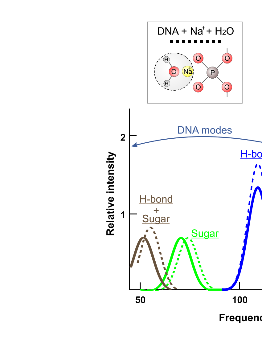
As the result the low-frequency Raman spectra of DNA with attached H2O2-Na+ complex is calculated as well as the spectra of DNA with H2O-Na+ complex (Fig. 6). The internal vibrations of the double helix (lower than 120 cm-1) are connected with the vibrations of H-bond stretching in nucleic bases and the vibrations in nucleosides due to the flexibility of sugar ring. These modes do not sense essentially the presence of hydrogen peroxide molecule. At the higher frequencies the modes, characterizing the vibrations of the complexes with respect to the phosphate groups, are determined about 155 cm-1 and 140 cm-1, respectively. The mode intensity is higher in the case of complex with hydrogen peroxide molecule. Thus, our estimates show that hydrogen peroxide molecule, attached to DNA phosphate groups, may induce the visible changes in the low-frequency Raman spectra.
6 Discussion
The calculations show that the complexes including hydrogen peroxide and water molecules are the most stable under the presence of sodium counterion. Without Na+ the stability of the complexes decreases for about one order. That is due to the dominant role of electrostatic interactions in the complex formation. The stability of H2O2–Na+–PO is about the same as H2O–Na+–PO. Therefore, hydrogen peroxide molecule may be expected to incorporate into the hydration shell of sodium counterion already tethered to the oxygen atoms of DNA phosphate group.
The formation of stable complex of H2O2–Na+ with the phosphate group may be detected by the low-frequency Raman spectra of DNA. According to our estimations the vibrations of counterion with hydrogen peroxide molecule with respect to the phosphate group of the double helix backbone should be about 10 cm-1 lower than the frequencies of vibrations of the complex with water molecule. Taking into consideration the determined frequency shift the lifetimes of H2O2–Na+–PO and H2O–Na+–PO complexes may be compared. According to the Arrhenius equation the ratio between characteristic lifetimes may be written in the following form:
| (5) |
where , and , are the lifetimes and frequencies of the complex with water and hydrogen peroxide molecules, respectively; and are the energies of the complexes with hydrogen peroxide and water molecules, respectively. The energies of the considered complexes are rather close (Fig. 5), therefore the value of the energy difference can be equal to zero. As the result the expected lifetime of the complex with H2O2 is higher than in the complex with H2O molecule.
Due to the larger lifetime of H2O2–Na+–PO complex the hydrogen peroxide molecules may be accumulated near DNA double helix. At significant amounts the H2O2 molecules can influence DNA biological functioning by two mechanisms. Firstly H2O2 can induce double strand breaks in double helix backbone by decaying into radicals, and secondly H2O2 molecules may break nucleic-protein recognition, stopping the processes of translation of genetic information. Thus, the hydrogen peroxide molecule, interacting with DNA double helix, can deactivate cell genetic apparatus and induce cell death.
7 Conclusions
The hydrogen peroxide molecules appear in the cell medium after the passage of high-energy ions. To study the possible effect of H2O2 molecule in the functioning of the living cell the complexes of hydrogen peroxide molecules with the phosphate groups of DNA backbone has been considered. Using the method of atom-atom potential functions the stability of different complexes, consisting of DNA phosphate group, H2O2 (or H2O) molecule, and Na+ metal ion, have been studied. The results show that the complexes with H2O2 molecule and phosphate group of DNA are no less stable than respective complexes with H2O molecule. The sodium counterion neutralizing the charge of phosphate group plays the key role in stabilization of these complexes. For the case of most stable complexes of hydrogen peroxide with the phosphate groups the low-frequency Raman spectra of DNA have been calculated. The vibrations of the complex as whole with respect to DNA phosphate group has been found near 150 cm-1. The observation of this mode may be considered as a test revealing the complex formation. Taking into consideration the determined frequencies of vibrations the lifetimes of H2O2–Na+–PO and H2O–Na+–PO complexes have been compared, and the lifetime of the complex with hydrogen peroxide has been found higher than in the case of water molecule. Taking this into consideration the hydrogen peroxide molecules are expected to be accumulated near DNA macromolecule. The action of H2O2 on DNA structure and dynamics may be sufficient for the braking the processes of DNA biological functioning. The influence of hydrogen peroxide molecules on DNA can be regarded as an individual channel in heavy ion therapy.
References
- [1] L. Gravitz, Nature Outlook 491, (2012) 7425.
- [2] A. Brown, S. Herman, Radiology and Oncology 73, (2004) 265-268.
- [3] G. Kraft, Progress in Particle and Nuclear Physics 45, (2000) S473-S544.
- [4] H. Suit, et all., Radiotherapy and Oncology, 95, (2010), 3-22.
- [5] C.D. Schlaff, A. Krauze, A. Belard, J.J. O’Connell, K.A. Camphausen, Radiation Oncology 9, (2014) 88.
- [6] N. W. Timofeeff-Ressovsky, A. V. Savich, and M. I. Shal’nov, Introduction to Molecular Radiobiology: Physico-Chemical Foundations (Medicina, Moscow, 1981) 320. [In Russian]
- [7] W. Saenger, Principles of Nucleic Acid Structure (Springer, New York, 1984) 584.
- [8] B. Boudaoiffa, P. Cloutier, D. Hunting, M.A. Huels, L. Sanche, Science, 287, (2000) 1658-1660.
- [9] N. Hamada, J. Radiat. Res., 50, (2009) 1-9.
- [10] A.V. Solov’yov, E. Surdutovich, E. Scifoni, I. Mishustin, W. Greiner, Phys. Rev. E, 79, (2009) 011909.
- [11] A.V. Yakubovich, E. Surdutovich, A.V. Solov’yov, Nuclear Instruments and Methods in Physical Research B, 279, (2012) 135-139.
- [12] E. Surdutovich, A.V. Yakubovich, A.V. Solov’yov, Scientific Reports, 3, (2013) 1289.
- [13] E. Surdutovich, A.V. Solov’yov, J. Phys.: Conf. Series, 438, (2013) 012014.
- [14] I. Pshenichnov, A. Botvina, I. Mishustin, W. Greiner, Nuclear Instruments and Methods in Physics Research, B 268, (2010) 604-615.
- [15] E. Haettner, H. Iwase, D. Schardt, Radiation Protection Dosimetry 122 (2006) 485-487.
- [16] J. Soltani-Nabipour, M A. Popovici, Gh. Cata-Danil , 62, (2010) 37-46.
- [17] B. Pastina, J.A. LaVerne, J. Phys. Chem. A 103, (1999) 1592-1597.
- [18] S. Le Caer, Water 3, (2011) 235-253.
- [19] V. Wasselin-Trupin, G. Baldacchino, S. Bouffard, B. Hickel, Radiation Physics and Chemistry, 65, (2002) 53-61.
- [20] M.S. Kreipl, W. Friedland, H.G. Paretzke, Radiat Environ Biophys, 48, (2009) 11-20.
- [21] S. Uehara, H. Nikjoo, J. Radiat. Res., 47, (2006) 69-81.
- [22] J.D. Watson, Molecular Biology of the Gene (W.B. Benjamin. Inc., Menlo Park, 1978) 706.
- [23] G. Manda, M.T. Nechifor, T.M. Neagu, Current Chemical Biology, 3, (2009) 342-366.
- [24] V.V. Zhurkin, V.I. Poletaev, V.L. Florentiev, Molecular Biology (Moscow). 14, (1980) 1116-1130.
- [25] V.I. Poltev, N.V. Shuliupina, Molecular Biology (Moscow). 18, (1984) 1549-1561.
- [26] Brief Chemical Encyclopedia. V. 1. (Soviet Encyclopedia, Moscow, 1961). [In Russian]
- [27] C. Kittel, Introduction to Solid State Physics (John Wiley and Sons, Inc., New York, 1954) 696.
- [28] S.M. Perepelytsya and S.N. Volkov, Ukr. J. Phys., 49, (2004) 1074-1080.
- [29] S.M. Perepelytsya and S.N. Volkov, Eur. Phys. J. E, 24, (2007) 261-269.
- [30] S.M. Perepelytsya and S.N. Volkov, Eur. Phys. J. E, 31, (2010) 201-205.
- [31] L.A. Bulavin, S.N. Volkov, S.Yu. Kutovy and S.M. Perepelytsya, Reports of National Academy of Science of Ukraine, No. 11, (2007) 69-73. arXiv:0805.0696.
- [32] S.N. Volkov, A.M. Kosevich, Mol. Biol. 21, (1987) 797-806. [Moscow]
- [33] S.N. Volkov, A.M. Kosevich, G.E. Weinreb, Biopolimery i Kletka 5, (1989) 32-39. [Kiev]
- [34] S.N. Volkov, A.M. Kosevich, J. Biomolec. Struct. Dyn. 8, (1991) 1069-1083.
- [35] A.M. Kosevich, S.N. Volkov, in Nonlinear Excitations in Biomolecules, edited by M. Peyrard (Springer, 1995) 118-128.