Duplex formation and secondary structure of -PNA observed by NMR and CD
Abstract
Peptide Nucleic Acids (PNA) are non-natural oligonucleotides mimics, wherein the phosphoribose backbone has been replaced by a peptidic moiety (N-(2-aminoethyl)glycine). This peptidic backbone lends itself to substitution and the -position has proven to yield oligomers with enhanced hybridization properties. In this study, we use Nuclear Magnetic Resonance (NMR) and Circular Dichroism (CD) to explore the properties of the supramolecular duplexes formed by these species. We show that standard Watson-Crick base pair as well as non-standard ones are formed in solution. The duplexes thus formed present marked melting transition temperatures substantially higher than their nucleic acid homologs. Moreover, the presence of a chiral group on the -peptidic backbone increases further this transition temperature, leading to very stable duplexes.
PNA duplexes with a chiral backbone present a marked chiral secondary structure, observed by CD, and showing a common folding pattern for all studied structures. Nevertheless small differences are observed depending on the details of the nucleobase sequence.
keywords:
-PNA , NMR Spectroscopy , imino proton , Circular Dichroism , secondary structureIntroduction
Peptide Nucleic Acid (PNA) are synthetic oligonucleotides first reported by Nielsen[1] with a backbone that recapitulate DNA’s inter-nucleobase distances. PNAs were developed as DNA mimics in order to recognize DNA double helix[2] and to form hybrid duplexes with DNA[3] or homoduplexes[4]. PNAs duplexes have high thermal stability, are not sensitive to nucleases or protases, and are metabolically stable. However, as polyamide backbone has no charge, no Coulomb repulsion is reported and PNA aggregates may exists in solution, and their solubility in water is limited[5]. PNAs have attracted interest in molecular biology and numerous applications have been reported, we can cite beacons for duplex DNA[6], and PNA as a diagnostic tool[7], a genomic tool[8], or a supramolecular barcodes[9, 10].
These applications were leveraged on the unique properties of PNA hybridizing to DNA or RNA. The enhanced hybridization properties of -modified PNA have been recently harnessed for gene editing applications[11]. PNA have also been used to tag small or macromolecules and program their assemblies based on hybridization[9] [10]. PNA homoduplexes have also been used in programmed assemblies as recently illustrated for the programmed pairing of PNA-tagged protein fragments[12]. While interactions of PNA with DNA or RNA have been extensively studied, homoduplex formation of PNA are not well characterized, particularly for modified PNAs.
PNA-DNA hybridization by Watson-Crick base pairing are well studied[3]. They reveal a helix formed by the -carbon and the nitrogen of the tertiary amide of the PNA backbone[13], stabilized by the sequential base stacking[14]. PNA neutral backbone bring thermal stability in PNA/DNA duplex compared to DNA/DNA duplexes, and present a better specificity[15]. Because of the absence of charge on the PNA backbone, the PNA/DNA hybrids are not dependent on the ionic strength of the solvent in contrast to DNA homoduplexes. Because of a flexible backbone, Watson-Crick, Hoogsteen, reverse Hoogsteen, or Wobble interactions can be formed by PNA/DNA duplexes[16].
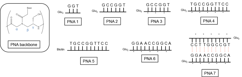
Unlike DNA which has a helical conformation due to its chiral centers, standard PNAs, with no chiral carbon, have no defined structural conformation in solution. Adding a chiral center on the PNA backbone forces left or right helix[17], and increases PNA/DNA hybridization. Adding a Lysine improves water solubility[14], however the Serine is less disruptive to hybridization and –OH groups can form hydrogen bonds with water in order to increase solubility. With the help of chiral centers, PNA homoduplexes should also be observed, and eventually present secondary helical structures. In this study, we explore the biophysical properties of PNAs built with a chiral center. The chiral center chosen here, consists in a L-Serine substitution of the -position on the PNA backbone[18], see insert in figure 1.
In order to determine homo-hybridization of PNAs through a structural pre-organization, a series of -PNAs were analyzed by Circular Dichroism (CD) and Liquid State Nuclear Magnetic Resonance (NMR). NMR allows to spotlight hydrogen bounds formed by nucleic acid base-pairing. Working in neutral conditions, specific NMR experiments bring information about the imino hydrogens engaged in the Watson-Crick, Hoogsteen, or Wobble bounds. Melting transitions () of the highlighted PNA homoduplexes were determined by CD and absorbance experiments. Circular Dichroism is finally used to assess the secondary structure displayed by the homoduplexes. All the experimental results show unambiguously that PNAs duplexes are formed, and that the chiral centers improve their stability, and drive the duplexes into a chiral secondary structure, probably organized as a left-handed helix.
Experimental
Materials
PNAs were synthesized as previously reported and purified by HPLC[19]. Six PNAs (see figure 1 with a schematic view) were chosen according to their nucleobases and the presence or absence of modification on the backbone. PNA 1 is a small one, with three nucleobases GGT and has a standard backbone. PNA 2 and 3 have the same six nucleobases: GCCGGT, differing by the presence of the L-Serine on PNA 3 schematically represented by a star. PNA 4 and 5 are also similar PNAs, their backbone contain ten nucleobases TGCCGGTTCC, and differ only by the presence of the L-Serine on PNA 4. PNA 6 is the complementary strand of PNA 4 and 5, and presents also chiral centers.
First, PNAs were dissolved in Milli-Q water, and diluted into phosphate buffer (pH=6.8). 10% of deuterated water was added and 10% of deuterated DMSO was also added to PNA 5 solution to insure solubility. Before study, all solutions were exposed to several annealing cycles by using a solid bath, and raising the temperature from room temperature to 95 ∘C and slowly cooled back to room temperature.
NMR spectroscopy
PNAs were analyzed in 3 mm tubes on a 700MHz Bruker spectrometer equipped with a Z-grad triple resonance cryoprobe. PNAs were prepared as above, with 10% D2O added for lock. Watergate or Excitation Sculpting water suppression experiments were not used because of the concern that the exchangeable imino protons would be suppressed too. The 1D suppression experiments used here is the Jump & Return (JR) excitation sequence[20], followed by a W5 water removal sequence[21]. The JR sequence suppresses the water signal by not exciting the water spin, in consequence the water magnetization is unperturbed and exchangeable protons like the imino protons appear fully in the 1D 1H NMR spectrum. The W5 sequence cleans the spectrum from residual water signal, with no perturbation on the other signals.
NMR experiments were run at a various temperatures, between 288 K and 318 K, as noted in the figure captions. In order to investigate the structural conformation and to assign spectra, JR-NOESY, HSQC, TOSCY and DOSY spectra were recorded on each compound. Spectra not presented in the text can be found in the Supplementary Materials. PNA concentrations range from 0.88 mM to 1.71 mM depending on the experiments and the studied PNA.
Optical Measure
Absorbance
PNA 3, 4 and 5 were analyzed at 60 M (PNA 3) and 50 M (PNA 4 - 5) by Absorbance on a Jasco J-815 spectropolarimeter in 1 mm path length quartz UV cells.
determinations were performed by absorbance measurement as a function of the temperature. From an initial absorbance spectrum, five wave-lengths were chosen and followed during the temperature variation. Absorbance curves were recorded at several positions between 200 nm and 350 nm. corresponding to the highest absorbance, or the lowest absorbance (blank signal). Absorbance was monitored during heating, the temperature varying from 308 K to 368 K with a 1 K/mn variation rate, and a cooling experiment performed at the same rate. The were determined by a fitting procedure of the various absorbance and CD curves obtained, using a python program (see Supplementary Materials). The program is freely available at https://github.com/delsuc/Melting-curve-analysis
Circular Dichroism
PNA 3, 4, 6, and 7 were analyzed at 60 M (PNA 3) and 50 M (PNA 4 - 6 - 7) by Circular Dichroism on the same Jasco equipment.
Samples were heated from 308 K to 363 K, and cooled down from 363 K to 308 K with the same temperature slope. CD spectra were first measured at 308 K before the temperature variation, then at 363 K, and finally again at the low 308 K after the cooling procedure.
Melting transitions were compared to prediction obtained for RNA presenting the same sequence, using the DINAMelt Web Server (Di-Nucleic Acid hybridization and Melting prediction). [22] This tool predicts nucleic acid pairing and melting transitions for DNA or RNA strands. It is found at http://mfold.rna.albany.edu/?q=dinamelt. Schematic view of PNA homo-hybridization proposed by the program are shown in Supplementary Materials (Figure S14).
Results and Discussion
NMR solution analysis of PNAs
The various PNA constructs were first studied by NMR. The NMR spectrum of PNA 3 is shown in figure 2. Because of the JR sequence used, the spectrum presents two domains, a positive one and a negative one, on each side of the location of the water signal. The use of the JR sequence allows the observation of the exchangeable protons which would be lost using a standard 1D 1H excitation.
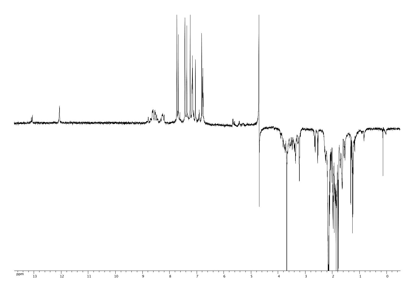
By homology with the NMR signals of DNA duplexes, the 13 ppm resonance signals are assigned to the imino proton of G-C Watson-Crick base pairs. Signals at 12 ppm are assigned to the imino proton of a Wobble base pair between a G nucleobase and a T nucleobase. From the NMR signals alone, it is not possible to determine whether the spectrum corresponds to a duplex formation or to a folding of the PNA on itself in a hair-pin conformation. The DOSY experiment performed on this molecule has been inconclusive (see Supplementary Data S3). However, the presence of two G-C base pairing seems incompatible with a hair-pin geometry, and probably indicates that PNA 3 is in a homoduplex form in solution.
The comparison of the integrals of the imino and amino protons is difficult because the excitation sequence used here has a non-uniform excitation profile over the spectrum, however it can be estimated from the respective integrals that there is a default of imino signals in the spectrum. As imino protons are specific to stable base-pairs, this is indicative that some amount of the PNA material is still in a single strand form in solution.
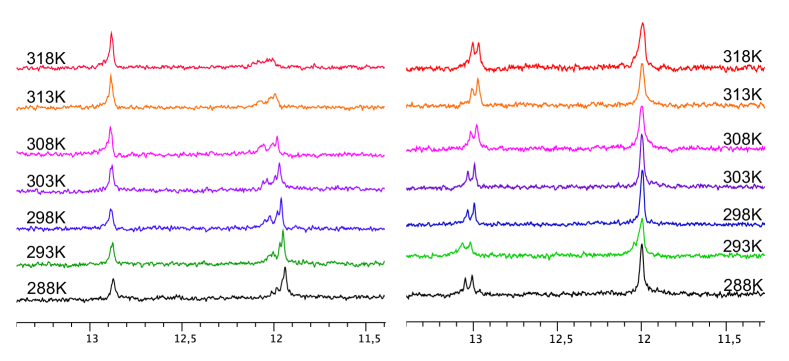
NMR spectra of PNA 2 and 3 for a temperature variation are shown in figure 3. The figure presents a zoom of the imino protons area, and spectra are recorded for temperatures ranging from 288 K to 318 K. For PNA 3, NMR spectra at various temperatures do not present any differences, in contrast to PNA 2 for which differences are seen for the T-G imino proton signal with a decrease of the signal at high temperature. For PNA 2 the T-G signal disappears completely at 318 K, unlike the G-C signals, which are still present. This indicates a stronger binding for the G-C base pair compared to the T-G base pair. As PNA 2 and PNA 3 contain the same nucleobase sequence, this indicates that the presence of the L-Serine in the position strengthen the Wobble T-G pair and increases the overall stability of the PNA homoduplex. The same temperature variation experiment has been made for PNA 4 and 5, (see figures S12 and S13 Supplementary Material) and lead to the same conclusion.
The NMR instrument cannot go higher in temperature due to the limit on the equipment, and the melting transition is not visible in this experiment. Optical approach were thus utilized to determine precisely the melting transition of the different PNAs.
Secondary structural analysis
The melting-transition temperature of the various -PNA studied here have been determined. Melting temperature were determined by temperature variation, monitoring both the absorbance and the ellipticity at several different wavelengths (see Supplementary Materials S17–S35). The experimental values are presented in the table 1. They are compared to theoretical values that nucleic acid strands with the same sequence would present, as computed using the DINAMelt Web Server[22]. Values computed for RNA were considered, because the enhanced flexibility of the RNA backbone better matches the flexibility of the -PNA structure.
Except for PNA 1 which do not present any duplex formation, the different PNAs studied here present strong base pairing, with rather high melting temperatures. The addition of a chiral center, in the form of a L-Serine substitution in the position on the PNA backbone has a stabilizing effect. This substitution has no net effect in the case of PNAs 2 and 3 but raises by 14 degrees the longer PNAs 5 and 4. The duplex 7, with 10 potential base-pairs and a chiral backbone for both strands, presents an high stability, with a transition temperature well above 100 ∘C. Such an extreme stability implies that we cannot be sure of the complete pairing of the strands in this sample, as the annealing cycles, performed in water, cannot completely disrupt the duplex in order to allow a complete sampling of all the possible conformations.
For all the PNAs tested here, the experimental melting temperatures are quite higher than the computed values of their RNA counterpart. This is probably due to the absence of the strong Coulombic repulsion observed in RNA because of the charged backbone. It should be noted that the differences between the experimental and computed values present important differences for the considered PNAs. In the case of PNAs 2 and 3, with four Watson-Crick G-C and 2 Wooble T-G base pairs, this difference is 30 to 35 ∘C. PNAs 4 and 5 have the same potential base-paring, located on a longer strand. They present differences on the same order of magnitude, except may be for PNA 5 which lacks chiral centers, and for which the entropic effect of the long non paired strand may have some destabilization effect. PNA 6 on the other hand is puzzling. While it is expected to form only 4 Watson-Crick G-C base pairs, it presents a melting transition equivalent to PNA 4, 58 ∘C above its theoretical RNA counterpart. This cannot be explained without the formation of additional non-standard base pair, such as the CA or AA base pairs, as observed in some RNA secondary structures[23]. These non-standard pairs are not isosteric to the standard Watson-Crick ones, but their formation might be possible here thanks to the greater flexibility on the PNA backbone.
| Exp | computed | Diff | |
|---|---|---|---|
| PNA 2 | 341 K 2.0 | 308.8 K | 32.2 |
| PNA 3 | 341 K 1.8 | 308.8 K | 32.2 |
| PNA 4 | 359 K 3.4 | 323.8 K | 35.2 |
| PNA 5 | 345 K 1.9 | 323.8 K | 21.2 |
| PNA 6 | 357 K 2.8 | 298.4 K | 58.6 |
| PNA 7 | 383 K | 342.0 K | 40 |
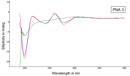
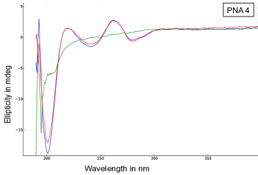
Full circular dichroism spectra of PNA 3 and 4 recorded at 308 K and 368 K are presented in figure 4. The ellipticity signals are very different at low and high temperatures. At 308 K (red curve) the complex signal observed for both PNAs is characteristic of a secondary structure existing in solution. The dichroic signal varies according to the chirality of the molecule and of the secondary structure of the molecule. This shows a pronounced Cotton effects, characteristic of a helix as show by Dragulescu-Andrasi et al[17]. This secondary structure is disrupted at high temperature (green curve), as at 368 K the ellipticity signal presents much less structure and remains mostly flat, probably dominated only by the chirality of the backbone Cγ carbons. This absence of secondary structure at high temperature is characteristic of the melting of the duplex, as was already indicated by the hyperchromicity observed at high temperatures (see Supplementary Material figure S17). The curves recorded at 308 K before and after (blue curve) the high temperature denaturation are fully superimposed, indicating that the secondary structure of PNA in solution is fully reversible. All these results are indicative of the formation of a chiral supramolecular organization, probably in the form of a helical secondary structure of the double strand.
Comparison of the overall shape of the CD spectra with the literature[24], and considering an antiparallel duplex, the maxima around 262 and 217 nm, and the minima around 277, 238, and 200 nm indicate that the formed duplex is probably a left handed helix.
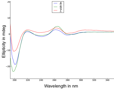
In the same manner, ellipticity curves for PNA 4, 6 and 7 are presented in figure 5. All three structures display similar curves, indicating similar supramolecular organization. However upon detailed analysis, we can observe that the maxima of the CD spectra occur at slightly different wavelengths, characteristic of slightly different structural organizations.
Conclusion
In conclusion, we have shown that PNA can adopt homo- and hetero-duplex conformations in solution through the formation of standard Watson-Crick as well as non-standard base pairing. These duplexes are extremely stable and present melting transitions at temperatures higher than their nucleic acid homologues. An extreme melting transition well above 100 ∘C was observed for a 10 bases complementary duplex, more than 40 ∘C higher than predicted for an RNA equivalent duplex.
PNAs duplexes with a chiral backbone present a marked chiral secondary structure, indicating that they are organized in supramolecular helices. Circular Dichroism spectra indicate a common folding pattern for all studied structures, with nevertheless small differences depending on the details of the nucleobase sequence.
Acknowledgements
This work was financially supported by the NMRTEC company (Illkirch - France), and the Association Nationale de la Recherche et de la Technologie, CIFRE number 376/2010. IGBMC is acknowledged for access to the NMR and CD spectrometers. JV want to thank Yves Nominé for his help with CD measurements.
Supplementary Data
Supplementary data associated with this article can be found in the online version.
-
1.
Document 1 : Figures S11–S14; NMR spectra of the different PNAs
-
2.
Document 2 : Programs and Figures S15–S35; Analysis of the Absorbance and CD data.
References
References
- Nielsen et al. [1991] P. E. Nielsen, M. Egholm, H. R. Berg, O. Buchardt, Sequence-Selective Recognition of DNA by Strand Displacement with a Thymine-Substituted Polyamide, Science 254 (1991) 1497–1500.
- Nielsen and Egholm [1999] P. E. Nielsen, M. Egholm, An Introduction to Peptide Nucleic Acid, Current issue Molec. Biol. 1 (2) (1999) 89–104.
- Egholm et al. [1993] M. Egholm, O. Buchardt, L. Christensen, C. Behrens, S. M. Freier, D. A. Driver, R. H. Berg, S. K. Kim, B. Norden, P. E. Nielsen, PNA hybridizes to complementary oligonucleotides obeying the Watson Crick hydrogen-bonding rules, Nature 365 (6446) (1993) 566–568.
- Wittung et al. [1994] P. Wittung, P. E. Nielsen, O. Buchardt, M. Egholm, B. Nordén, DNA-like double helix formed by peptide nucleic acid, Nature 368 (6471) (1994) 561–563, URL http://dx.doi.org/10.1038/368561a0.
- Uhlmann et al. [1998] E. Uhlmann, A. Peyman, G. Breipohl, D. W. Will, PNA: Synthetic polyamide nucleic acids with unusual binding properties, Angewandte Chemie-International Edition 37 (20) (1998) 2797–2823.
- Kuhn et al. [2001] H. Kuhn, V. V. Demidov, B. D. Gildea, M. J. Fiandaca, J. C. Coull, M. D. Frank-Kamenetskii, PNA beacons for duplex DNA., Antisense & nucleic acid drug development 11 (4) (2001) 265–270.
- Stender [2003] H. Stender, PNA FISH: an intelligent stain for rapid diagnosis of infectious diseases, Expert Review of Molecular Diagnostics 3 (5) (2003) 649–655.
- Demidov [2001] V. V. Demidov, PD-loop technology: PNA openers at work, Expert Review of Molecular Diagnostics 1 (3) (2001) 343–351.
- Pianowski and Winssinger [2008] Z. Pianowski, N. Winssinger, Nucleic acid encoding to program self-assembly in chemical biology, Chem Soc Rev 37 (7) (2008) 1330–1336.
- Barluenga and Winssinger [2015] S. Barluenga, N. Winssinger, PNA as a Biosupramolecular Tag for Programmable Assemblies and Reactions, Acc. Chem. Res. 48 (5) (2015) 1319–1331, URL http://dx.doi.org/10.1021/acs.accounts.5b00109.
- Bahal et al. [2014] R. Bahal, E. Quijano, N. McNeer, Y. Liu, D. Bhunia, F. Lopez-Giraldez, R. Fields, W. Saltzman, D. Ly, P. Glazer, Single-Stranded -PNAs for In Vivo Site-Specific Genome Editing via Watson-Crick Recognition, Current Gene Therapy 14 (5) (2014) 331–342, URL http://dx.doi.org/10.2174/1566523214666140825154158.
- Kazane et al. [2013] S. A. Kazane, J. Y. Axup, C. H. Kim, M. Ciobanu, E. D. Wold, S. Barluenga, B. A. Hutchins, P. G. Schultz, N. Winssinger, V. V. Smider, Self-Assembled Antibody Multimers through Peptide Nucleic Acid Conjugation, J.Am.Chem.Soc. 135 (1) (2013) 340–346.
- Avitabile et al. [2012] C. Avitabile, L. Moggio, G. Malgieri, D. Capasso, S. Di Gaetano, M. Saviano, C. Pedone, A. Romanelli, sulphate PNA (PNA S): Highly Selective DNA Binding Molecule Showing Promising Antigene Activity, PLoS ONE 7 (5) (2012) 1–10.
- Yeh et al. [2011] J. I. Yeh, B. Shivachev, S. Rapireddy, M. J. Crawford, R. R. Gil, S. Du, M. Madrid, D. H. Ly, Crystal Structure of Chiral PNA with Complementary DNA Strand: Insights into the Stability and Specificity of Recognition and Conformational Preorganization, J.Am.Chem.Soc. 132 (31) (2011) 10717–10727.
- Demidov and Frank-Kamenetskii [2004] V. V. Demidov, M. D. Frank-Kamenetskii, Two sides of the coin: affinity and specificity of nucleic acid interactions, Trends in Biochemical Sciences 29 (2) (2004) 62–71.
- Faccini et al. [2008] A. Faccini, A. Tortori, T. Tedeschi, S. Sforza, R. Tonelli, A. Pession, R. Corradini, R. Marchelli, Circular dichroism study of DNA binding by a potential anticancer peptide nucleic acid targeted against the MYCN oncogene, Chirality 20 (3-4) (2008) 494–500.
- Dragulescu-Andrasi et al. [2006] A. Dragulescu-Andrasi, S. Rapireddy, B. M. Frezza, C. Gayathri, R. R. Gil, D. H. Ly, A Simple -Backbone Modification Preorganizes Peptide Nucleic Acid into a Helical Structure, J.Am.Chem.Soc. 128 (31) (2006) 10258–10267.
- He et al. [2010] W. He, M. J. Crawford, S. Rapireddy, M. Madrid, R. R. Gil, D. H. Ly, C. Achim, The structure of a c-modified peptide nucleic acid duplex, Molecular BioSystems 6 (9) (2010) 1619.
- Chouikhi et al. [2012] D. Chouikhi, M. Ciobanu, C. Zambaldo, V. Duplan, S. Barluenga, N. Winssinger, Expanding the scope of PNA-encoded synthesis (PES): Mtt-protected PNA fully orthogonal to fmoc chemistry and a broad array of robust diversity-generating reactions, Chemistry 10 (40) (2012) 12698–12704.
- Plateau and Guéron [1982] P. Plateau, M. Guéron, Exchangeable proton NMR without base-line distorsion, using new strong-pulse sequences, J.Am.Chem.Soc. 104 (25) (1982) 7310–7311.
- Liu et al. [1998] M. Liu, X. Mao, C. Ye, J. K. Nicholson, J. C. Lindon, Improved WATERGATE pulse sequences for solvent suppression in NMR spectroscopy, J Magn Reson 132 (1998) 125––129.
- Markham and Zuker [2005] N. R. Markham, M. Zuker, DINAMelt web server for nucleic acid melting prediction, Nucleic Acids Research 33 (suppl 2) (2005) W577–W581.
- Leontis et al. [2002] N. B. Leontis, J. Stombaugh, E. Westhof, The non-Watson-Crick base pairs and their associated isostericity matrices, Nucleic Acids Res 30 (16) (2002) 3497–3531.
- Tedeschi et al. [2005] T. Tedeschi, S. Sforza, A. Dossena, R. Corradini, R. Marchelli, Lysine-based peptide nucleic acids (PNAs) with strong chiral constraint: control of helix handedness and DNA binding by chirality., Chirality 17 Suppl (2005) S196–204.
See pages - of SupMat_I.pdf See pages - of SupMat_II.pdf