Direct spectroscopic evidence for completely filled Cu shell in BaCu2As2 and -BaCu2Sb2
Abstract
We use angle-resolved photoemission spectroscopy to extract the band dispersion and the Fermi surface of BaCu2As2 and -BaCu2Sb2. While the Cu bands in both materials are located around 3.5 eV below the Fermi level, the low-energy photoemission intensity mainly comes from As states, suggesting a completely filled Cu shell. The splitting of the As core levels and the lack of pronounced three-dimensionality in the measured band structure of BaCu2As2 indicate a surface state likely induced by the cleavage of this material in the collapsed tetragonal phase, which is consistent with our observation of a Cu+1 oxydation state. However, the observation of Cu states at similar energy in -BaCu2Sb2 without the pnictide-pnictide interlayer bonding characteristic of the collapsed tetragonal phase suggests that the short interlayer distance in BaCu2As2 follows from the stability of the Cu+1 rather than the other way around. Our results confirm the prediction that BaCu2As2 is an metal with weak electronic correlations.
pacs:
74.70.Xa, 74.25.Jb, 79.60.-i, 71.20.-bEven today, the cuprate superconductors constitute the family of unconventional superconductors exhibiting the highest critical temperatures. The parent compounds of the cuprates are strongly correlated materials with Cu in the configuration (Cu2+) bridged by oxygen. Chalcogen and pnictogen atoms also serve as bridges in the layered Fe-based superconductors, for which typical electronic band renormalization factors of 2-5 are usually found P. Richard, T. Sato, K. Nakayama, T. Takahashi and H. Ding (2011). Naively, one would expect that a Cu-based material crystallizing in the same crystal structure as a Fe-based superconductor should also show strong electronic correlations and possible superconductivity, thus motivating the study of such compounds. Not only superconductivity is not observed in BaCu2As2 B. Saparov, A. S. Sefat (2012), this material has been predicted to be an metal with a filled shell Singh (2009). Unfortunately, there is still no spectroscopic evidence supporting this scenario.
Here we report an angle-resolved photoemission spectroscopy study of the electronic band dispersion and the Fermi surface of BaCu2As2 and -BaCu2Sb2. Our photon energy-dependent study reveals that the Cu states are located around 3.5 eV below the Fermi level (), whereas the intensity around is mainly derived from As 4 states. The experimental Fermi surfaces of both materials are very similar and qualitatively consistent with that of -BaCu2Sb2 derived from generalized gradient approximation (GGA) calculations. Except for the lack of three-dimensionality in BaCu2As2 associated with a surface state following the cleavage of samples in the collapsed tetragonal phase, the experimental band dispersion are also consistent with non-renormalized GGA calculations, suggesting the absence of strong electronic correlations. Our ARPES results indicate that BaCu2As2 and -BaCu2Sb2 have a fully filled Cu shell and that the stability of this electronic configuration favors the collapsed tetragonal phase of BaCu2As2.
Large single-crystals of BaCu2As2 and -BaCu2Sb2 were grown by the self-flux method B. Saparov, A. S. Sefat (2012). ARPES measurements were performed at the PGM and APPLE-PGM beamlines of the Synchrotron Radiation Center (Wisconsin) equipped with a VG-Scienta R4000 analyzer and a SES 200 analyzer, respectively. The energy and angular resolutions for the angle-resolved data were set at 10-30 meV and 0.2∘, respectively. The samples were cleaved in situ and measured at 20 K in a working vacuum better than Torr. In the following, we label the momentum values with respect to the 1 Cu/unit cell Brillouin zone, and use as the distance between two Cu planes. Raman scattering measurements were performed using Argon-Krypton laser lines in a back-scattering micro-Raman configuration with a triple-grating spectrometer (Horiba Jobin Yvon T64000) equipped with a nitrogen-cooled CCD camera. In this manuscript, we define and as the directions along the or axes, oriented 45∘ from the Cu-Cu bounds, while and are defined as the Cu-Cu directions. We performed first-principles calculations of the electronic band structure by using the full-potential linearized-augmented plane-wave (FP-LAPW) method implemented in the WIEN2K package for the crystal structures of BaCu2As2 and -BaCu2Sb2 B. Saparov, A. S. Sefat (2012). The exchange-correlation potential was treated using the generalized gradient approximation (GGA) based on the Perdew-Burke-Ernzerhof (PBE) approach J. P. Perdew, K. Burke, and M. Ernzerhof (1996).
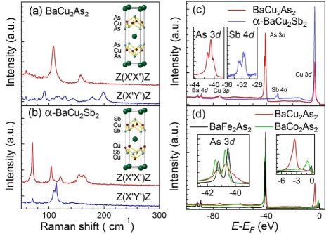
The crystal structures of BaCu2As2 and -BaCu2Sb2 are closely related, as illustrated by the insets of Figs. 1(a) and 1(b). While BaCu2As2 has the same crystal structure as BaFe2As2, described by the space group D (I4/mmm), -BaCu2Sb2, characterized by the space group D(P4/nmm), has the relative positions of the Sb and Cu atoms exchanged in two successive layers. To confirm that our samples of BaCu2As2 and -BaCu2Sb2 have different crystal structures, we recorded Raman spectra at room temperature with incident and scattered light polarized in the plane. Our analysis predicts 4 Raman active modes in BaCu2As2 (A1g+B1g+2Eg) and 9 in -BaCu2Sb2 (3A1g+2B1g+4Eg). While the in-plane polarization used does not allow us to detect Eg modes, the A1g (related to the vibration of the pnictogens along the -axis) and B1g (related to the vibration of Cu) channels are accessible in the and configurations, respectively. The experimental results for BaCu2As2 and -BaCu2Sb2 are shown in Figs. 1(a) and 1(b), respectively. We observe an A1g mode at 108.9 cm-1 and a B1g mode at 198.2 cm-1 in BaCu2As2, whereas 3 A1g modes at 70.4 cm-1, 104 cm-1 and 121.4 cm-1 and 2 B1g modes at 108.7 cm-1 and 113.4 cm-1 are detected in -BaCu2Sb2. Although additional weak and unidentified peaks are also detected, the spectra of our BaCu2As2 and -BaCu2Sb2 samples are clearly different, thus proving that they correspond to different crystal phases.
We then confirm the elemental composition of our samples by showing the core level spectra of BaCu2As2 and -BaCu2Sb2 in Figs. 1(c). In both materials we detect the Ba and Cu core levels around 90 and 75 eV of binding energy (), respectively. As expected, the spectrum of BaCu2As2 exhibits additional peaks around 41 eV corresponding to the As states that do not appear in the spectrum of -BaCu2Sb2. However, 4 features instead of 2 are observed, suggesting a surface state similar to the one reported previously for the EuFe2As2-xPx system P. Richard, C. Capan, J. Ma, P. Zhang, N. Xu, T. Qian, J. D. Denlinger, G.-F. Chen, A. S. Sefat, Z. Fisk and H. Ding (2014). The spectra of -BaCu2Sb2 exhibits 4 peaks around eV assigned to the Sb electronic states. In this case though, the existence of four Sb peaks does not indicate the presence of a surface state since the crystal structure of -BaCu2Sb2 itself contains 2 inequivalent Sb sites.
A rigid band shift is the simplest assumption one can make when describing the effect of doping. When considering only the shape of the quasi-particle dispersions, this assumption works surprisingly well in the (Ba,K)Fe2As2 and Ba(Fe,Co)2As2 systems M. Neupane, P. Richard, Y.-M. Xu, K. Nakayama, T. Sato, T. Takahashi, A. V. Federov, G. Xu, X. Dai, Z. Fang, Z. Wang, G.-F. Chen, N.-L. Wang, H.-H. Wen and H. Ding (2011), despite a strong dependence of the strength of correlations and coherence on the filling P. Werner, M. Casula, T. Miyake, F. Aryasetiawan, A. J. Millis and S. Biermann (2012). It is also still valid when the filling of the -shell reaches 7 electrons in BaCo2As2 Xu et al. (2013). As illustrated in the left inset of Fig. 1(d) that compares the As core levels of BaFe2As2, BaCo2As2 and BaCu2As2, the As core levels in BaCo2As2 are downshifted in energy compared to their energy position in BaFe2As2, indicating an upward shift of the chemical potential. Assuming that the Cu shell in BaCu2As2 contains 9 electrons as in the cuprates, a large downshift of the core level position should be observed. In contrast, the center of gravity of the As core levels in BaCu2As2 is found nearly at the same energy as in BaFe2As2. This indicates clearly that the rigid band shift approximation is no longer valid and that other effects, such as the electronic valency of As, must be considered. A direct corollary is that the valency of the Cu atoms, which have As as ligands, may also be strongly affected.
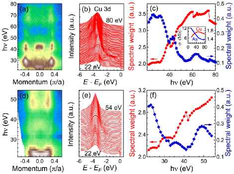
To check this latter scenario, we performed ARPES measurements of the valence states. In Fig. 2(a) we display the photon energy () dependence of the ARPES intensity at recorded along -M in BaCu2As2. Although the intensity is modulated along , the momentum position of the features observed varies very little with , suggesting a quasi-two-dimensional electronic structure. Thus, we can tentatively assign the values 41 eV and 36 eV to and , but we cannot unambiguously determine which one is which. Despite a different crystal structure, very similar results are observed for -BaCu2Sb2, as shown in Fig. 2(d), except that the values associated with eV and 41 eV are exchanged.
The dependence of the normal emission energy distribution curve (EDC) in BaCu2As2 and -BaCu2Sb2 are displayed in Figs. 2(b) and 2(e), respectively. Unlike the ferropnictide materials, for which strong spectral weight associated to the Fe states is observed near H. Ding, K. Nakayama, P. Richard, S. Souma, T. Sato, T. Takahashi, M. Neupane, Y.-M. Xu, Z.-H. Pan, A. V. Fedorov, Z. Wang, X. Dai, Z. Fang, G. F. Chen, J. L. Luo and N. L. Wang (2011), BaCu2As2 and -BaCu2Sb2 exhibit only very small intensity at . On the other hand, the Cu-pnictides show a very strong peak around 3.5 eV below . In Fig. 2(c) we compare the spectral intensity of the normal emission EDCs integrated in the [-4, -3] eV and [-1, 0] energy ranges. The dependences of these spectral intensities show different trends, thus indicating that their origin is different. While the spectral intensity in the [-4, -3] eV range increases with increasing from 22 eV to about 60 eV and then slowly decreases, the spectral intensity near is at its highest at low values and drops with increasing. Besides additional features, like the small peaks around 41 eV and 64 eV in the [-1, 0] energy range that are likely induced by the effect on the photoemission matrix elements, the intensity of the [-4, -3] eV and [-1, 0] eV ranges are qualitatively in reasonable agreement with the photoemission cross sections Yeh and Lindau (1985) of the Cu and As states, respectively, which are reproduced in the inset of 2(c). A similar observation is made for -BaCu2Sb2, as illustrated by Figs. 2(e)-(f).
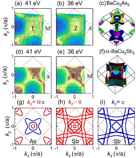
The results discussed above indicate clearly that most of the Cu states are located at about 3.5 eV below . We thus conclude that unlike in the cuprates, the shell of Cu is completely filled, an experimental conclusion consistent with a previous prediction based on a local density approximation (LDA) study of BaCu2As2 and SrCu2As2 Singh (2009). Experimentally, a Fermi surface different from that of the ferropnictides, essentially made of pnictogen states, is thus expected. In Figs. 3(a) and 3(b), we compare the Fermi surface intensity maps obtained on BaCu2As2 using eV and 36 eV, respectively. In agreement with the dependence of the intensity plot shown in 2(a) and despite a redistribution of spectral intensity along the Fermi surface, the measured Fermi surfaces for these two set of data are surprisingly similar, indicating a quasi-two-dimensional electronic structure. This is in sharp contrast with our GGA calculation of the three-dimensional Fermi surface of this material, which are displayed in Fig. 3(c), as well as with the calculated Fermi surface cuts at and , shown in Fig. 3(g). Even more surprising is the strong resemblance between the results recorded on BaCu2As2, and those recorded on -BaCu2Sb2, which are displayed in Figs. 3(d) and 3(e), although the features obtained for -BaCu2Sb2 are broader, which we attribute to a bad surface quality due to the difficulty of cleaving this material with strong Cu-Sb inter-layer bonding. Unlike BaCu2As2, -BaCu2Sb2 is predicted to have a two-dimensional Fermi surface, as illustrated in Fig. 3(f) and by the Fermi surface cuts at and given in Figs. 3(h) and 3(i), respectively.
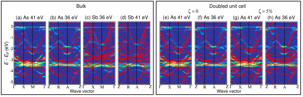
In Fig. 4, we display the ARPES curvature Zhang et al. (2011) intensity plots of BaCu2As2 and -BaCu2Sb2 recorded at 20 K along some high-symmetry lines with 41 eV and 36 eV photons. As mentioned above, the quasi-two-dimensional nature of the experimental Fermi surfaces prevents us from identifying unambiguously which one corresponds to and which one corresponds to , but our dependence data suggest that the roles are exchanged in BaCu2As2 and -BaCu2Sb2. For these plots we tentatively assume that 41 eV corresponds to () in BaCu2As2 (-BaCu2Sb2). Except for a few obvious discrepancies, like the absence of electron pocket at in the experimental data and the position of the hole-like dispersion around , the GGA calculations at capture well the main features observed experimentally, without any renormalization. A similar comment can be made for -BaCu2Sb2, although in that case the agreement of the experimental data and the GGA predictions for the location of the Cu bands is slightly worse.
Given the indications of the presence of a surface state, we have tried to understand the striking resemblance of the measured BaCu2As2 spectral function with the one of -BaCu2Sb2, by assuming that the ARPES spectra display a substantial contribution from the surface layer. In BaFe2As2, a surface relaxation that consists essentially in an elongation of the As-height at the surface has been studied in Ref. V. B. Nascimento, Ang Li, Dilushan R. Jayasundara, Yi Xuan, J. O’Neal, Shuheng Pan, T. Y. Chien, Biao Hu, X. B. He, Guorong Li, A. S. Sefat, M. A. McGuire, B. C. Sales, D. Mandrus, M. H. Pan, Jiandi Zhang, R. Jin and E. W. Plummer (2). The result of such a structural surface relaxation is that the two FeAs layers of the surface unit cell are no longer equivalent, putting the surface crystal structure in the same symmetry group as the bulk of -BaCu2Sb2 – namely P4/nmm – where two inequivalent layers appear due to the inversion of Cu and Sb. To the best of our knowledge, the precise surface crystal structure of BaCu2As2 has not been characterized yet, but it seems plausible that the “collapsed” character of this phase rather enhances such a structural distortion at the surface, and it is safe to postulate that a substantial distortion, leaving the two upper-most CuAs surface layers inequivalent, occurs.
Now, we can speculate that chemically, substituting Sb by As does not result in a drastic difference in the spectra, as long as the crystal symmetry is preserved. We have checked this hypothesis by calculating the band structure of a hypothetical BaCu2As2 compound where we have lowered the crystal symmetry as if the two layers were inequivalent. The resulting bands are overlaid to the experimental data in Figs. 4e and 4f. We also show the band structure that we obtain if we increase the As height by 5% on one of the layers, in Figs. 4g and 4h. One of the consequences of such a doubled unit cell is that the number of bands in the plane is multiplied by two due to folding (Figs. 4e and 4g), and the dispersion corresponds to a superposition of the bands in the and plane of the I4/mmm structure (Figs. 4a and 4b). In the plane of the doubled unit cell, the number of bands is also multiplied by two but they are degenerate if the two layers have the same structure (Fig. 4f), while a difference appears if we introduce a distortion (Fig. 4h). While the knowledge of the precise surface structure would be needed to obtain quantitative agreement, Figs. 4e-4h strongly suggest that the similarity of the BaCu2As2 spectra to the -BaCu2Sb2 ones can indeed be attributed to a lowering of the surface crystal symmetry of the kind we describe.
Finally, we comment on the 3 configuration of Cu. Singh already pointed out Singh (2009) that the much shorter parameter in BaCu2As2 as compared to BaNi2As2 and BaFe2As2 is suggestive of different bonding in BaCu2As2, which is confirmed by our As core level spectra. In fact, comparison of the lattice parameters places BaCu2As2 in the collapsed tetragonal phase Anand et al. (2012), which is associated with a stronger As-As interlayer bonding R. Hoffmann and C. Zheng (1985); T. Yildirim (2009). However, our results suggest that the strong As-As interlayer bonding cannot be the cause of the 3 configuration of Cu in BaCu2As2. Indeed, the 3 Cu states in -BaCu2Sb2 are located practically at the same energy as in BaCu2As2, despite the absence of Sb-Sb direct interlayer bonding in the former material, thus reinforcing previous arguments by Anand et al. based on the resemblance of the physical properties of SrCu2As2 and -SrCu2Sb2 Anand et al. (2012). Interestingly, the Cu dopant states in Cu-substituted BaFe2As2 are also found around 3-4 eV below J. A. McLeod, A. Buling, R. J. Green, T. D. Boyko, N. A. Skorikov, E. Z. Kurmaev, M. Neumann, L. D. Finkelstein, Ni Ni, A. Thaler, S. L. Bud’ko, P. C. Canfield and A. Moewes (2012); Ideta et al. (2013), suggesting that Cu-dopants are already in a +1 oxidation state. This is consistent with the absence of any shift in the Cu core level spectra as a function of doping Y. J. Yan, P. Cheng, J. J. Ying, X. G. Luo, F. Chen, H. Y. Zou, A. F. Wang, G. J. Ye, Z. J. Xiang, J. Q. Ma and X. H. Chen (2013), as well as with the suppression of magnetic moment and the observation of superconductivity only for a very narrow Cu substitution range E. D. Mun, S. L. Bud’ko, Ni Ni, A. N. Thaler and P. C. Canfield (2009); Ni et al. (2010), in contrast to substitution of Fe by Co A. S. Sefat, R. Jin, M. A. McGuire, B. C. Sales, D. J. Singh and D. Mandrus (2008); N. Ni, M. E. Tillman, J.-Q. Yan, A. Kracher, S. T. Hannahs, S. L. Bud’ko and P. C. Canfield (2008) and Ni L. J. Li, Y. K. Luo, Q. B. Wang, H. Chen, Z. Ren, Q. Tao, Y. K. Li, X. Lin, M. He, Z. W. Zhu, G. H. Cao and Z. A. Xu (2009). However, the clear observation of added electron carriers in Ba(Fe1-xCux)xAs2 E. D. Mun, S. L. Bud’ko, Ni Ni, A. N. Thaler and P. C. Canfield (2009); Ni et al. (2010) indicates that the Cu substitution is electron doping nevertheless, suggesting that the local As-As and As-transition metal bondings are strongly affected, a situation that could be stabilized at high substitution levels by the collapsed tetragonal phase. Our findings support the hypothesis Anand et al. (2012) that it is the stability of the 3 configuration of Cu (Cu+1) that favors the collapsed tetragonal phase of BaCu2As2, rather than the other way around.
In summary, we have performed angle-resolved photoemission spectroscopy measurements on BaCu2As2 and -BaCu2Sb2 to extract their electronic band dispersions and Fermi surfaces. We found that most of the Cu 3 spectral weight locates around 3-4 eV below , whereas the intensity around mainly comes from As 4 bands, suggesting a filled Cu shell. The observation of split As core levels and the absence of pronounced three-dimensionality in the measured electronic structure of BaCu2As2 is compatible with a surface state emerging from the cleavage of this material in the collapsed tetragonal phase. However, the observation of similar Cu states in -BaCu2Sb2 without pnictide-pnictide interlayer bonding suggests that the stability of the Cu+1 configuration favors the collapsed tetragonal phase rather than the other way around. Our study indicates that BaCu2As2 and -BaCu2Sb2 are metals with weak electronic correlations.
We acknowledge W.-L. Zhang for useful discussions. This work was supported by grants from MOST (2010CB923000 and 2011CBA001000, 2011CBA00102, 2012CB821403) and NSFC (10974175, 11004232, 11034011/A0402, 11234014 and 11274362) from China, the Cai Yuanpei program, the French ANR via project PNICTIDES, IDRIS/GENCI under project 091393 and the European Research Council under Project No. 617196. This work is based in part on research conducted at the Synchrotron Radiation Center, which was primarily funded by the University of Wisconsin-Madison with supplemental support from facility Users and the University of Wisconsin-Milwaukee. The work at ORNL was supported by the Department of Energy, Basic Energy Sciences, Materials Sciences and Engineering Division.
References
- P. Richard, T. Sato, K. Nakayama, T. Takahashi and H. Ding (2011) P. Richard, T. Sato, K. Nakayama, T. Takahashi and H. Ding, Rep. Prog. Phys. 74, 124512 (2011).
- B. Saparov, A. S. Sefat (2012) B. Saparov, A. S. Sefat, J. Solid State Chem. 191, 213 (2012).
- Singh (2009) D. J. Singh, Phys. Rev. B 79, 153102 (2009).
- J. P. Perdew, K. Burke, and M. Ernzerhof (1996) J. P. Perdew, K. Burke, and M. Ernzerhof, Phys. Rev. Lett. 77, 3865 (1996).
- P. Richard, C. Capan, J. Ma, P. Zhang, N. Xu, T. Qian, J. D. Denlinger, G.-F. Chen, A. S. Sefat, Z. Fisk and H. Ding (2014) P. Richard, C. Capan, J. Ma, P. Zhang, N. Xu, T. Qian, J. D. Denlinger, G.-F. Chen, A. S. Sefat, Z. Fisk and H. Ding, J. Phys.: Condens. Matter 26, 035702 (2014).
- M. Neupane, P. Richard, Y.-M. Xu, K. Nakayama, T. Sato, T. Takahashi, A. V. Federov, G. Xu, X. Dai, Z. Fang, Z. Wang, G.-F. Chen, N.-L. Wang, H.-H. Wen and H. Ding (2011) M. Neupane, P. Richard, Y.-M. Xu, K. Nakayama, T. Sato, T. Takahashi, A. V. Federov, G. Xu, X. Dai, Z. Fang, Z. Wang, G.-F. Chen, N.-L. Wang, H.-H. Wen and H. Ding, Phys. Rev. B 83, 094522 (2011).
- P. Werner, M. Casula, T. Miyake, F. Aryasetiawan, A. J. Millis and S. Biermann (2012) P. Werner, M. Casula, T. Miyake, F. Aryasetiawan, A. J. Millis and S. Biermann, Nature Phys. 8, 331 (2012).
- Xu et al. (2013) N. Xu, P. Richard, A. van Roekeghem, P. Zhang, H. Miao, W.-L. Zhang, T. Qian, M. Ferrero, A. Sefat, S. Biermann, et al., Phys. Rev. X 3, 011006 (2013).
- H. Ding, K. Nakayama, P. Richard, S. Souma, T. Sato, T. Takahashi, M. Neupane, Y.-M. Xu, Z.-H. Pan, A. V. Fedorov, Z. Wang, X. Dai, Z. Fang, G. F. Chen, J. L. Luo and N. L. Wang (2011) H. Ding, K. Nakayama, P. Richard, S. Souma, T. Sato, T. Takahashi, M. Neupane, Y.-M. Xu, Z.-H. Pan, A. V. Fedorov, Z. Wang, X. Dai, Z. Fang, G. F. Chen, J. L. Luo and N. L. Wang, J. Phys: Condens. Matter 23, 135701 (2011).
- Yeh and Lindau (1985) J. Yeh and I. Lindau, At. Data Nucl. Data Tables 32, 1 (1985).
- Zhang et al. (2011) P. Zhang, P. Richard, T. Qian, Y.-M. Xu, X. Dai, and H. Ding, Rev. Sci. Instrum. 82, 043712 (2011).
- V. B. Nascimento, Ang Li, Dilushan R. Jayasundara, Yi Xuan, J. O’Neal, Shuheng Pan, T. Y. Chien, Biao Hu, X. B. He, Guorong Li, A. S. Sefat, M. A. McGuire, B. C. Sales, D. Mandrus, M. H. Pan, Jiandi Zhang, R. Jin and E. W. Plummer (2) V. B. Nascimento, Ang Li, Dilushan R. Jayasundara, Yi Xuan, J. O’Neal, Shuheng Pan, T. Y. Chien, Biao Hu, X. B. He, Guorong Li, A. S. Sefat, M. A. McGuire, B. C. Sales, D. Mandrus, M. H. Pan, Jiandi Zhang, R. Jin and E. W. Plummer, Phys. Rev. Lett. 103, 076104 (2009).
- Anand et al. (2012) V. K. Anand, P. K. Perera, A. Pandey, R. J. Goetsch, A. Kreyssig, and D. C. Johnston, Phys. Rev. B 85, 214523 (2012).
- R. Hoffmann and C. Zheng (1985) R. Hoffmann and C. Zheng, J. Phys. Chem. 89, 4175 (1985).
- T. Yildirim (2009) T. Yildirim, Phys. Rev. Lett. 102, 037003 (2009).
- J. A. McLeod, A. Buling, R. J. Green, T. D. Boyko, N. A. Skorikov, E. Z. Kurmaev, M. Neumann, L. D. Finkelstein, Ni Ni, A. Thaler, S. L. Bud’ko, P. C. Canfield and A. Moewes (2012) J. A. McLeod, A. Buling, R. J. Green, T. D. Boyko, N. A. Skorikov, E. Z. Kurmaev, M. Neumann, L. D. Finkelstein, Ni Ni, A. Thaler, S. L. Bud’ko, P. C. Canfield and A. Moewes, J. Phys.: Condens. Matter 24, 215501 (2012).
- Ideta et al. (2013) S. Ideta, T. Yoshida, I. Nishi, A. Fujimori, Y. Kotani, K. Ono, Y. Nakashima, S. Yamaichi, T. Sasagawa, M. Nakajima, et al., Phys. Rev. Lett. 110, 107007 (2013).
- Y. J. Yan, P. Cheng, J. J. Ying, X. G. Luo, F. Chen, H. Y. Zou, A. F. Wang, G. J. Ye, Z. J. Xiang, J. Q. Ma and X. H. Chen (2013) Y. J. Yan, P. Cheng, J. J. Ying, X. G. Luo, F. Chen, H. Y. Zou, A. F. Wang, G. J. Ye, Z. J. Xiang, J. Q. Ma and X. H. Chen, Phys. Rev. B 87, 075105 (2013).
- E. D. Mun, S. L. Bud’ko, Ni Ni, A. N. Thaler and P. C. Canfield (2009) E. D. Mun, S. L. Bud’ko, Ni Ni, A. N. Thaler and P. C. Canfield, Phys. Rev. B 80, 054517 (2009).
- Ni et al. (2010) N. Ni, A. Thaler, J. Q. Yan, A. Kracher, E. Colombier, S. L. Bud’ko, P. C. Canfield, and S. T. Hannahs, Phys. Rev. B 82, 024519 (2010).
- A. S. Sefat, R. Jin, M. A. McGuire, B. C. Sales, D. J. Singh and D. Mandrus (2008) A. S. Sefat, R. Jin, M. A. McGuire, B. C. Sales, D. J. Singh and D. Mandrus, Phys. Rev. Lett. 101, 117004 (2008).
- N. Ni, M. E. Tillman, J.-Q. Yan, A. Kracher, S. T. Hannahs, S. L. Bud’ko and P. C. Canfield (2008) N. Ni, M. E. Tillman, J.-Q. Yan, A. Kracher, S. T. Hannahs, S. L. Bud’ko and P. C. Canfield, Phys. Rev. B 78, 214515 (2008).
- L. J. Li, Y. K. Luo, Q. B. Wang, H. Chen, Z. Ren, Q. Tao, Y. K. Li, X. Lin, M. He, Z. W. Zhu, G. H. Cao and Z. A. Xu (2009) L. J. Li, Y. K. Luo, Q. B. Wang, H. Chen, Z. Ren, Q. Tao, Y. K. Li, X. Lin, M. He, Z. W. Zhu, G. H. Cao and Z. A. Xu, New J. Phys. 11, 025008 (2009).