Translation with frameshifting of ribosome along mRNA transcript
Abstract
Translation is an important process for prokaryotic and eukaryotic cells to produce necessary proteins for cell growth. Numerious experiments have been performed to explore the translational properties. Diverse models have also been developed to determine the biochemical mechanism of translation. However, to simplify the majority of the existing models, the frameshifting of ribosome along the mRNA transcript is neglected, which actually occurs in real cells and has been extensively experimentally studied. The frameshifting of ribosome evidently influences the efficiency and speed of translation, considering that the peptide chains synthesized by shifted ribosomes will not fold into functional proteins and will degrade rapidly. In this study, a theoretical model is presented to describe the translational process based on the model for totally asymmetric simple exclusion process. In this model, the frameshifting of the ribosome along the mRNA transcript and the attachment/detachment of the ribosome to/from the main body of mRNA codons during translation elongation process, are explicitly included. The results show that, with ribosome frameshifing, the speed of correctly synthesized peptide chains may increase first and then decrease with both the translation initiation rate and the ribosome detachment rate . This results indicates that regulating the translation process to reach maximal synthesized speed of proteins is theoretically feasible. Traffic-related problems of ribosome motion along the mRNA transcript are also addressed theoretically. Depending on parameter values, shock wave (or domain wall) may exist for ribosome probabilities along the mRNA.
I Introduction
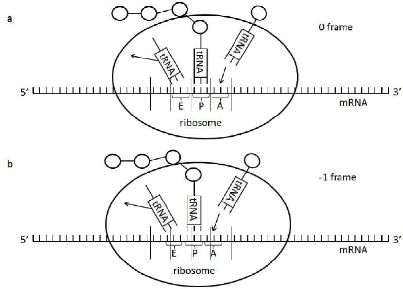
In living cells, proteins are synthesized by ribosome through the translation process with the information coded in the template messenger RNA (mRNA). The mRNA is composed of a sequence of codons, and each codon consists of three nucleotides. Given the four kinds of nucleotides in mRNAs, a total of kinds of codons exits. Except for the stop codon, which is used to finish the translation process, each kind of codon corresponds to a certain kind of amino acid, although each kind of amino acid may correspond to several kinds of codons Turanov2009 . During translation, an amino acid is transferred by the transfer RNA (tRNA). With the binding of tRNA to mRNA through one of its end, which has complementary anticodon sequence to that of the mRNA, the amino acid carried by the other end of tRNA is then chained together into a polypeptide as the mRNA passes through. A schematic description of this process is found in Fig. 1.
Translation is followed by molecular machine ribosome, which includes a three step process, that is, initiation, elongation, and termination Racle2013 . The 30S subunit of the ribosome contains three tRNA binding sites, which are denoted by A, P, and E (Fig. 1). In the initial step, the first tRNA and the start codon of mRNA will form a codon-anticodon duplex in the P site of the 30S subunit, with the Shine-Dalgarno (SD) sequence on the 5’ end of the mRNA binding with the 3’ end of the 16S rRNA (an anti-SD sequence in the 30S subunit of a ribosome) Shine1974 ; Czernilofskya1975 ; Yusupova2001 ; Korostelev2007 ; Duval2013 . Meanwhile, the 50S subunit of ribosome will attach to the mRNA from the other side. During translation, mRNA will be kept in the channel between these two subunits. During elongation, each binding site of the 30S subunit of ribosome will be bound by one mRNA codon, while the tRNA anticodon end will bind to the mRNA codon in the A site. When the ribosome steps forward along the mRNA, the codon from the A site, together with the tRNA attached to it, will move to the P site. Then, the amino acid carried by the tRNA will be added to the tail of the synthesizing peptide chain in the P site. Finally, when the complex of mRNA codon and tRNA moves to the E site, the tRNA will detach from the mRNA codon and the peptide chain (Fig. 1). No tRNA that contains an anticodon that is complementary to the stop codon exists. In the termination process, the stop codon will be bound by one release factor, which helps the release of the peptide chain and ribosome from the mRNA, which completes the translation process.
The translation process described above is only applicable for ideal cases, in which the ribosome always moves forward codon by codon, and it always reads three nucleic acids from the same codon in each elongation step. However, in actual cells, the ribosome may shift to one nucleic acid upstream or downstream along the template mRNA in a low frequency Atkins2010 . This phenomenon is called frameshifting. Two mRNA codons are considered to be in the same frame if and only if the number of nucleic acids between theses codons is divisible by 3. In the following, the mRNA start codon is called a 0 frame codon (or a codon in 0 frame). Thus, all codons that are in the same frame with the start codon are also 0 frame codons (or codons in 0 frame). A codon that is one nucleic acid upstream to a 0 frame codon is called a frame codon (or a codon in frame). Meanwhile, a codon that is one nucleic acid downstream to a 0 frame codon is called a frame codon (or a codon in frame). Considering that every codon consists of three nucleic acids, any mRNA codon must belong to one of the three frames, i.e 0, , and frames. If one mRNA codon read by ribosome belongs to the 0 frame (or 1 frame), then the mRNA is said to be read in the 0 frame (or 1 frame). Therefore, each mRNA has three reading frames. With frameshifting of ribosome, the reading frames of mRNAs are distinct. To obatin intuitive impressions of frameshifting, one schematic diagram of frameshifting is depicted in Fig. 1.
Translation is generally initialized from the start codon, so, in the initiation of translation, mRNA is usually in the 0 frame. During the elongation period, frameshifting may occur at some codons, so the mRNA will change into the frame or frame. Regardless of the reading frame, translation will continue until the ribosome meets the stop codon. However, peptide chains produced by ribosome in reading frames are different from those produced by correct translations in the 0 reading frame, and these chains are usually nonfunctional, and will degrade rapidly.
Experimental findings have shown that the frequency of frameshifting is only to per codon. However, the frequency of frameshifting increases for a slippery sequence with the motif X XXY YYZ Chen2014 . The 3’ secondary structure and the 5’ internal SD sequence in the mRNA can further increase the frameshift frequency Larsen1994 ; Larsen1997 . Numerous studies have been performed to describe the property and mechanism of frameshifting Tinoco2013 ; Plant2003 ; Baranov2004 ; Horsfield1995 ; Lopinski2000 ; Jacks1988 ; Namy2006 ; Weiss1989 ; Leger2007 . A general mechanistic and conformational framework for frameshifting is recently presented in Chen2014 to attempt to elucidate the mechanism of frameshifting. Structural insights into frameshifting are discussed in Maehigashi2014 , and some possible mechanisms of frameshifting can also be found in Belew2014 .
Biophysically, translation can be roughly regarded as a totally asymmetric simple exclusion process (TASEP) see Derrida1993 ; Parmeggiani2003 ; Zhang20101 ; Zhang2012 . TASEP is one statistical physics model to describe the unidirectional hopping process of general particles along a one-dimensional lattice. In this model, the particle at site of the main body of lattice hops forward to site provided that site is not occupied. The particle at the last site of lattice leaves the lattice into the environment at a given rate constant, and particles in the environment enter the first site of lattice provided that this site is unoccupied. If one mRNA with codons is considered as a one-dimensional lattice of length , then the initiation process of translation corresponds to the entrance of the ribosome into the first lattice site. The elongation process of translation corresponds to the forward hopping process of ribosome along the main body of lattice, and finally the termination of translation corresponds to the leaving of ribosome from the last lattice site. Therefore, to a certain extent, TASEP is a reasonable model to describe the translation process in gene expression.
Different types of TASEP models have been conceptualized in various kinds to describe corresponding biophysical or biochemical processes. In these models, particles may be allowed to detach from or attach to any site of lattice, particles may have multiple internal states, there may be different types of particles which hop along the same track, particles may be allowed to hop to the nonadjacent lattice sites, and particles may hop along multiple parallel lattices Krug1991 ; Kolomeisky1998 ; Schutz2003 ; Lipowsky2006 ; Raguin2013 ; Popkov2013 ; Bressloff2013 ; Zhang20113 ; Zhang20131 ; Gupta2014 . Nevertheless, no theoretical model to date has been presented to study the translational process with occasional frameshifting of ribosome along mRNA. Frameshifting influences on both the speed of correct translation (i.e., the product speed of correct peptide chain) and the accuracy of translation. Therefore, to determine the detailed translational properties, and explore the mechanism to regulate gene expression, a more reasonable model that includes frameshifting should be presented. In this study the translation process, which includes initiation, elongation and termination, will also be regarded as one TASEP of particle hopping along a one-dimensional lattice. Contrary to the usual TASEP, particles (i.e., ribosomes) in the translation elongation period may make one frameshifting stochastically. The corresponding peptide chains produced by shifted ribosomes will be nonfunctional and degrade quickly. Given the difference of ribosomes in correct and incorrect translation states, the proposed model is similar to the TASEP with two particle species. However, the proposed model is different because, during forward hopping, particles may change from one species to the other. Additionally, due to frameshifting, ribosomes in incorrect translation state do not stop at the supposed end of lattice. This is because that for ribosomes that translocate in the wrong mRNA frames (i.e., 1 frame), the stop codon cannot be recognized as usual. This study was primarily designed to discuss the translational properties of by the modified TASEP model, which includes the frameshifting of ribosome, especially the dependence on model parameters of correct translation speed and the ratio of correctly produced peptide chains in all peptide chains.
This study is organized as follows. The theoretical model will be presented in the next section, and then the numerical results on the translational properties with ribosome frameshifting will be provided in Section III. Finally, concluding remarks will be presented in the last section.
II Modified TASEP with ribosome frameshifting
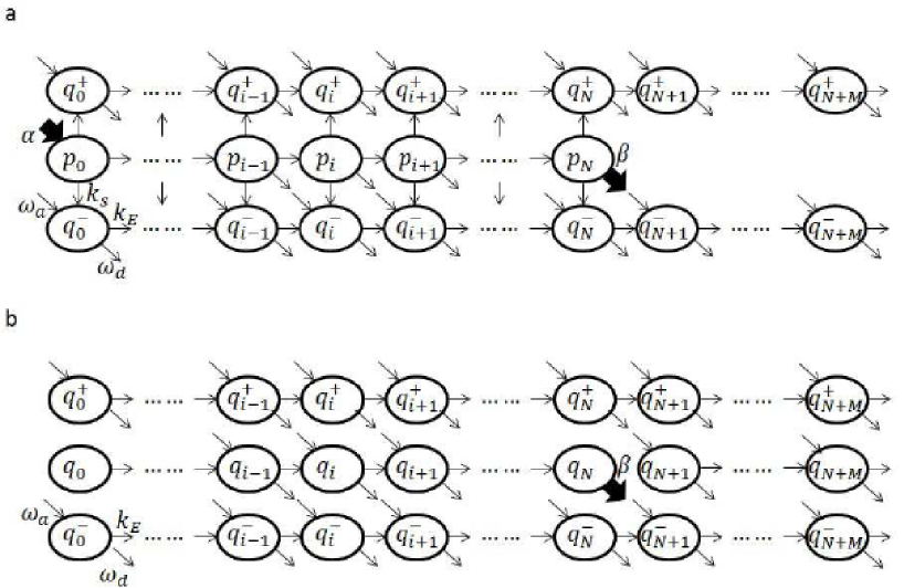
In the usual TASEP without ribosome frameshifting and attachment/detachment to/from the main body of mRNA, the model parameters that influence translation speed are as follows: (i) entrance rate of particles from the environment to the first lattice site, i.e., the rate of ribosome binding to the mRNA start codon. This value depends on the concentration of free ribosomes, and the sequence of ribosome binding site (RBS); (ii) length of lattice (mRNA), which is usually denoted by ; (iii) leaving rate of particles (ribosomes) from the last lattice site (the stop codon) to environment; and (iv) forward hopping rate of particles in the main body of lattice, i.e., the elongation rate of translation. For simplicity, the particles are usually assumed to be able to hop forward along the track, and their forward hopping rates at any lattice site are assumed to be the same. In this study, the usual TASEP had been modified to include frameshifting of ribosome as shown in the schematic depiction in Fig. 2.
Given that the frequency of frameshifting is usually low, this study assumes that throughout the translocation along mRNA, each ribosome can only frameshift at most once. This assumption implies that only ribosomes in the 0 frame can shift one nucleotide forward to 1 frame or backward to frame. General models without this restriction can be presented similarly, but those models will complicate the following analysis. Actually, with more possible frameshitings or additional ribosome frameshiftings in the frame, the results obtained in this study will not change essentially. In this study, the codon with one nucleotide upstream or downstream of codon is denoted by or . The rates of frameshifting from codon to codon and codon are denoted by and , respectively.
Considering that ribosomes only have one chance of framshifting, the ribosomes in frame will not shift back into the 0 frame. Thus, no stop codon will generally exist in the frame, and ribosomes in the frame will come across the stop codon and continue moving forward to the 3’ end of mRNA. This study denotes the number of codons between the usual stop codon and the 3’ end of mRNA by , and they are numbered by . Thus, the length of track for ribosomes translocating in frame is , which is codons longer than that for ribosomes translocating in the 0 frame (Fig. 2). The hopping rate of particles (elongation rate of translation) from codon to codon are denoted by and the probabilities of finding one ribosome at codon are denoted by .
In addition to start and stop codons, this study also allows the attachment/detachment of ribosomes to/from any mRNA codon with rates denoted by . Attachment and detachment of ribosomes are also allowed at codon for any with rates denoted by and respectively. Therefore, ribosomes at codon for can be divided into two classes, namely, ribosomes with correctly and incorrectly synthesized peptide chains. If a ribosome reaches codon from the start codon without frameshifting, then the synthesized peptide chain will be correct. On the contrary, if a ribosome binds to mRNA at codon for , then the synthesized peptide chain will be incorrect. For , the probability of finding a ribosome at codon with one correct or incorrect peptide chain is denoted by or . This study assumes that only ribosomes in the 0 frame with a correct peptide chain can frameshift, and the probability is always equals to zero. Considering that frameshifting of ribosomes with incorrect peptide chain will not change the correct translation speed essentially. Without loss of generality, except for the normal initiation and termination, this study assumes the absence of ribosome detachment/attachment at the start/stop codon.
For simplicity, this study assumes that and . This condition indicates that for a ribosome regardless of frames, either in the 0 or in frame, its forward translocation rate and detachment/attachment rates are the same. Meanwhile, the rates of upstream and downstream frameshifting are also equal.
At any time, only one of the three codons , and can be occupied by ribosome. Thus, if codon is occupied by one ribosome, then the codons will be empty. This phenomenon indicates that the ribosome at codon always has the possibility to frameshift and then translocate to either codon or codon . For convenience, denotes the probability that position is unoccupied. One can show that (see Fig. 2),
| (1) |
The probabilities of finding ribosome with correctly synthesized peptide chain at codon are governed by the following equations:
| (2) |
Similarly, the equations for probabilities of finding ribosome at codon are as follows:
| (3) |
Where . If then the probabilities of finding ribosome at codon but with incorrectly synthesized peptide chain satisfy
| (4) |
and similar equations can be easily obtained for . With the values of probabilities and , the effective rate of translation initiation can be obtained by , and the effective translation rate, i.e. the rate of synthesizing correct peptide chains, can be obtained by . The ratio of to , , is one index to describe the effectiveness of translation along given mRNA template. Without ribosome frameshifting, attachment and detachment, the steady state value of is equal to 1. For such simple cases, each ribosome that successfully begins its translation process from the start codon will finally complete its translation at the stop codon with one correctly synthesized peptide chain.
III Results
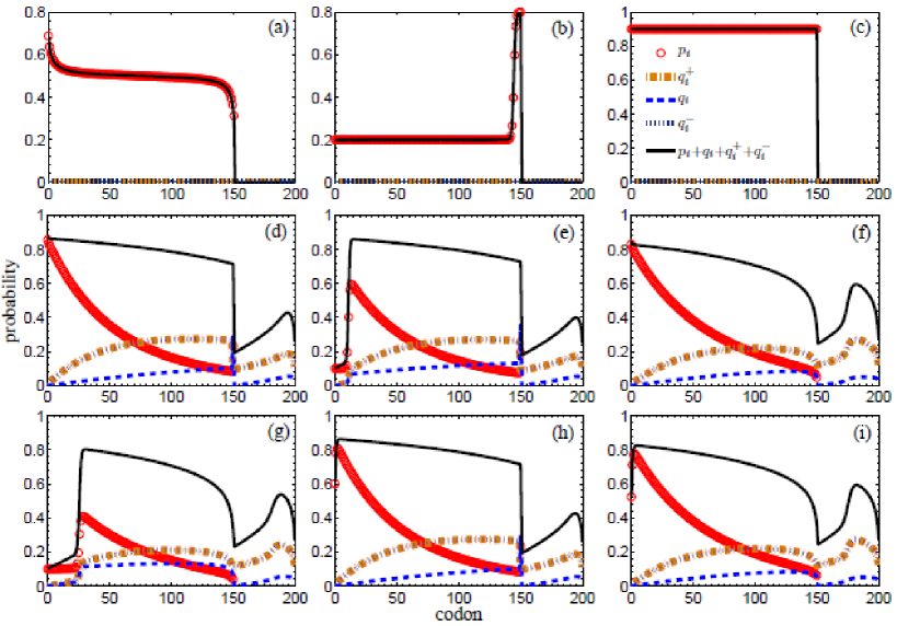
To illustrate the modified TASEP used in this study for the description of ribosome translocation along mRNA with frameshifting, typical examples of the probabilities and their summation are plotted in Fig. 3. Where is the probability that site is occupied by one ribosome. These examples indicate that, from the viewpoint of total probability of ribosome, the modified TASEP can be regarded as a combination of two usual TASEPs for ribosome translocation along mRNA but without frameshifting. Where one involves ribosome translocation from lattice site 0 to lattice site , and the other involves for ribosome translocation from lattice site to lattice site .
For TASEP without ribosome frameshifting, attachment or detachment, i.e., , the theoretical studies in Schutz1993 ; Derrida19931 show a total of three possible phases, namely, low ribosome density phase, high ribosome density phase, and maximal current phase. The plots in Figs. 3(a-c) show that, such three phases also exist between lattice site and lattice site in the modified TASEP. An example for maximal current cases is found in Fig. 3(a), in which the total probability is almost equals to 1/2 except for the sharp changes near sites and . The maximal current phase occurs when both and are bigger than 1/2. In TASEP, the current is defined by . The plots in Figs. 3(b) and 3(c) correspond to low and high density phases, respectively, with only sharp change at site or site . For these special cases, considering the absence of ribosome frameshifting, attachment or detachment, probabilities are equal to zero, and . The current of ribosome with correctly synthesized peptide chain is conversed along the mRNA region between the start and stop codons. Thus the effectiveness of translation is equal to 1, and actually and are both equal to the current .
For general TASEP, i.e., , previous studies have shown that domain wall (or shock wave) and sharp changes at both or only one of the boundaries may exist Parmeggiani2003 ; Zhang20101 . The examples plotted in Figs. 3(d-i) show that, with frameshifting of ribosome, in the two regions and , domain wall and sharp change of probability at one or both of the two boundaries may be found. Meanwhile, the examples plotted in Fig. 3 show that, similar to the total probability , three different phases may also exist for probability (for the cases , see Figs. 3(a-c)), domain wall and sharp change at boundaries may also exist (for general cases with nonzero attachment and detachment rates, see Figs. 3(d-i)). In all these figures, the total probability decreases suddenly at codon . This phenomenon is due to the existence of the stop codon at site . Instead of moving forward to codon , ribosomes at the stop codon will leave the mRNA template, and consequently the total probability at codon will decrease rapidly.
For translation with ribosome frameshifting, the main properties that should be determined are the influences of frameshifting on the rate of translation initiation and the rate of synthesizing correct peptide chain , as well as their ratio . Generally, shifted ribosomes, i.e., ribosomes in frame of mRNA, will not leave the mRNA from the stop codon. The probability of establishing a ribosome at one codon, even if the codon is upstream of the stop codon (i.e., its codon number is less than ), may be influenced by the ribosome probabilities between the stop codon and the 3’ end of the mRNA. Meanwhile, previous studies about usual TASEP show that ribosome probabilities between sites and depend on the length of the untranslated mRNA region Zhang2012 . Thus, the ribosome probability at any mRNA codon may also depend on parameter , and consequently, any properties of the modified TASEP model for translation with ribosome frameshifting obtained from the probabilities , and may be parameter -dependent. Considering the lack of experimental measured values for parameter , one strategy used in this study is the selection of a sufficiently large value of , such that all the interested properties of translation, especially the effective initiation rate and termination rate , reach their steady values. Or in other words, and will not change with further increase in parameter , i.e., the length of the 3’ untranslated mRNA region is large enough to have as little influence as possible to the ribosome translocation in the translated region. The numerical calculations show that, for the parameter values used in this study, the selection of one value of that is larger than 20 is sufficient (Fig. S1 in supplemental ). In Fig. 3, is used, and in Figs. 4-6, is used.
Without frameshifting, attachment, or detachment, ribosomes initiate their translations from the mRNA start codon and terminate their translations at the stop codon with correctly synthesized peptide chains. On the contrary, with frameshifting, attachment, and detachment, ribosomes may begin/end their translations at any mRNA codon. However, only ribosomes that initiate their translations from the start codon and leave the mRNA from the stop codon synthesize correct peptide chains. Thus, the synthesizing rate of correct peptide chain, which is called the effective translation rate , will be less than the effective translation initiation rate . Their ratio will be one important biophysical index to describe the effectiveness of gene translation.
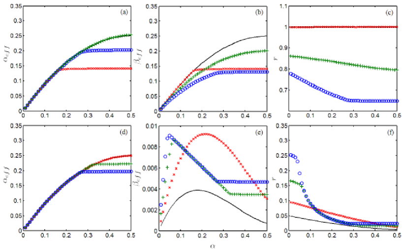
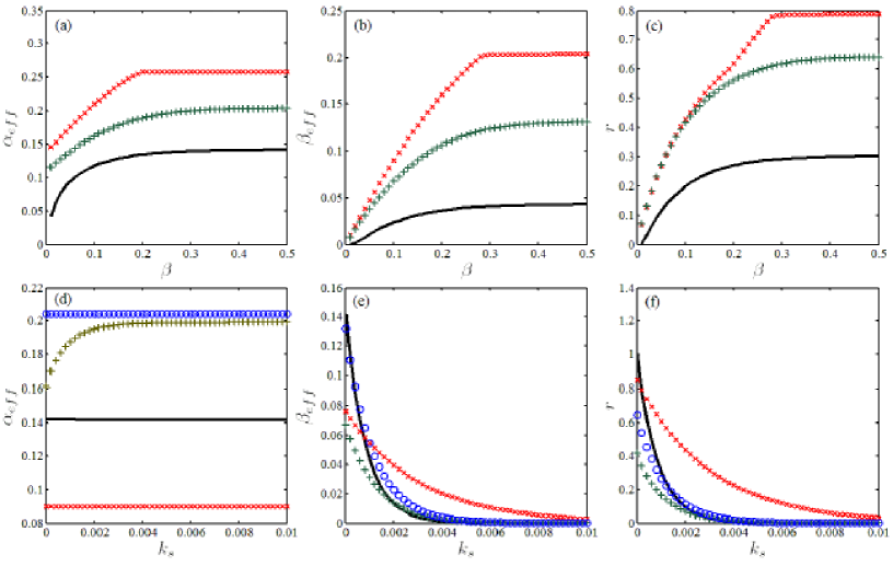
In Fig. 4, the effective translation initiation rate , effective translation speed , and their ratio are plotted with the change in translation initiation rate . The plots in Figs. 4(a,b) show that, without frameshifting , both and increase with . By contrast, for nonzero attachment rate , rates and will reach their up limits rapidly. Given that the new ribosome attachment will quicken the saturation of the ribosome on the mRNA. Fig. 4(c) shows that, if , then the ratio is equal to 1. In such cases, all ribosomes that begin their translations from the start codon will finally end their translations at the stop codon. Generally, holds, and decreases with initiation rate and finally tends to one low limit value. For , i.e., translation with ribosome frameshifting, the plots in Fig. 4(d) indicate that the effective translation initiation rate increases also with , while the plots in Fig. 4(e) show that the effective translation rate increases first and then decreases with rate . The reason that decreases with large values of is that for large , the probability of finding a ribosome with incorrect peptide chain will increase, so the translocation speed of ribosome with correct peptide chain will decrease because of the block of ribosome with incorrect peptide chain. Finally, the plots in Fig. 4(f) indicate that for , the ratio will always be less than 1 and decreases with translation initiation rate . The plots in Fig. 4 show that the main differences that resulted from ribosome frameshifting are as follows: the effective translation rate may decrease with translation initiation rate (see Fig. 4(b,e)), and the effectiveness of translation decreases much rapidly with rate (see Fig. 4(c,f)).
Meanwhile, the plots in Figs. 5(a-c) show that , and their ratio increase with the translation termination rate , and tend to corresponding limit constants with large . These results imply that, with high leaving rate of ribosome from the stop codon, more correct peptide chains is synthesized, and the effective initiation rate also increases. Given the increase in leaving rate but fixed rates of frameshifting and detachment, ribosomes with correct peptide chain rapidly translocate along the mRNA template and have less chance to frameshift or detach from the mRNA before they reach the stop codon. On the contrary, the plots in Figs. 5(e,f) indicate that, both and ratio decrease with the rate of frameshifting. Considering large rate , ribosomes will have less chance to complete their translation processes correctly, but have more chances to frameshift. Figs. 6(b,c) show that, and ratio also decrease with ribosome attachment rate . With the increase in attachment rate , the mRNA template is bound by more ribosomes with incorrect peptide chains, and then the translocation speed of ribosome with correct peptide chain is reduced. Thus, ribosomes with correct peptide chains translocate along the mRNA with lower speed and then have more chances to detach from the mRNA or frameshift before they reach the stop codon. The plots in Fig. 6(e) show that may not change monotonically with the ribosome detachment rate . When the detachment rate is small, the increase in the rate may help decrease the overall ribosome density along the mRNA, and consequently, increase the translocation speed of ribosomes with correct peptide chain. For large values of , the detachment rate of ribosome with correct peptide chain may increase rapidly, and then the synthesizing rate of correct peptide chain will decrease. This phenomenon implies that, one optimal ribosome detachment rate may exist, with which the synthesizing rate of correct peptide chain reaches its maximum.
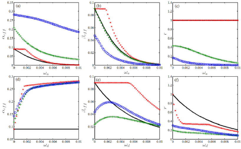
The plots in Figs. 5(d), Fig. 6(a,d) show that the effective initiation rate increases with the rate of frameshifting and the rate of detachment, but decreases with the attachment rate . With large detachment/attachment rate, the start mRNA codon has more chances to be unoccupied/occupied, therefore is enlarged/reduced correspondingly. Fig. 5(d) also shows that with large values of frameshifting rate , the effective initiation rate tends to approach one limit constant. With large frameshifting rate , the entrance rate of ribosome to mRNA is only determined by the detachment rate , the attachment rate , and the termination rate . The overall density of the ribosome along the mRNA, with either correct peptide chain or incorrect peptide chain, is completely determined by , and , see Derrida1993 ; Parmeggiani2003 ; Zhang20101 . The plots in Fig. 6 show that, although the translation effectiveness always decreases with attachment rate and detachment , for some special cases the effective (correct) translation rate may increase by increasing the ribosome detachment rate , see Fig. 6(e). Therefore, detachment maybe one possible mechanism used by cells to increase the synthesizing rate of needed peptide chains.
IV Conclusions
Recent experiments found that, during translation, ribosome may frameshift along the mRNA, and the reasons of frameshifting have been experimentally studied thoroughly Weiss1989 ; Larsen1994 ; Horsfield1995 ; Larsen1997 ; Jacks1988 ; Plant2003 ; Baranov2004 ; Namy2006 ; Leger2007 ; Atkins2010 ; Tinoco2013 ; Gupta2013 ; Chen2014 ; Belew2014 . However, so far, no theoretical model has been designed to describe the gene translation process with ribosome frameshifting, especially the influence of frameshifting on translation, such as the effective translation rate and the efficiency (or effectiveness) of translation. In this study, a modified TASEP model is presented to describe the translation process with possible ribosome frameshifting. Similar to the usual translation process, i.e., translation without ribosome frameshifting, the probability density of ribosome along the mRNA template may have shock wave (or domain wall) and boundary layers. The effective translation rate, i.e., the rate of synthesizing correct peptide chain, increases with the translation termination rate , and decreases with the rate of frameshifting , but may not change monotonically with the translation initiation rate . Meanwhile, the translation effectiveness, which is defined as the ratio of effective synthesizing rate of correct peptide chain to the effective initiation rate of translation, increases with the termination rate, but decreases with initiation rate and the frameshifting rate of ribosome. At the same time, the influences of attachment/detachment of ribosomes to/from the main body of mRNA were also discussed. The results will be beneficial for the understandings of actual translation processes in cells, and the model presented may also be useful for the prediction of translation rate with reasonable chosen parameters.
Acknowledgements.
This study was supported by the Natural Science Foundation of China (Grant No. 11271083), and the National Basic Research Program of China (National “973” program, project No. 2011CBA00804).References
- (1) Anton A. Turanov, Alexey V. Lobanov, Dmitri E. Fomenko, Hilary G. Morrison, Mitchell L. Sogin, Lawrence A. Klobutcher, Dolph L. Hatfield, and Vadim N. Gladyshev. Genetic code supports targeted insertion of two amino acids by one codon. Science, 323(5911):259–261, January 2009.
- (2) Julien Racle, Flora Picard, Laurence Girbal, Muriel Cocaign-Bousquet, and Vassily Hatzimanikatis. A genome-scale integration and analysis of lactococcus lactis translation data. PLOS Computational Biology, 9(10), October 2013.
- (3) J. Shine and L. Dalgarno. The 3’-terminal sequence of Escherichia coli 16s ribosomal RNA: Complementarity to nonsense triplets and ribosome binding sites. Proceedings of the National Academy of Sciences of the United States of America, 71(4):1342–1346, April 1974.
- (4) A. P. Czernilofskya, C. G. Kurlanda, and G. Stöfflerb. 30s ribosomal proteins associated with the 3’-terminus of 16s RNA. FEBS Letters, 58(1):281–284, October 1975.
- (5) Gulnara Zh. Yusupova, Marat M. Yusupov, J. H. D. Cate, and Harry F. Noller. The path of messenger RNA through the ribosome. Cell, 106(2):233–241, July 2001.
- (6) Andrei Korostelev and Harry F. Noller. The ribosome in focus: new structures bring new insights. Trends in Biochemical Sciences, 32(9):434–441, September 2007.
- (7) Mélodie Duval, Alexey Korepanov, Olivier Fuchsbauer, Pierre Fechter, Andrea Haller, Attilio Fabbretti, Laurence Choulier, Ronald Micura, Bruno P. Klaholz, Pascale Romby, Mathias Springer, and Stefano Marzi. Escherichia coli ribosomal protein S1 unfolds structured mRNAs onto the ribosome for active translation initiation. PLOS Biology, 11(12), December 2013.
- (8) John F. Atkins and Raymond F. Gesteland, editors. Recoding: Expansion of Decoding Rules Enriches Gene Expression, volume 24 of Nucleic Acids and Molecular Biology. Heidelberg: Springer, New York, 2010.
- (9) Jin Chen, Alexey Petrov, Magnus Johansson, Albert Tsai, Seán E. O’Leary, and Joseph D. Puglisi. Dynamic pathways of translational frameshifting. Nature, page to appear, 2014.
- (10) Bente Larsen, Norma M. Wills, Raymond F. Gesteland, and John F. Atkins. rRNA-mRNA base pairing stimulates a programmed ribosomal frameshift. Journal of Bacteriology, 176(22):6842–6851, November 1994.
- (11) Bente Larsen, Raymond F. Gesteland, and John F. Atkins. Structural probing and mutagenic analysis of the stem-loop required for Escherichia coli dnaX ribosomal frameshifting: programmed efficiency of 50%. Journal of Molecular Biology, 271(1):47–60, August 1997.
- (12) Ignacio Tinoco Jr., Hee-Kyung Kim, and Shannon Yan. Frameshifting dynamics. Biopolymers, 99(12):1147–1166, December 2013.
- (13) Ewan P. Plant, Kristi L. Muldoon Jacobs, Jason W. Harger, Arturas Meskauskas, Jonathan L. Jacobs, Jennifer L. Baxter, Alexey N. Petrov, and Jonathan D. Dinman. The 9-Å solution: how mRNA pseudoknots promote efficient programmed ribosomal frameshifting. RNA, 9(2):168–174, February 2003.
- (14) Pavel V. Baranov, Raymond F. Gesteland, and John F. Atkins. P-site tRNA is a crucial initiator of ribosomal frameshifting. RNA, 10(2):221–230, February 2004.
- (15) Julie A. Horsfield, Daniel N. Wilson, Sally A. Mannering, Frances M. Adamski, and Warren P. Tate. Prokaryotic ribosomes recode the HIV-1 gag-pol frameshift sequence by an E/P site post-translocation simultaneous slippage mechanism. Nucleic Acids Research, 23(9):1487–1494, 1995.
- (16) John D. Lopinski, Jonathan D. Dinman, and Jeremy A. Bruenn. Kinetics of ribosomal pausing during programmed translational frameshifting. Molecular and Cellular Biology, 20(4):1095–1103, February 2000.
- (17) Tyler Jacks, Hiten D. Madhani, Frank R. Masiarz, and Harold E. Varmus. Signals for ribosomal frameshifting in the rous sarcoma virus gag-pol region. Cell, 55(3):447–458, November 1988.
- (18) Olivier Namy, Stephen J. Moran, David I. Stuart, Robert J. C. Gilbert, and Ian Brierley. A mechanical explanation of RNA pseudoknot functionin programmed ribosomal frameshifting. Nature, 441(7090):244–247, May 2006.
- (19) R. B. Weiss, D. M. Dunn, M. Shuh, J. F. Atkins, and R. F. Gesteland. E. coli ribosomes re-phase on retroviral frameshift signals at rates ranging from 2 to 50 percent. The New Biologist, 1(2):159–169, December 1989.
- (20) Mélissa Léger, Dominic Dulude, Sergey V. Steinberg, and Léa Brakier-Gingras. The three transfer RNAs occupying the A, P and E sites on the ribosome are involved in viral programmed ribosomal frameshift. Nucleic Acids Research, 35(16):5581–5592, 2007.
- (21) Tatsuya Maehigashi, Jack A. Dunkle, Stacey J. Miles, and Christine M. Dunham. Structural insights into frameshifting promoted by expanded or modification-deficient anticodon stem loops. Proceedings of the National Academy of the Sciences of the United States of America, August 2014.
- (22) Ashton Trey Belew, Arturas Meskauskas, Sharmishtha Musalgaonkar, Vivek M. Advani, Sergey O. Sulima, Wojciech K. Kasprzak, Bruce A. Shapiro, and Jonathan D. Dinman. Ribosomal frameshifting in the CCR5 mRNA is regulated by miRNAs and the NMD pathway. Nature, 512(7514):265–269, August 2014.
- (23) B. Derrida, S. A. Janowsky, J. L. Lebowitz, and E. R. Speer. Exact solution of the totally asymmetric simple exclusion process: Shock profiles. Journal of Statistical Physics, 73:813–842, 1993.
- (24) A. Parmeggiani, T. Franosch, and E. Frey. Phase coexistence in driven one-dimensional transport. Physical Review Letters, 90:086601, 2003.
- (25) Yunxin Zhang. Domain wall of the totally asymmetric exclusion process without particle number conservation. Chinese Journal of Physics, 48:607–618, 2010.
- (26) Yunxin Zhang. Microtubule length dependence of motor traffic in cells. Eur. Phys. J. E, 35:101, 2012.
- (27) Joachim Krug. Boundary-induced phase transitions in driven diffusive systems. Phys. Rev. Lett., 67:1882–1885, 1991.
- (28) A. Kolomeisky, G.M. Schutz, E.B. Kolomeisky, and J.P. Straley. Phase diagram of one-dimensional driven lattice gases with open boundaries. J. Phys. A: Math. Gen., 31:6911–6919, 1998.
- (29) Gunter M Schütz. Critical phenomena and universal dynamics in one-dimensional driven diffusive systems with two species of particles. J. Phys. A: Math. Gen., 36:R339–R379, 2003.
- (30) R. Lipowsky, Y. Chai, S. Klumpp, S. Liepelt, and M. J. I. Muller. Molecular motor traffic: From biological nanomachines to macroscopic transport. Physica A, 372:34–51, 2006.
- (31) Adélaïde Raguin, Andrea Parmeggiani, , and Norbert Kern. Role of network junctions for the totally asymmetric simple exclusion process. Physical Review E, 88:042104, 2013.
- (32) V. Popkov, J. Schmidt, and G. M. Schütz. Superdiffusive modes in two-species driven diffusive systems. Physical Review Letters, 112:200602, 2013.
- (33) Paul C. Bressloff and Jay M. Newby. Stochastic models of intracellular transport. Reviews Of Modern Physics, 85:135–196, 2013.
- (34) Yunxin Zhang. Periodic one-dimensional hopping model with transitions between nonadjacent states. Phys. Rev. E, 84:031104, 2011.
- (35) Yunxin Zhang. Theoretical analysis of kinesin KIF1A transport along microtubule. J. Stat. Phys., 152:1207 C1221, 2013.
- (36) Arvind Kumar Gupta and Isha Dhiman. Asymmetric coupling in two-lane simple exclusion processes with langmuir kinetics: Phase diagrams and boundary layers. Physical Review Letters, 89:022131, 2014.
- (37) G. Schutz and E. Domany. Phase transitions in an exactly soluble one-dimensional exclusion process. Journal of Statistical Physics, 72:277–296, 1993.
- (38) B. Derrida, M.R. Evans, V. Hakim, and V. Pasquier. Exact solution of a 1d asymmetric exclusion model using a matrix formulation. J. Phys A: Math. Gen., 26:1493–1517, 1993.
- (39) The supplementary material including one figure is available at {URL to be provided}.
- (40) Pulkit Gupta, Krishna Kannan, Alexander S. Mankin, and Nora Vázquez-Laslop. Regulation of gene expression by macrolide-induced ribosomal frameshifting. Molecular cell, 52(5):629–642, December 2013.
| figures | |||||
|---|---|---|---|---|---|
| Fig. 3(a) | 0.8 | 0.8 | 0 | 0 | 0 |
| Fig. 3(b) | 0.2 | 0.2 | 0 | 0 | 0 |
| Fig. 3(c) | 0.9 | 0.1 | 0 | 0 | 0 |
| Fig. 3(d) | 0.9 | 0.1 | 0.001 | 0.001 | 0.001 |
| Fig. 3(e) | 0.1 | 0.1 | 0.001 | 0.001 | 0.001 |
| Fig. 3(f) | 0.9 | 0.9 | 0.001 | 0.001 | 0.001 |
| Fig. 3(g) | 0.1 | 0.9 | 0.001 | 0.001 | 0.001 |
| Fig. 3(h) | 0.3 | 0.1 | 0.001 | 0.001 | 0.001 |
| Fig. 3(i) | 0.3 | 0.7 | 0.001 | 0.001 | 0.001 |
| figures | label | |||||
|---|---|---|---|---|---|---|
| - | 1 | 0 | 0 | 0 | ||
| Fig. 4(a,b,c) | 1 | 0 | 0.001 | 0 | ||
| + | 1 | 0 | 0 | 0.001 | ||
| 1 | 0 | 0.001 | 0.001 | |||
| - | 1 | 0.01 | 0 | 0 | ||
| Fig. 4(d,e,f) | 0.05 | 0.007 | 0 | 0 | ||
| + | 0.01 | 0.004 | 0 | 0 | ||
| 0.01 | 0.003 | 0 | 0 | |||
| Fig. 5(a,b,c) | - | 1 | 0.001 | 0.001 | 0 | |
| 1 | 0 | 0 | 0.001 | |||
| + | 1 | 0 | 0.001 | 0.001 | ||
| Fig. 5(d,e,f) | - | 1 | 1 | 0.001 | 0 | |
| 0.1 | 1 | 0 | 0.001 | |||
| + | 1 | 0.1 | 0.001 | 0.001 | ||
| 1 | 1 | 0.001 | 0.001 | |||
| Fig. 6(a,b,c) | - | 1 | 0.1 | 0 | 0 | |
| 0.1 | 1 | 0 | 0 | |||
| + | 1 | 0.1 | 0 | 0.001 | ||
| 1 | 0.1 | 0 | 0.01 | |||
| Fig. 6(d,e,f) | - | 0.1 | 0.1 | 0 | 0 | |
| 1 | 0.1 | 0 | 0 | |||
| + | 1 | 0.1 | 0.001 | 0.001 | ||
| 1 | 1 | 0.001 | 0.001 |