Highly coherent electron beam from a laser-triggered tungsten needle tip
Abstract
We report on a quantitative measurement of the spatial coherence of electrons emitted from a sharp metal needle tip. We investigate the coherence in photoemission using near-ultraviolet laser triggering with a photon energy of eV and compare it to DC-field emission. A carbon-nanotube is brought in close proximity to the emitter tip to act as an electrostatic biprism. From the resulting electron matter wave interference fringes we deduce an upper limit of the effective source radius both in laser-triggered and DC-field emission mode, which quantifies the spatial coherence of the emitted electron beam. We obtain nm in laser-triggered and nm in DC-field emission mode, revealing that the outstanding coherence properties of electron beams from needle tip field emitters are largely maintained in laser-induced emission. In addition, the relative coherence width of 0.36 of the photoemitted electron beam is the largest observed so far. The preservation of electronic coherence during emission as well as ramifications for time-resolved electron imaging techniques are discussed.
Coherent electron sources are central to studying microscopic objects with highest spatial resolution. They provide electron beams with flat wavefronts that can be focused to the fundamental physical limit given by matter wave diffraction Spence2013. Currently, time-resolved electron based imaging is pursued with large efforts, both in real-space microscopy Zewail2010a; King2005 and in diffraction Sciaini2011; Baum2007b. However, the spatial resolution in time-resolved electron microscopy is about two orders of magnitude worse than its DC counterpart Yurtsever2012, which reaches below Å Erni2009. Combining highest spatial resolution with time resolution in the picosecond to (sub-) femtosecond range requires spatially coherent electron sources driven by ultrashort laser pulses. Although laser-driven metal nanotips promise to provide coherent electron pulses with highest time resolution, a quantitative study of their spatial coherence has been elusive. Here we demonstrate that photoemitted electrons from a tungsten nanotip are highly coherent.
So far no time-resolved electron based imaging instrument fully utilizes the coherence capabilities provided by nanotip electron sources. Meanwhile, nanotips operated in DC-field emission are known and employed in practical applications for almost half a century for their paramount spatial coherence properties Crewe1968. Thence, highest resolution microscopy as well as coherent imaging, such as holography and interferometry, have long been demonstrated in DC-field emission Spence2013; Lichte2008; Hasselbach2010. Here we investigate whether these concepts can be inherited to laser-driven nanotip sources by comparing the spatial coherence of photoemitted electron beams to their DC counterparts. This would enable time-resolved high resolution imaging, but may also herald fundamental studies based on the generation of quantum degenerate electron beams Lougovski2011.
The spatial coherence of electron sources is commonly quantified by means of their effective source radius . It equals the radius of a virtual incoherent emitter that resembles the coherence properties of the real emitter. As discussed later, is inversely proportional to the transverse coherence length of the electron beam. A virtual source is formed in a finite area where electron trajectories intersect when extrapolating their paths back into the metal tip (Fig. 1d). For tungsten field emitters typical values for are on the order of nm and the smallest reported down to nm, significantly smaller than the geometrical tip radius that is typically in the range of a few tens of nanometers Spence2013; Cho2004.
DC-field and laser-driven emission occur due to fundamentally different emission processes (Fig. 1a) Jensen2010. The former is a tunnelling process through a static potential barrier, covered within the Fowler-Nordheim-theory Fowler1928, whereas a variety of laser-driven emission processes exist. They are distinguishable into linear one-photon emission and nonlinear multi-photon and tunneling processes, with the respective prominent examples of Einstein’s photoelectric effect and multi-photon emission Delone1994. The effective source radius is highly sensitive to the shape of the electron trajectories in close vicinity of the tip apex Cook2010, and hence to the emission process. As a result, the coherence properties in photoemission might be drastically different from DC-field emission.
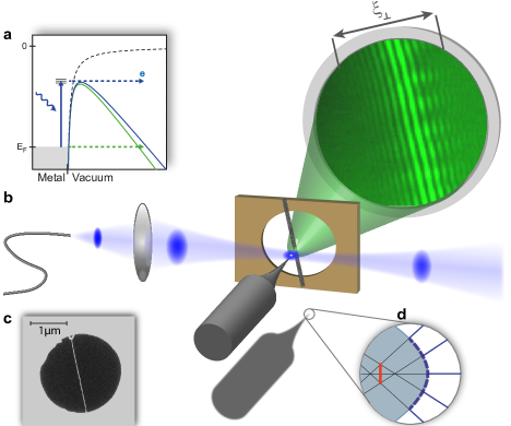
To compare the coherence properties of a monocrystalline tungsten tip electron emitter with a radius of 10 nm in laser-triggered and DC-field emission we record electron matter wave interference images in both emission modes. We use a freestanding carbon nanotube (CNT) as an electron beam splitter, which acts as a biprism filament with nanometer radius Hasselbach2010. It splits the wavefront of the electron matter wave in two parts, which are then overlapped at the electron detector, giving rise to interference fringes on the detector screen Cho2004. A scanning electron microscope image of a single, freestanding CNT on a holey silicon nitride membrane is shown in Fig. 1c (see Supplementary Material for details). The electrically grounded CNT is brought into the electron beam path at a typical distance of less than one micrometer from the tip, resembling a point projection microscopy configuration, that is also commonly used for electron holography Beyer2010; Longchamp2013. CNT and the gold coated holey silicon nitride membrane act as a counter electrode for the biased tip. Electron interference can be observed in conventional DC-field emission as well as in laser-triggered mode when a near-UV laser beam is focused on the tip’s apex (see Fig. 1b and Supplementary Material for details).
An upper bound for is obtained by measuring the full width of coherent illumination at the detector screen. It can be deduced by identifying the distance between the outermost interference fringes, observed perpendicular to the orientation of the CNT Spence1994. The van Cittert-Zernicke theorem relates and the effective source radius for an incoherent emitter with Gaussian intensity profile Pozzi1987; Spence1994:
| (1) |
Here is the electron de Broglie wavelength and the source-detector distance.
Ideally, an electron source should exhibit a narrow (longitudinal) momentum distribution to reduce chromatic effects in subsequent electron optics. Hence, in order to achieve efficient electron emission with little excess momentum we match the Schottky-lowered barrier at the metal-vacuum interface, which is tunable by means of the tip voltage, to the photon energy (eV) of the focused laser light. Here the barrier height is set to eV, closely above the onset of DC-field emission and yielding the highest photoemission probability with negligible DC component. The experiments are performed with a pulsed laser (second harmonic of fs long pulses derived from a 2.7 MHz repetition rate long-cavity Ti:sapphire oscillator) and a high power cw-diode laser source (mW at 405 nm) to boost the electron current for the effective source radius measurements.
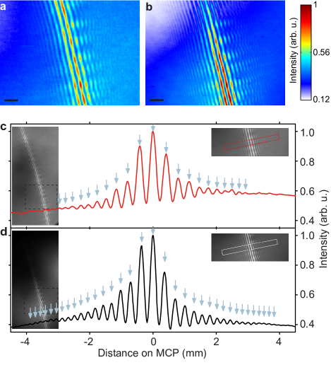
Electron interference patterns in laser-triggered and DC-field emission are shown in Fig. 2a and b, respectively, recorded at the identical CNT position with a tip-sample distance of less than micrometer. Clearly, interference fringes that are aligned parallel to the CNT are observed in both modes. A tip voltage of V is chosen in laser-induced emission, such that the barrier is lowered for efficient photoemission. For DC-field emission a voltage of V is applied, leading to a comparable field emission electron current as in photoemission.
The panels in Fig. 2c, d show line profiles obtained from integrating the count rate parallel to the fringes in the marked area. The spatial coherence width is obtained from these line profiles. For photoemission we obtain mm at a CNT-screen distance of mm. With Å, the effective source radius equals nm.
In DC-field emission mode a very comparable value of the coherence width is deduced with mm, albeit slightly larger (Fig. 2d). With Å the effective source radius equals nm, in line with previously published values Spence1994; Cho2004.
Clearly, the source radii in laser-triggered and DC-field emission mode differ only slightly, even though the emission process is qualitatively different. Furthermore, in both cases the effective source radius is about an order of magnitude smaller than the geometrical source radius. For comparison, the record resolution laser-triggered electron microscope employs a fully illuminated flat cathode of a few tens of microns in diameter Barwick2008; NoteCommercialLasertriggeredSEM. In this configuration the effective source radius equals the geometric one, given by the smaller of either the laser spot size or the dimensions of the cathode.
The relative coherence width , namely the ratio of to the electron beam radius , is a conserved quantity in electron optics Pozzi1987. Thus, it allows calculating for any given beam size, in particular for arbitrary focusing conditions at a sample. With mm (-radius of the electron beam) at the detector the relative coherence width of the photoemitted beam equals , representing the highest value reported for of a laser-triggered electron source to date. It benefits largely from the use of a monocrystalline tip, which exhibits a low divergence of the emitted beam of (half angle) in photoemission and in DC-field emission.
With increasing electron current it can be expected that the effective source size increases due to space charge and stochastic Coulomb electron-electron repulsion Cook2010. Strictly, these effects come into play for more than one electron per pulse emitted from the tip. Hence most conservatively, the maximum current attainable with highest spatial coherence is set by the repetition rate of the laser. For instance, laser pulses with MHz inducing emission of one electron per pulse yield a time averaged current of pA. Even though this value is low compared to the electron current emitted from standard field emission guns electron imaging with a stably aligned laser beam, as demonstrated here, remains well possible as demonstrated in time-resolved scanning electron microscopy Yang2010. The restriction to one electron per pulse, however, also prevents other unwanted detrimental effects such as temporal electron pulse broadening due to Coulomb repulsion Kirchner2014. In this experiment, the required minimum peak fluence to obtain one electron per pulse without DC contributions equals , with the pulse energy and the -beam waist radius . Note that many more than one electron per pulse can be drawn from the tip for most settings without detrimental effects on the beam quality, especially after propagation to a sample. This, however, depends on various parameters such as tip radius, laser pulse duration, acceleration field and electron beam path.
Next to the transverse coherence, quantified by , the energy spread of the electron beam is crucially important for most applications. Here of the photoemitted beam equals eV (FWHM), less than twice as much as in DC-field emission Spence2013. This implies that the longitudinal coherence length is smaller by a factor of about 2 in photoemission Lichte2008, likely causing the reduced visibility of the interference pattern in Fig. 2c (see Supplementary Material). We find that in principle the energy spread can be made as low as in DC-field emission with constant electron current by decreasing the DC-field at the tip and simultaneously increasing the laser power (see Supplementary Material). For instance, here eV is feasible with an increased barrier height of eV and tripled laser fluence.
We conclude that the coherence of the electron beam in one-photon photoemission close to the threshold is almost as good as that of a DC-field emitted beam. It has been previously shown that the initial electronic states inside the metal from which the electrons originate affect the coherence of the emitted electron beam Cho2004. Our measurements demonstrate that the coherence of the original electronic states inside the metal is maintained in photoemission. One may thus expect that a cooled tip also provides a fully coherent beam under laser irradiation, as demonstrated in DC-field emission Cho2004.
By virtue of the excellent coherence properties of a DC-field emitted electron beam it was shown that the combination of point projection holography and coherent electron diffraction allows for Å resolution in imaging of graphene Longchamp2013. Very recently, first time-resolved results have been obtained in femtosecond point projection microscopy Quinonez2013, ultrafast low-energy electron diffraction Gulde2014 and combinations of both Muller2014 based on femtosecond laser-triggered tungsten field emission tips as electron sources Hommelhoff2006; Hommelhoff2006a; Ropers2007. In this context our findings clearly show that electron imaging devices equipped with field emission guns can be laser-triggered to obtain highest temporal resolution without losing their supreme coherence and imaging properties. The excellent source properties will also be of great interest for novel laser-based electron acceleration schemes as recently demonstrated Peralta2013; Breuer2013.
The authors thank H. Kaupp for discussions on electron beam coherence measurements prior to the experiment, S. Stapfner, L. Ost and E. Weig for discussions on CNT fabrication and J.P. Kotthaus for cleanroom access. This research is funded in part by the the Gordon and Betty Moore Foundation, the DFG Cluster of Excellence Munich Centre for Advanced Photonics and the ERC Grants NearFieldAtto and QuantumCANDI.