Low-Background In-Trap Decay Spectroscopy with TITAN at TRIUMF
Abstract
An in-trap decay spectroscopy setup has been developed and constructed for use with the TITAN facility at TRIUMF. The goal of this device is to observe weak electron-capture (EC) branching ratios for the odd-odd intermediate nuclei in the decay process. This apparatus consists of an up-to 6 Tesla, open-access spectroscopy ion-trap, surrounded radially by up to 7 planar Si(Li) detectors which are separated from the trap by thin Be windows. This configuration provides a significant increase in sensitivity for the detection of low-energy photons by providing backing-free ion storage and eliminating charged-particle-induced backgrounds. An intense electron beam is also employed to increase the charge-states of the trapped ions, thus providing storage times on the order of minutes, allowing for decay-spectroscopy measurements. The technique of multiple ion-bunch stacking was also recently demonstrated, which further extends the measurement possibilities of this apparatus. The current status of the facility and initial results from a 116In measurement are presented.
1 Introduction
1.1 Decay Nuclear Matrix Elements (NMEs)
Recent evidence of massive neutrinos has generated great interest in exotic nuclear decay modes [1, 2]. As a part of these studies, searches for the mode of decay are particularly interesting [2], since an observation of this mode would establish the neutrino as a Majorana particle. If this decay mode is observed, the effective Majorana mass of the neutrino can be extracted if the nuclear matrix element (NME) that connects the initial and final states is known [3]. The calculation of decay NMEs is the focus of current theoretical efforts and includes several different model descriptions. These calculations can be constrained from experimental data such as measurements of the and electron-capture (EC) branching ratios of the intermediate nuclei in the -decay process. Typically, EC transitions are several orders of magnitude weaker than the dominant decays from the same parent nucleus, making them difficult to measure.
In EC decay, the characteristic X-ray originates from the filling of the vacated atomic -shell electron, and typically has an energy less than 100 keV. For the cases of interest to -decay studies [4] the X-rays are much lower in energy, and are generally less than 40 keV. To observe weak EC branches at these energies, it is important that effects such as positron-annihilation, Compton, and charged-particle induced backgrounds are minimized. Reducing photon backgrounds at these energies requires a high level of control over the decay environment which can be provided using ion traps. Therefore a low-background, high-sensitivity decay-spectroscopy tool has been developed using the TITAN ion traps for measuring characteristic X-rays from weak EC decays [4, 5].
2 TITAN at TRIUMF-ISAC
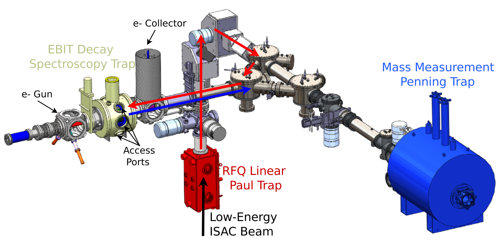
TRIUMF’s Ion Trap for Atomic and Nuclear Science (TITAN) [6] consists of three ion traps; (i) an RFQ linear Paul trap [7] for buffer-gas cooling and bunching the low-energy ion beam, (ii) a 3.7 T, high-precision mass-measurement Penning-trap (MPET) [14], and (iii) an electron-beam ion trap (EBIT) which provides highly charged ions (HCIs) [9]. TITAN resides at the Isotope Separator and Accelerator (ISAC) facility at TRIUMF, which employs a high-intensity (up to 100 A) beam of 500 MeV protons to produce RIBs using the isotope separation on-line (ISOL) technique [10]. The mass-selected, continuous beam of radioactive singly charged ions (SCIs) is delivered at low energies ( keV) to a suite of experimental facilities for both cooled- and stopped-beam experiments [15]. The TITAN facility is primarily used to perform high-precision Penning-trap mass spectrometry on short-lived radioactive nuclides [11, 12, 13, 14], however this article describes a decay-spectroscopy setup using the TITAN ion traps (Fig. 1), and described in detail below.
2.1 Decay Spectroscopy with TITAN
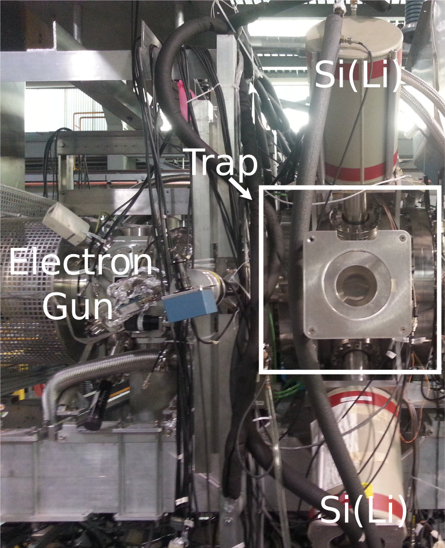
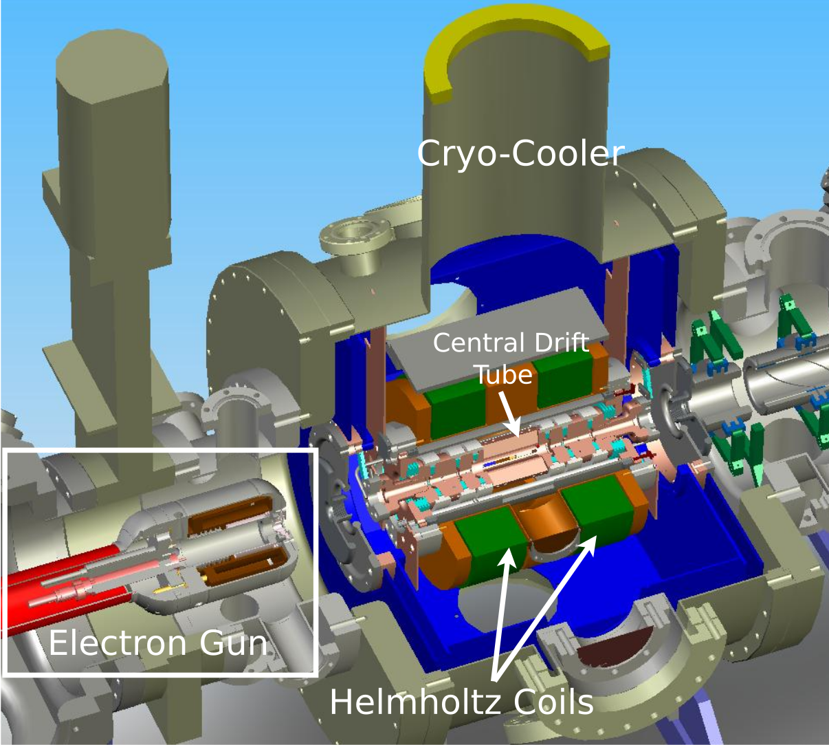
The TITAN EBIT confines ions (i) axially by an electrostatic square-well potential, and (ii) radially by the electron-beam space-charge potential and magnetic field [9]. HCIs are generated by successive electron-impact ionization using a the 100 mA, 1.7 keV electron beam compressed by a strong magnetic field. The up-to 6 Tesla magnetic field is produced by two superconducting Nb3Sn coils in a Helmholtz-like configuration [9]. To observe the characteristic photons from the EC decays, each of the EBIT’s seven external ports is occupied by a 5 mm thick Si(Li) crystal, which is optimized for the detection of X-rays [5]. A schematic view and an image of the TITAN decay-spectroscopy setup are displayed in Fig. 2.
3 116In Experiment
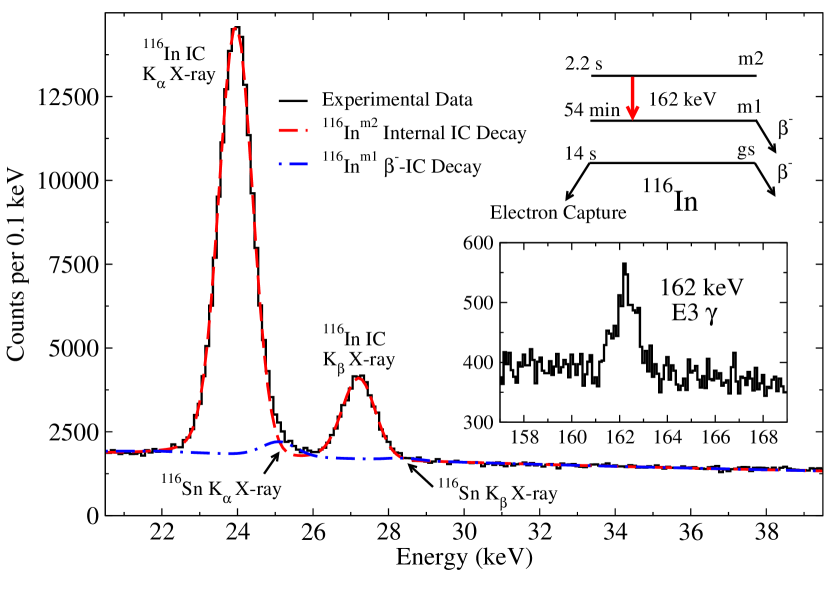
As a continued commissioning experiment, and a first attempt at one of the ECBR measurements relevant for -decay nuclear matrix elements, the -decay of 116In was observed in the TITAN EBIT. 116In is the intermediate nucleus in the decay of 116Cd and decays primarily by with a half-life of 14.1 s [16]. The 20 keV 116In ion beam from ISAC delivered to TITAN’s RFQ consisted of roughly 104:106:105 ions/s of 116Ings:m1:m2, respectively. The ion bunches were subsequently injected into the EBIT (cycles are described below), and the summed photon spectrum that resulted from all 15 s decay cycles is displayed in Fig. 3. The dominant component in the spectrum is the Kα X-ray from the internal conversion of the 162 keV E3 transition from the 2.2 s 116Inm2. Due to limited experimental time, and the large amount of isomeric contamination, the total collected statistics were insufficient to observe the weak () electron-capture branch to 116Cd from the 116Ings.
3.1 Multiple Ion-Bunch Stacking
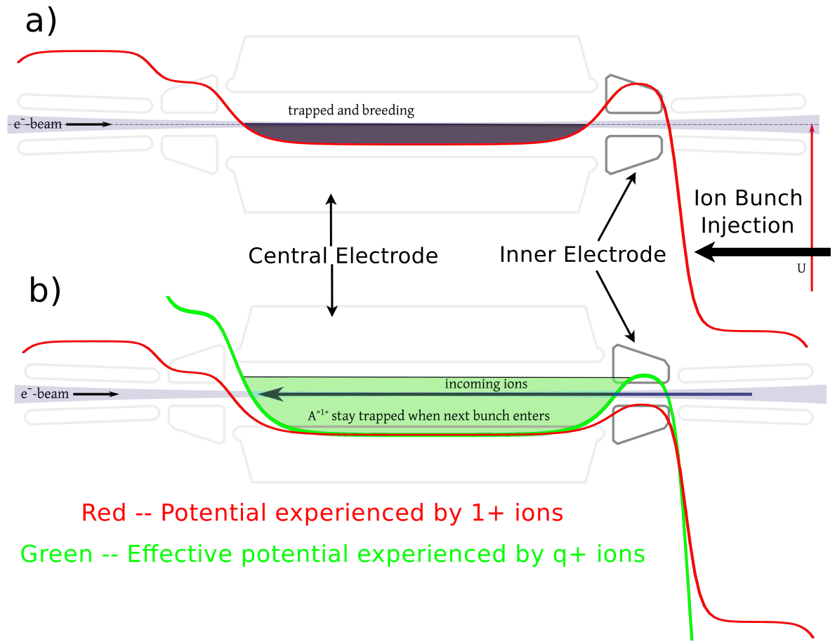
Since the space-charge limit of the RFQ is - [5], high ISAC beam intensities for some species cannot be fully used and thus high production rates are wasted. This is of particular concern for this experimental program, as the EC branching ratios relevant for the -decay cases [4] are weak, ranging from to , and require a large number of decays to observe them with any statistical significance. A method for overcoming this space-charge limit was therefore tested using the decay of 116In by injecting many ion bunches into the EBIT without extraction [20]. To achieve this, the inner electrode potential is lowered for first ion-bunch injection and subsequently raised to confine the first ion bunch. Following this, the injected ion bunch(es) quickly reach and remain confined during subsequent injections due to the increased effective potential experienced by the highly-charged ions. The ions are then ejected, and the cycle is repeated. This ion-bunch stacking technique is schematically displayed in Fig. 4(b).
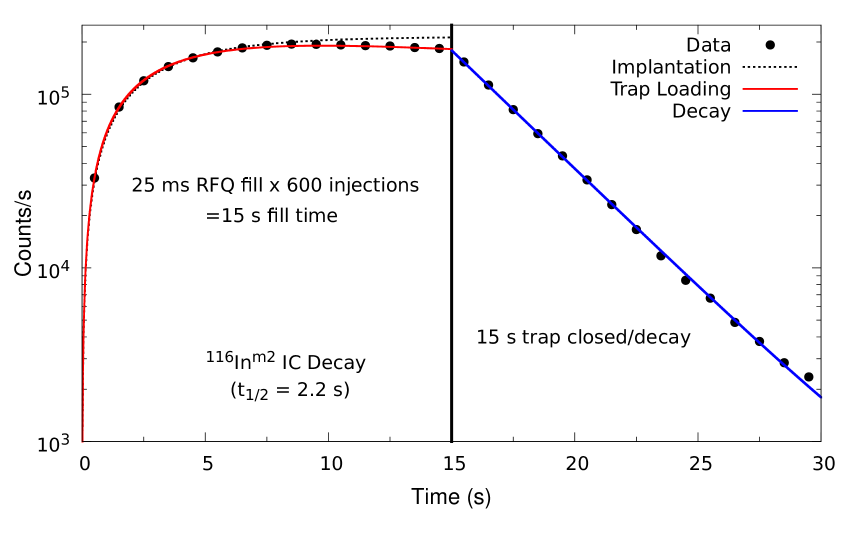
In order to investigate these new trapping effects, the cycles consisted of 15 s of bunch accumulation in the EBIT (from 600 ion-bunch injections at 25 ms RFQ fills), a 15 s decay measurement portion while the trap is closed, ion-bunch extraction from the trap, 5 s of background counting, and the cycles were repeated. The observed injection and decay cycle is displayed in Fig. 5 for all data acquired during the experiment. The trapping efficiency is nearly 100% relative to ion-beam implantation until roughly 9 s, where saturation of the space-charge appears to limit injection of subsequent ion bunches. Assuming a space-charge limit of the RFQ of , this cycle time corresponds to a maximum of roughly ions stored in the EBIT (at a charge-state of ).
4 Conclusions
In summary, an in-trap decay spectroscopy tool has been developed at TRIUMF using TITAN’s electron-beam ion-trap. The goal of this facility is to provide a low-background environment for the observation of weak EC branching ratios of the intermediate nuclei for decay. The ion-trap environment allows for the detection of low-energy photons by providing backing-free storage, while simultaneously guiding charged decay particles away from the trap center via the strong (up to 6 T) magnetic field. The highly-charged ions also allow multiple ion-bunch stacking, whereby ion-bunches are injected into the EBIT in quick succession, without extraction, thus circumventing the space-charge limit of TITAN’s RFQ. The first demonstration of this technique with the EBIT was performed during the decay measurement of 116In, and displayed a near 100% efficiency for trapping up to the space charge limit of the EBIT. Loss mechanisms after this point cause a slight decrease in the overall injection performance, which are not yet fully understood. The successful employment of the multiple injection technique opens the avenue for high-sensitivity experiments which were previously unfeasible due to statistical limitations.
References
- [1] V. Cirigliano, S. Gardner, B.R. Holstein, Prog. Part. Nucl. Phys. 71, (2013) 93-118.
- [2] F.T. Avignone, S.R. Elliott, J. Engel, Rev. Mod. Phys. 80, (2008) 481.
- [3] J. Barea, J. Kotila, and F. Iachello, Phys. Rev. Lett. 109, (2012) 042501.
- [4] D. Frekers, J. Dilling, and I. Tanihata, Can. J. Phys. 85, (2007) 57-75.
- [5] K.G. Leach et al., Nucl. Instrum. Methods Phys. Res. A (submitted); arXiv:1405.7209 (2014).
- [6] J. Dilling, R. Baartman, P. Bricault, et al., Int. Journ. Mass Spec. 251, (2006) 198.
- [7] T. Brunner, M. Smith, M. Brodeur, et al., Nucl. Instrum. Methods Phys. Res. A 676, (2012) 32.
- [8] M. Brodeur, V. Ryjkov, T. Brunner, et al., Int. Journ. Mass Spec. 310, (2012) 20.
- [9] A. Lapierre et al., Nucl. Instrum. Methods Phys. Res. A 624, (2010) 54.
- [10] Y. Blumenfeld, T. Nilsson, and P. Van Duppen, Phys. Scr. T152, (2013) 014023.
- [11] A.T. Gallant et al., Phys. Rev. Lett. 113, (2014) 082501.
- [12] D. Frekers et al., Phys. Lett. B 722, (2013) 233.
- [13] A.T. Gallant et al., Phys. Rev. Lett. 109, (2012) 032506.
- [14] M. Brodeur et al., Phys. Rev. Lett. 108, (2012) 212501.
- [15] J. Dilling, R. Krücken, and G. Ball, Hyper. Inter. 225, (2014) 1.
- [16] J. Blachot, Nucl. Data Sheets 111, (2010) 717.
- [17] A. Lennarz et al., Phys. Rev. Lett. 113, (2014) 082502.
- [18] J. Dilling, R. Krücken, and L. Merminga, Hyper. Inter. 225, (2014) 235.
- [19] R. Klawitter et al., AIP Conf. Proc., EBIS/T 2014.
- [20] M. Rosenbusch et al., Appl. Phys. B 114, (2013) 147-155.