State-of-the-Art in Retinal Optical Coherence Tomography Image Analysis
Abstract
Optical Coherence Tomography (OCT) is one of the most emerging imaging modalities that has been used widely in the field of biomedical imaging. From its emergence in 1990’s, plenty of hardware and software improvements have been made. Its applications range from ophthalmology to dermatology to coronary imaging etc. Here, the focus is on applications of OCT in ophthalmology and retinal imaging. OCT is able to non-invasively produce cross-sectional volume images of the tissues which are further used for analysis of the tissue structure and its properties. Due to the underlying physics, OCT images usually suffer from a granular pattern, called speckle noise, which restricts the process of interpretation, hence requiring specialized noise reduction techniques to remove the noise while preserving image details. Also, given the fact that OCT images are in the -level, further analysis in needed to distinguish between the different structures in the imaged volume. Therefore the use of different segmentation techniques are of high importance. The movement of the tissue under imaging or the progression of disease in the tissue also imposes further implications both on the quality and the proper interpretation of the acquired images. Thus, use of image registration techniques can be very helpful. In this work, an overview of such image analysis techniques will be given.
1 Introduction
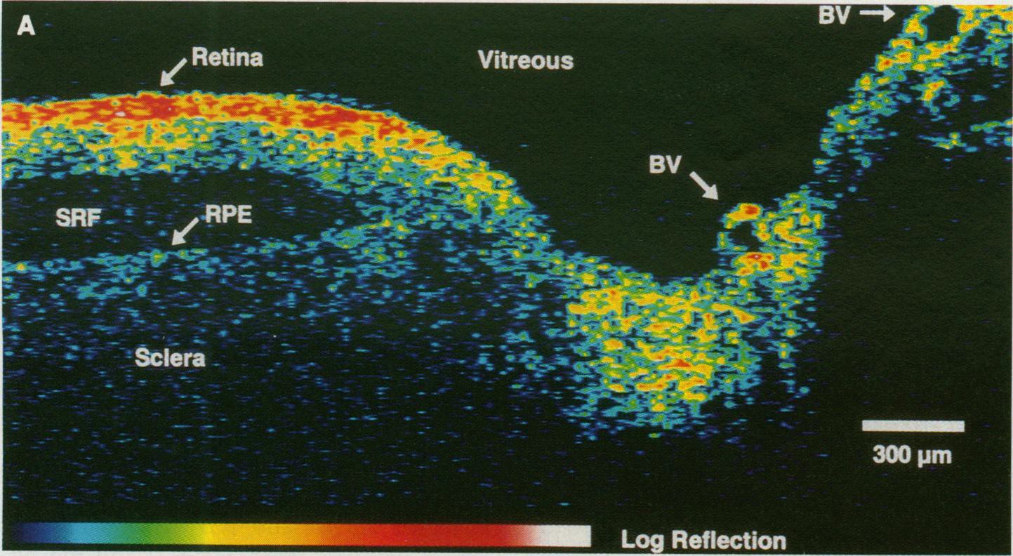
Optical Coherence Tomography (OCT) is a powerful imaging system for acquiring 3D volume images of tissues non-invasively. In simple terms, OCT can be considered as echography with light [1]. Unlike the echography which is done by sound waves, OCT imaging is not time-of-flight based and instead produces the image based on the interference patterns. Fig. 1 shows a typical retinal OCT image with false color. Throughout the past 2 decades, many improvements have been achieved regarding the OCT imaging system which not only improved the acquisition time but also the quality of the acquired images. Nowadays taking -level volume images of the tissues is very common especially in ophthalmology and retinal imaging. Therefore the need for specialized OCT image analysis techniques is of high interest, making this by far the most attractive area in biomedical imaging [2].
From a practical point of view, several aspects of image processing techniques are of high demand in analyzing OCT images which will be discussed in more detail in this article. As a pre-processing step, noise reduction techniques are usually applied to OCT images. Due to the physical implications of coherent imaging, OCT images usually suffer from granular patterns which block fine details in the tissue images. Plenty of techniques are proposed to remedy this issue some of which will be mentioned in the following sections.
Use of image segmentation methods is another area which is widely explored in OCT image analysis, especially in ophthalmology. For example, layered-structure of retina has been the subject of plenty of papers in the past few years. The methods that are used are usually specialized versions of the corresponding methods in general image segmentation, ranging from graph-based techniques to active contour methods etc. They will be further discussed later in this article.
Image registration also has its own place among the many image processing techniques used in OCT image analysis. This becomes more obvious when one realizes that OCT imaging is a -level imaging system which means even a small movement in the subject can have a big impact on the result. Even though the OCT imaging systems are very fast, still, the volume to be imaged is very dense so having an informative and comprehensive volume image requires hundreds of cross-sections which makes the imaging modality vulnerable to small movements. Also with the progress of degeneration in the tissue due to illness, image registration can be very useful in tracking the changes over time.
Based on the above-mentioned notes, the article is organized as follows: In Section 2 an overview of OCT imaging and different techniques that are used to acquire and reconstruct the images is given. Section 3 focuses on noise reduction techniques. In Section 4 a few techniques for OCT image segmentation, with a focus on retinal layer segmentation are reviewed. Section 5 contains an overview of the use of image registration techniques in OCT image analysis. Section 6 concludes the article with pointers on some of the future paths that can be taken for further investigations.
2 Optical Coherence Tomography (OCT) Imaging
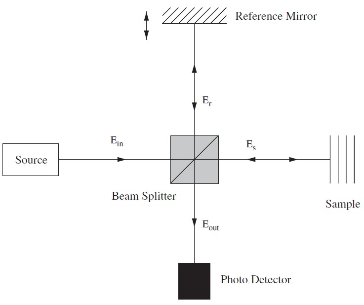
OCT imaging works based on the interference between a split and later re-combined broadband optical field [2]. Fig. 1 displays a typical OCT imaging system. The light beam from the source is exposed to a beam splitter and travels in two paths: one toward a moving reference mirror and the other to the sample to be imaged. The reflected light from both the reference mirror and the sample will be fed to a photo detector in order to observe the interference pattern. The sample usually contains particles (or layers) with different refractive indexes and the variation between neighbor refractive indexes causes intensity peaks in the interference pattern detected by the photo detector. By translating the reference mirror, a time domain interference pattern can be obtained. Also, by frequency domain measurements of the output spectrum, depth information can be derived. The result will be a line scan (aka A-scan) of depth of the 3D volume to be imaged. Using multiple A-scans, 2D cross-sections and 3D volumes can be constructed.
The most popular OCT imaging system is called time-domain OCT (TD-OCT) in which a reference mirror is translated to match the optical path from reflections within the sample [3]. Unlike the TD-OCT, in Fourier-domain OCT (FD-OCT) there is no need for moving parts in the design of the imaging system to obtain the axial scans [4]. In FD-OCT, the reference path length is fixed and the detection system is replaced with a spectrometer. Because of not having any moving parts, the speed of FD-OCT systems in acquiring images is very high in comparison to TD-OCT.
Moving away from the classical behavior of light and going toward the quantum nature of light, Quantum OCT (Q-OCT) is a new imaging modality [5]. Full field OCT (FF-OCT) is another optical coherence imaging technique in which a CCD camera is placed at the output instead of the single detector of the TD-OCT to capture 2D en face image in a single exposure [6]. Taking into account the polarization state of the light, polarization-sensitive OCT (PS-OCT) is another technique for imaging the birefringence within a biological sample [7]. Doppler OCT (D-OCT) which is also called optical Doppler tomography (ODT) is a combination of OCT imaging system with laser Doppler flowmetry. This system allows for the quantitative imaging of fluid flow in a highly scattering medium; such as monitoring in vivo blood flow beneath the skin [8].
In the following sections more focus will be given to the software based image analysis techniques rather than hardware of OCT systems.
3 Noise Reduction
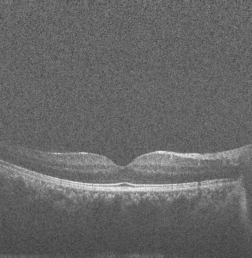
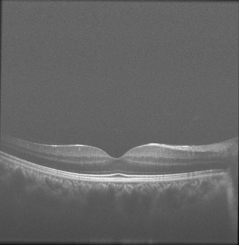
Speckle is a fundamental property of the signals and images acquired by narrow-band detection systems like SAR, ultrasound and OCT. In OCT, not only the optical properties of the system, but also the motion of the subject to be imaged, size and temporal coherence of the light source, multiple scattering, phase deviation of the beam and aperture of the detector can affect the speckle [9]. Two main processes affect the spatial coherence of the returning light beam which is used for image reconstruction: 1) multiple back-scattering of the beam, and 2) random delays for the forward-propagating and returning beam caused by multiple forward scattering. In the case of tissue imaging, since the tissue is packed with sub-wavelength diameter particles which act as scatterers, both of these phenomena contribute to the creation of speckle. As stated in [9], two types of speckle are present in OCT images: signal-carrying speckle which originates from the sample volume in the focal zone; and signal-degrading speckle which is created by multiple-scattered out-of-focus light. The latter kind is what that is considered as speckle noise. Fig. 3 displays the common scene in retinal OCT imaging: a highly noisy image.
The distribution of the speckle can be represented with a Rayleigh distribution. Speckle is considered as a multiplicative noise, in contrast to Gaussian additive noise. Due to the limited dynamic range of displays, OCT signals are usually compressed by a logarithmic operator applied to the intensity information. After this, the multiplicative speckle noise is transformed to additive noise and can be further treated [22].
OCT noise reduction techniques can be divided into two major classes: 1) methods of noise reduction during the acquisition time and 2) post-processing techniques. In the first class, aka compounding techniques, multiple uncorrelated recordings are averaged. Among them, spatial compounding [11], angular compounding [12], polarization compounding [13] and frequency compounding [14] techniques can be mentioned.
Usually the methods from the first class are not preferred since they require multiple scanning of the same data which extends the acquisition time. Plus, these techniques can be very restrictive in terms of different OCT imaging systems in use. Therefore the use of more general post-processing techniques is more favorable.
In the literature, plenty of post-processing methods are proposed for speckle noise reduction of OCT images. There are a few classic de-speckling techniques for noise reduction that are successfully used in OCT images too: Lee [15], Kuan [16] and Frost [17] filters.
One of the very interesting groups of methods for speckle reduction use the well-known anisotropic diffusion method [18]. Due to its poor performance in the case of very noisy images, there are different variations of this method in the literature [19, 20, 21]. In [22] a complete comparison is provided for regular anisotropic diffusion and complex anisotropic diffusion approaches for denoising of OCT images. Another example of using anisotropic diffusion is in [23] where an Interval type-II fuzzy approach is used for determining the diffusivity function of the anisotropic diffusion.
Another important group of widely used methods for de-speckling of OCT images take advantage of multiscale/multiresolution geometric representation techniques [1, 24]. The main reason for this is that these representations of the images compress the essential information of the image into a few large coefficients while noise is spread among all of the coefficients. Generally, for such methods a transform domain that provides a sparse representation of images and an optimal threshold is needed. Using the optimal threshold, hard- or soft-thresholding can be done for coefficient shrinkage in order to reduce the noise. This threshold can be a constant or even better, sub-band adaptive or spatially adaptive. For example, in OCT images, since it is known that most of the features are horizontal, higher thresholds can be applied to the vertical coefficients. Wavelet transform is one example of such methods which has been widely used in this area [25]. Even though wavelet transform has shown promising results, its lack of directionality imposes some limitations in properly denoising OCT images. This can be further improved by use of dual-tree complex wavelet transform (DT-CWT) which doubles the directional information of wavelet transform [26]. Still, there are better transform domains which offer more proper representations for OCT images. In the past few years use of Curvelet transform [27, 28], Contourlet transform [29, 30] and Ripplet transform [31] showed promising results in the denoising of speckle noise. Fig. 4 shows the result of curvelet coefficients’ shrinkage for speckle noise reduction.

Compressive sensing and sparse representation are new directions that are taken for image acquisition and noise reduction. Very recent examples are [32, 33] which use dictionary learning and sparse representation for noise reduction and image reconstruction of OCT. The process for these methods usually involves dividing the images into overlapping patches and training dictionaries. This is usually done by taking patches from both high-SNR-high-resolution (HH) and low-SNR-low-resolution (LL) samples from a volume data. The trained dictionary is then used in the process of denoising or reconstruction of images which acquired using random and incomplete patterns of data reading.
4 Image Segmentation
Image segmentation is a very active area in the field of medical image analysis. The most difficult step in any medical image analysis system is the automated localization and extraction of the structures of interest [34]. A critical role for medical image segmentation in OCT images involves the segmentation and extraction of regions of the image in order to quantify areas and volumes of interest in imaged tissues for further analysis and diagnosis.
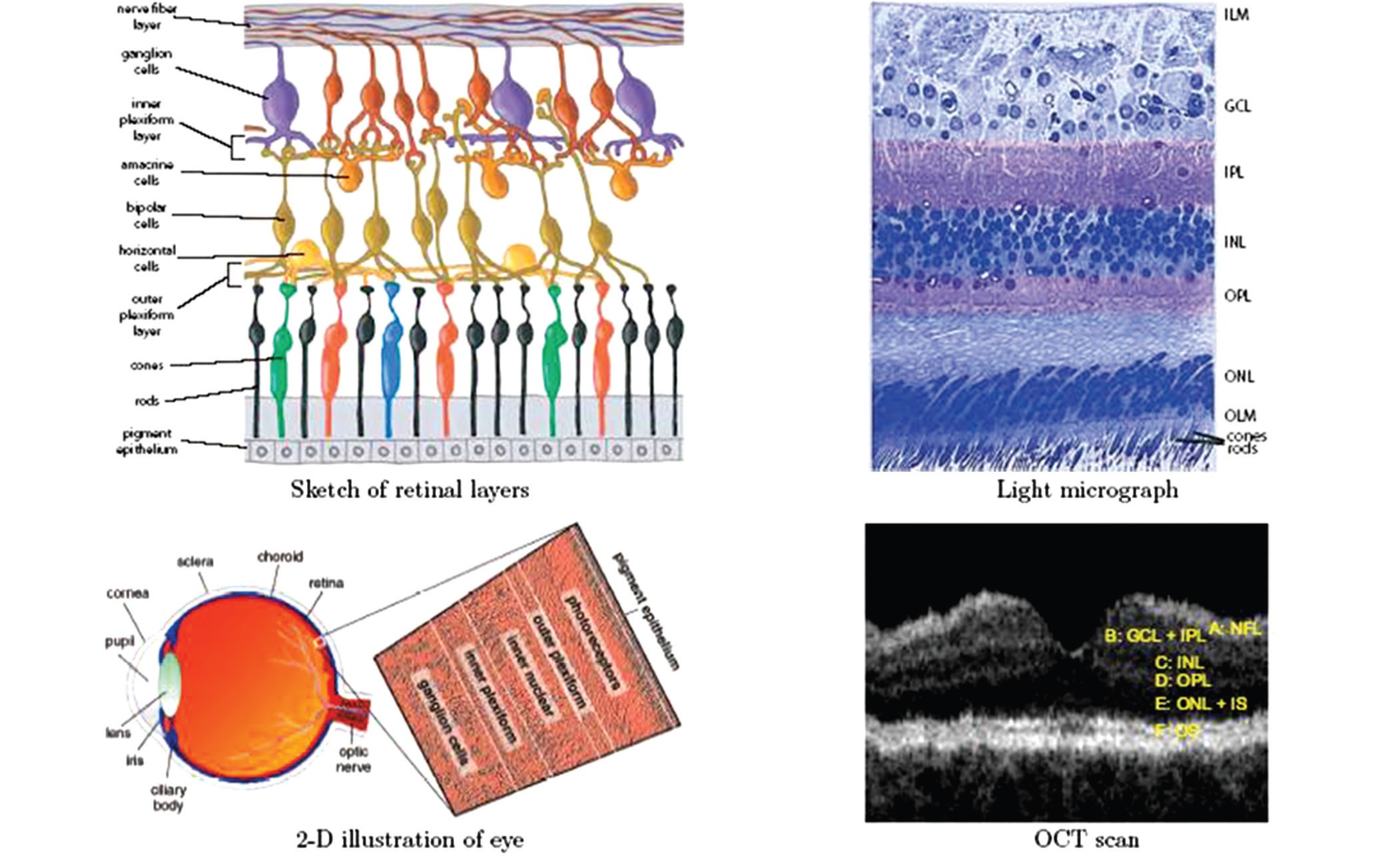
The signal strength in OCT images raises from the intrinsic differences in tissue optical properties. In retina, multiple layers of different types of cells can be seen in OCT images. Fig. 2 displays different views of retina [35].
During the last two decades plenty of methods are proposed for segmentation of OCT images. For a more complete review on the methods the reader is referred to [34] and [36]. Here an overview of the different classes of methods will be given.
Four major problems are encountered in the segmentation of OCT images:
-
1.
The presence of speckle noise complicates the process of the precise identification of the retinal layers, therefore most of the segmentation methods require pre-processing steps to reduce the noise.
-
2.
Intensity of homogeneous areas decreases with the increase of the depth of imaging. This is due to the fact that the intensity pattern in OCT images is the result of the absorption and scattering of light in the tissue.
-
3.
Optical shadows imposed by blood vessels also can affect the performance of segmentation methods.
-
4.
Quality of OCT images degrades as a result of motion artifacts or sub-optimal imaging conditions.
Methods of retinal segmentation can be categorized in five classes: 1) Methods applicable to A-scans, 2) intensity-based B-scans analysis, 3) active contour approaches, 4) analysis methods using artificial intelligence and pattern recognition techniques and 5) segmentation methods using 2D/3D graphs constructed from the 2D/3D OCT images [36, 37].
A-scan based methods consider the difference in the intensity levels to extract the edge information. Due to the effects of speckle noise, these methods are usually used for determining the most significant edges on the retina. Examples of such methods are presented in [3, 38, 39, 40, 41]. A-scan based methods lack the contribution from 2D/3D data while the computational time is hight and the accuracy is low. Taking advantage of better denoising methods, intensity-based B-scan analysis approaches work based on the intensity variations and gradients which are still too sensitive and make these methods case-dependent [42, 43, 44].
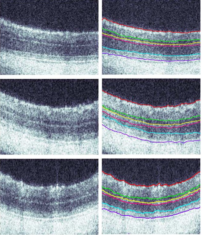
Active contour methods also provide promising results for segmentation of retinal layers in OCT images. Methods presented in [45], [46] and [47] are three well-known examples. Fig. 6 shows the results of layer segmentation using the active contour-based method proposed in [47] for three OCT images.
Use of pattern recognition based methods is a new direction that attracted researchers for more explorations. To name a few, in [48] a support vector machine (SVM) is used to perfom the semi-automatic segmentation of retinal layers. In [49] Fuzzy C-means clustering method is used for layer segmentation. The use of random forest classifier for segmentation is also reported in [50].
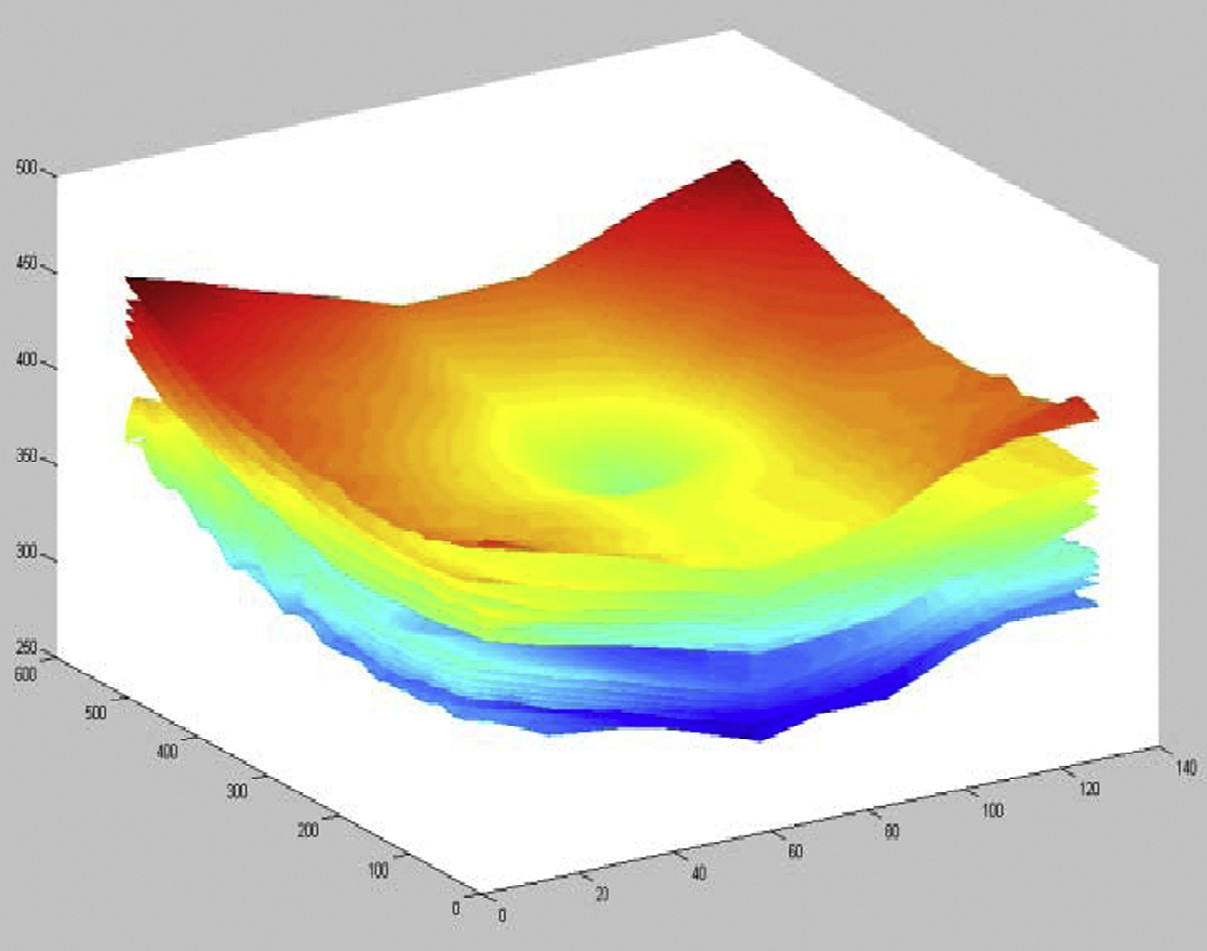
2D/3D graph based methods are probably the best among all of the segmentation methods. During the past few years several variations of this class are introduced in the literature. Examples of such methods can be found in [37, 51, 52, 53, 54]. The method in [37] is based on diffusion maps [55] and takes advantage of regional image texture rather than edge information. Therefore it is more robust in low contrast and poor layer-to-layer image gradients. On the other hand methods presented in [52, 53, 54] use the graph-cuts and shortest path method for segmentation of retinal layers. Fig. 7 shows the final 3D visualization of the segmented layers using the diffusion -based method proposed in [37].
Despite the plethora of different methods in this area, retinal layer segmentation is still an open problem with lots of room for improvement. Here, only a few of the well-known methods are shortly introduced and the reader is referred to the mentioned articles and references therein.
5 Image Registration
Having two input images, template image and reference image, image registration tries to find a valid optimal spatial transformation to be applied to the template image to make it more similar to the reference image [56]. Therefore the process of image registration consists of an optimization process fulfilling some imposed constraints. The applications of medical image registration range from mosaicing of retinal images [57] to slice interpolation [58] etc.
There are two different classes of image registration techniques: 1) Parametric approaches and 2) Non-parametric approaches [59]. In general, the process of finding the valid optimal spatial transformation can be formulated as in which and are reference and template images respectively, is the displacement field, is the similarity/dissimilarity measure (Mutual Information (MI), Normalized MI (NMI), Cross-Correlation (CC), Normalized CC, Sum of Squared Differences (SSD) and etc), is the regularization term (which imposes additional constraints on the deformation), is the coefficient that determines the amount of regularization and is the energy functional which depending on the problem either should be minimized or maximized [60].
There are several different applications for using image registration approaches in OCT image analysis. One of the basic applications is in speckle noise reduction. As mentioned in Section 3, hardware-based noise reduction techniques take the average of several uncorrelated scans in order to reduce the noise. The same thing can happen after finishing image acquisition too. The main idea is to gather several images of the same cross-section in retina, register them together and take the average. Having a small movement in the eye provides us with an uncorrelated pattern of speckle. Having B-scans, the SNR can be improved by a factor of . In [61] a dynamic programming based method is used for compensation of the movements between several B-scans and reducing the speckle noise. A hierarchical model-based motion estimation scheme based on an affine-motion model is used in [62] for registering multiple B-scans to be used for speckle reduction. Fig. 6 shows the result of affine registering the images from close locations in retina and averaging. Recently, the use of low-rank/sparse decomposition based batch alignment has been investigated for speckle noise reduction in OCT images too. Taking advantage of Robust Principal Component Analysis (RPCA) [63] and simultaneous decomposition and alignment of a stack of OCT images via linearized convex optimization, better performance is achieved in comparison to previous registration based denoising techniques. For more details on the method and results, the reader is referred to [64, 65]
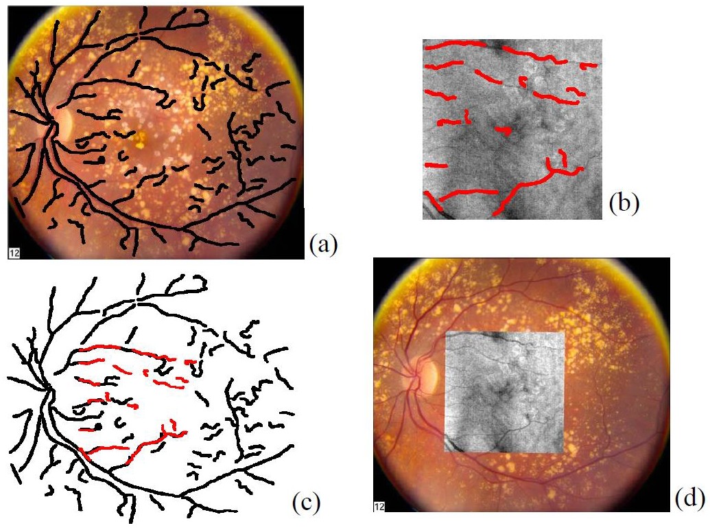
Another application is in the registration of fundus images with en face OCT images. This is very helpful to better correlate retinal features across different imaging modalities. In [66] an algorithm is proposed for registering OCT fundus images with color fundus photographs. In this paper, blood vessel ridges are taken as features for registration. A similar approach is taken in [67] too. Using the curvelet transform, the blood vessels are extracted for the two modalities and then image registration is done in two steps: 1) registration based on only scaling and translation and 2) registration based on a quadratic transformation. Fig. 9 shows different stages of the results of color fundus and en face OCT image registration of the method proposed in [66].
Motion correction of OCT images is another area in which image registration techniques are of high interest. There are several different involuntary eye movements that can happen during the fixation: tremor, drifts and micro-saccades [68]. One way to deal with this issue is a hardware solution which involves having eye tracking equipment to compensate the eye movement during image acquisition time. Usually a Scanning Laser Ophthalmoscopy (SLO) device is merged with the OCT for tracking the eye movements during imaging [69]. Taking a software approach can be more general and applicable without the need for additional imaging equipment. In [70] at first the vessels are detected and then using an elastic registration technique the tremor and drift motions are corrected. Micro-saccades are corrected in the next step by finding the horizontal shift at each pixel in the scan that best aligns the result of tremor and drift correction with the SLO image. In [71] a 3D aligning method for motion correction is proposed based on particle filtering. Fig. 10 shows one example of the use of the algorithm in [71] for correcting the motion in OCT images. Other techniques can be found in [72, 73] and references therein.
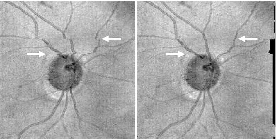
Image mosaicing is another example of using image registration methods in OCT image processing. OCT systems are capable of acquiring a huge amount of data in a very short period of time; but still the field of view is very limited. Stitching several volume data from a patient can improve the interpretation of data significantly. Usually in such methods, a set of overlapping OCT data is acquired and then stitched together using global/local image registration techniques. In [74] at first a set of 8 overlapping 3D OCT volume data over a wide area of retina is acquired. Then the OCT en face fundus images are registered together using blood vessel ridges as features of interest. Finally the 3D OCT data sets are merged together using cross-correlation as a metric. A relatively similar approach is taken in [75] too for motion correction and 3D volume mosaicing of OCT images. Fig. 11 shows a color encoded depth image of retina with three main vessel layers. One of the very recent works in this area can be found in [76] for bladder OCT images which is used with an integrated White Light Cystoscopy (WLC) system.

The use of image registration techniques in analysis and processing of OCT images has grown significantly in the past few years. Here only a few areas are mentioned and there are more to explore.
6 Conclusion
Even though the techniques introduced here don not represent a comprehensive list of all of the approaches that are used for analysis of OCT images, the variety of the techniques gives us some pointers on the possible directions for further explorations. Especially with the emergence of parallel computing and GPU programming in the past few years, it is possible to have real time pre-processing and analysis systems to help with the increasing amount of data produced by OCT imaging systems. In this article, an overview of 3 major problems in OCT image analysis is provided: noise reduction, image segmentation and image registration, and several different techniques in each category are introduced. Starting from noise reduction, both hardware and software techniques are discussed to some extent. Moving to image segmentation, different segmentation techniques with a focus on retinal layer segmentation are introduced. Finally some of the image registration methods that are used for noise reduction, motion correction and image mosaicing of OCT images are reviewed. Overall, the use of image analysis techniques for OCT is a rapidly growing field and there remain many areas for investigation.
References
- [1] Pizurica, Aleksandra, et al. ”Multiresolution denoising for optical coherence tomography: a review and evaluation.” Current Medical Imaging Reviews 4.4 (2008): 270-284.
- [2] Tomlins, Peter H., and R. K. Wang. ”Theory, developments and applications of optical coherence tomography.” Journal of Physics D: Applied Physics 38.15 (2005): 2519.
- [3] Huang, David, et al. ”Optical coherence tomography.” Science 254.5035 (1991): 1178-1181.
- [4] Fercher, Adolph F., et al. ”Measurement of intraocular distances by backscattering spectral interferometry.” Optics Communications 117.1 (1995): 43-48.
- [5] Nasr, Magued B., et al. ”Demonstration of dispersion-canceled quantum-optical coherence tomography.” Physical review letters 91.8 (2003): 083601.
- [6] Beaurepaire, E., et al. ”Full-field optical coherence microscopy.” Optics letters 23.4 (1998): 244-246.
- [7] de Boer J F, Srinivas S M, Nelson J S, Milner T E and Ducros M G et al 2002 Handbook of Optical Coherence Tomography ed B E Bouma and G J Tearney (New York: Dekker) pp 237-74
- [8] Chen, Zhongping, et al. ”Optical Doppler tomography: imaging in vivo blood flow dynamics following pharmacological intervention and photodynamic therapy.” Photochemistry and photobiology 67.1 (1998): 56-60.
- [9] Schmitt, Joseph M., S. H. Xiang, and Kin M. Yung. ”Speckle in optical coherence tomography.” Journal of biomedical optics 4.1 (1999): 95-105.
- [10] Salinas, Harry M., and D. Cabrera Fernández. ”Comparison of PDE-based nonlinear diffusion approaches for image enhancement and denoising in optical coherence tomography.” IEEE transactions on medical imaging 26.6 (2007): 761-771.
- [11] Avanaki, Mohammad RN, et al. ”Spatial compounding algorithm for speckle reduction of dynamic focus OCT images.” Photonics Technology Letters, IEEE 25.15 (2013): 1439-1442.
- [12] Schmitt, J. M. ”Array detection for speckle reduction in optical coherence microscopy.” Physics in Medicine and Biology 42.7 (1997): 1427.
- [13] Kobayashi, Masaru, et al. ”Polarization-independent interferometric optical-time-domain reflectometer.” Lightwave Technology, Journal of 9.5 (1991): 623-628.
- [14] Pircher, Michael, et al. ”Speckle reduction in optical coherence tomography by frequency compounding.” Journal of Biomedical Optics 8.3 (2003): 565-569.
- [15] Lee, Jong-Sen. ”Digital image enhancement and noise filtering by use of local statistics.” Pattern Analysis and Machine Intelligence, IEEE Transactions on 2 (1980): 165-168.
- [16] Kuan, Darwin T., et al. ”Adaptive noise smoothing filter for images with signal-dependent noise.” Pattern Analysis and Machine Intelligence, IEEE Transactions on 2 (1985): 165-177.
- [17] Frost, Victor S., et al. ”A model for radar images and its application to adaptive digital filtering of multiplicative noise.” Pattern Analysis and Machine Intelligence, IEEE Transactions on 2 (1982): 157-166.
- [18] Perona, Pietro, and Jitendra Malik. ”Scale-space and edge detection using anisotropic diffusion.” Pattern Analysis and Machine Intelligence, IEEE Transactions on 12.7 (1990): 629-639.
- [19] Yu, Yongjian, and Scott T. Acton. ”Speckle reducing anisotropic diffusion.” Image Processing, IEEE Transactions on 11.11 (2002): 1260-1270.
- [20] Abd-Elmoniem, Khaled Z., A. Youssef, and Yasser M. Kadah. ”Real-time speckle reduction and coherence enhancement in ultrasound imaging via nonlinear anisotropic diffusion.” Biomedical Engineering, IEEE Transactions on 49.9 (2002): 997-1014.
- [21] Krissian, Karl, et al. ”Oriented speckle reducing anisotropic diffusion.” Image Processing, IEEE Transactions on 16.5 (2007): 1412-1424.
- [22] Salinas, Harry M., and D. Cabrera Fernández. ”Comparison of PDE-based nonlinear diffusion approaches for image enhancement and denoising in optical coherence tomography.” IEEE transactions on medical imaging 26.6 (2007): 761-771.
- [23] Puvanathasan, Prabakar, and Kostadinka Bizheva. ”Interval type-II fuzzy anisotropic diffusion algorithm for speckle noise reduction in optical coherence tomography images.” Optics express 17.2 (2009): 733-746.
- [24] Jacques, Laurent, et al. ”A panorama on multiscale geometric representations, intertwining spatial, directional and frequency selectivity.” Signal Processing 91.12 (2011): 2699-2730.
- [25] Adler, Desmond C., Tony H. Ko, and James G. Fujimoto. ”Speckle reduction in optical coherence tomography images by use of a spatially adaptive wavelet filter.” Optics letters 29.24 (2004): 2878-2880.
- [26] Selesnick, Ivan W., Richard G. Baraniuk, and Nick C. Kingsbury. ”The dual-tree complex wavelet transform.” Signal Processing Magazine, IEEE 22.6 (2005): 123-151.
- [27] Jian, Zhongping, et al. ”Speckle attenuation in optical coherence tomography by curvelet shrinkage.” Optics letters 34.10 (2009): 1516-1518.
- [28] Jian, Zhongping, et al. ”Three-dimensional speckle suppression in optical coherence tomography based on the curvelet transform.” Optics express 18.2 (2010): 1024-1032.
- [29] Guo, Qing, et al. ”Image denoising algorithm based on contourlet transform for optical coherence tomography heart tube image.” Image Processing, IET 7.5 (2013): 442-450.
- [30] Xu, Jianbing, et al. ”Speckle reduction of retinal optical coherence tomography based on contourlet shrinkage.” Optics letters 38.15 (2013): 2900-2903.
- [31] Gupta, Deep, R. S. Anand, and Barjeev Tyagi. ”Ripplet domain non-linear filtering for speckle reduction in ultrasound medical images.” Biomedical Signal Processing and Control 10 (2014): 79-91.
- [32] Fang, Leyuan, et al. ”Sparsity based denoising of spectral domain optical coherence tomography images.” Biomedical optics express 3.5 (2012): 927-942.
- [33] L. Fang, S. Li, R. McNabb, Q. Nie, A. Kuo, C. Toth, J. A. Izatt, and S. Farsiu, “Fast acquisition and reconstruction of optical coherence tomography images via sparse representation,” IEEE Trans. Med. Imag., vol. 32, no. 11, pp. 2034–2049, Nov. 2013
- [34] DeBuc, D. Cabrera. ”A review of algorithms for segmentation of retinal image data using optical coherence tomography.” Image Segmentation (2011): 15-54.
- [35] Garvin, M., ”Automated 3D segmentation and analysis of retinal Optical Coherence Tomography images”, PhD thesis, University of Iowa, Iowa, USA, 2008
- [36] Kafieh, Raheleh, Hossein Rabbani, and Saeed Kermani. ”A Review of Algorithms for Segmentation of Optical Coherence Tomography from Retina.” Journal of medical signals and sensors 3.1 (2013): 45.
- [37] Kafieh, Raheleh, et al. ”Intra-retinal layer segmentation of 3D optical coherence tomography using coarse grained diffusion map.” Medical image analysis 17.8 (2013): 907-928.
- [38] Hee, Michael R., et al. ”Optical coherence tomography of the human retina.” Archives of ophthalmology 113.3 (1995): 325-332.
- [39] Dillenseger, George A., Weber A, Pechereau A. Optical coherence tomography image processing. Invest Ophthalmol Vis Sci. (2000);41:165–73
- [40] Koozekanani, Dara, Kim Boyer, and Cynthia Roberts. ”Retinal thickness measurements from optical coherence tomography using a Markov boundary model.” Medical Imaging, IEEE Transactions on 20.9 (2001): 900-916.
- [41] Shahidi, Mahnaz, Zhangwei Wang, and Ruth Zelkha. ”Quantitative thickness measurement of retinal layers imaged by optical coherence tomography.” American journal of ophthalmology 139.6 (2005): 1056-1061.
- [42] Baroni, Maurizio, Pina Fortunato, and Agostino La Torre. ”Towards quantitative analysis of retinal features in optical coherence tomography.” Medical engineering and physics 29.4 (2007): 432-441.
- [43] Tan, Ou, et al. ”Mapping of macular substructures with optical coherence tomography for glaucoma diagnosis.” Ophthalmology 115.6 (2008): 949-956.
- [44] Kajić, Vedran, et al. ”Robust segmentation of intraretinal layers in the normal human fovea using a novel statistical model based on texture and shape analysis.” Optics express 18.14 (2010): 14730-14744.
- [45] Fernández DC, Villate N, Puliafito CA, Rosenfeld PJ. Comparing total macular volume changes measured by optical coherence tomography with retinal lesion volume estimated by active contours. Invest Ophthalmol Vis Sci. (2004);45 E-Abstract 3072
- [46] Mishra, Akshaya, et al. ”Intra-retinal layer segmentation in optical coherence tomography images.” Optics express 17.26 (2009): 23719-23728.
- [47] Yazdanpanah, Azadeh, et al. ”Segmentation of intra-retinal layers from optical coherence tomography images using an active contour approach.” Medical Imaging, IEEE Transactions on 30.2 (2011): 484-496.
- [48] Fuller, Alfred R., et al. ”Segmentation of three-dimensional retinal image data.” Visualization and Computer Graphics, IEEE Transactions on 13.6 (2007): 1719-1726.
- [49] Mayer, M., Tornow, R., Bock, R., Hornegger, J., Kruse, F., 2008. Automatic nerve fiber layer segmentation and geometry correction on spectral domain OCT images using fuzzy c-means clustering. Investigative Ophthalmology and Visual Science 49, E-Abstract 1880.
- [50] Lang, Andrew, et al. ”Retinal layer segmentation of macular OCT images using boundary classification.” Biomedical optics express 4.7 (2013): 1133-1152.
- [51] Garvin, Mona Kathryn, et al. ”Intraretinal layer segmentation of macular optical coherence tomography images using optimal 3-D graph search.” Medical Imaging, IEEE Transactions on 27.10 (2008): 1495-1505.
- [52] Chiu, Stephanie J., et al. ”Automatic segmentation of seven retinal layers in SDOCT images congruent with expert manual segmentation.” Optics express 18.18 (2010): 19413-19428.
- [53] Chiu, Stephanie J., et al. ”Validated automatic segmentation of AMD pathology including drusen and geographic atrophy in SD-OCT images.” Investigative ophthalmology and visual science 53.1 (2012): 53-61.
- [54] Srinivasan, Pratul P., et al. ”Automatic segmentation of up to ten layer boundaries in SD-OCT images of the mouse retina with and without missing layers due to pathology.” Biomedical optics express 5.2 (2014): 348-365.
- [55] Coifman, Ronald R., and Stéphane Lafon. ”Diffusion maps.” Applied and computational harmonic analysis 21.1 (2006): 5-30.
- [56] Zitova, Barbara, and Jan Flusser. ”Image registration methods: a survey.” Image and vision computing 21.11 (2003): 977-1000.
- [57] Patton, Niall, et al. ”Retinal image analysis: concepts, applications and potential.” Progress in retinal and eye research 25.1 (2006): 99-127.
- [58] Baghaie, A., Yu, Z.: Curvature-Based Registration for Slice Interpolation of Medical Images. In: Y.J. Zhang, J.M.R.S. Tavares (Eds.): CompIMAGE 2014, LNCS 8641, pp. 69-80, Springer (2014)
- [59] Modersitzki, Jan.: Numerical methods for image registration. OUP Oxford, (2003)
- [60] Sotiras, Aristeidis, Christos Davatzikos, and Nikos Paragios. ”Deformable medical image registration: A survey.” Medical Imaging, IEEE Transactions on 32.7 (2013): 1153-1190.
- [61] Jorgensen, Thomas Martini, et al. ”Enhancing the signal-to-noise ratio in ophthalmic optical coherence tomography by image registration—method and clinical examples.” Journal of biomedical optics 12.4 (2007): 041208-041208.
- [62] Alonso-Caneiro, David, Scott A. Read, and Michael J. Collins. ”Speckle reduction in optical coherence tomography imaging by affine-motion image registration.” Journal of biomedical optics 16.11 (2011): 116027-1160275.
- [63] Candes, Emmanuel J., et al. ”Robust principal component analysis?.” Journal of the ACM, 58.3 (2011): 11.
- [64] Peng, Yigang, et al. ”RASL: Robust alignment by sparse and low-rank decomposition for linearly correlated images.”, IEEE Transactions on Pattern Analysis and Machine Intelligence, 34.11 (2012): 2233-2246.
- [65] Baghaie, Ahmadreza, Roshan M. D’souza, and Zeyun Yu. ”Sparse And Low Rank Decomposition Based Batch Image Alignment for Speckle Reduction of retinal OCT Images.” arXiv preprint arXiv:1411.4033 (2014).
- [66] Li, Ying, et al. ”Registration of OCT fundus images with color fundus photographs based on blood vessel ridges.” Optics express 19.1 (2011): 7-16.
- [67] Golabbakhsh, Marzieh, and Hossein Rabbani. ”Vessel-based registration of fundus and optical coherence tomography projection images of retina using a quadratic registration model.” IET Image Processing 7.8 (2013): 768-776.
- [68] Hart Jr., W.H.: Adler’s physiology of the eye: clinical application, 9th edn., Mosby-Year Book Inc., 11830 Westline Industrial Drive, St. Louis, Missouri 63146 (1992)
- [69] Pircher, Michael, et al. ”Simultaneous SLO/OCT imaging of the human retina with axial eye motion correction.” Optics express 15.25 (2007): 16922-16932.
- [70] Ricco, Susanna, et al. ”Correcting motion artifacts in retinal spectral domain optical coherence tomography via image registration.” Medical Image Computing and Computer-Assisted Intervention–MICCAI 2009. Springer Berlin Heidelberg, 2009. 100-107.
- [71] Xu, Juan, et al. ”Alignment of 3-D optical coherence tomography scans to correct eye movement using a particle filtering.” Medical Imaging, IEEE Transactions on 31.7 (2012): 1337-1345.
- [72] Kraus, Martin F., et al. ”Motion correction in optical coherence tomography volumes on a per A-scan basis using orthogonal scan patterns.” Biomedical optics express 3.6 (2012): 1182-1199.
- [73] Kraus, Martin F., et al. ”Quantitative 3D-OCT motion correction with tilt and illumination correction, robust similarity measure and regularization.” Biomedical Optics Express 5.8 (2014): 2591-2613.
- [74] Li, Ying, et al. ”Automatic montage of SD-OCT data sets.” Optics express 19.27 (2011): 26239-26248.
- [75] Hendargo, Hansford C., et al. ”Automated non-rigid registration and mosaicing for robust imaging of distinct retinal capillary beds using speckle variance optical coherence tomography.” Biomedical optics express 4.6 (2013): 803-821.
- [76] Lurie, Kristen L., Roland Angst, and Audrey K. Ellerbee. ”Automated mosaicing of feature-poor optical coherence tomography volumes with an integrated white light imaging system.”, Biomedical Engineering, IEEE Transactions on, 61(7):2141-53 (2014)