Phosphorene oxides: bandgap engineering of phosphorene by oxidation
Abstract
We show that oxidation of phosphorene can lead to the formation of a new family of planar (2D) and tubular (1D) oxides and sub-oxides, most of them insulating. This confers to black phosphorus a native oxide that can be used as barrier material and protective layer. Further, the bandgap of phosphorene oxides depends on the oxygen concentration, suggesting that controlled oxidation can be used as a means to engineer the bandgap. For the oxygen saturated composition, P2O5, both the planar and tubular phases have a large bandgap energy of about 8.5 eV, and are transparent in the near UV. These two forms of phosphorene oxides are predicted to have the same formation enthalpy as o′-P2O5, the most stable of the previously known forms of phosphorus pentoxide.
pacs:
73.20.At,73.61.Cw,73.61.NgI Introduction
Phosphorene, a single layer of black phosphorus, is a unique two-dimensional semiconductor with a nearly-direct gap in the visible range.Rodin et al. (2014); Tran et al. (2014) It stands out amongst the family of 2D materials for its orthorhombic waved structure with superior flexibility and small Young’s modulus,Wei and Peng (2014) allowing for strain-driven band structure engineering.Rodin et al. (2014) From the point of view of electronic and optoelectronic applications, it features high carrier mobility and on-off ratio,Koenig et al. (2014); Xia et al. as well as a giant photoresponse in the IR and UV.Buscema et al. (2014)
In contrast to graphene, phosphorene is prone to oxidation,A. Ziletti and Neto which usually leads to degradation of the structure and electronic properties.Doganov et al. ; Castellanos-Gomez et al. At present oxidation is avoided by encapsulating with polymers or other two-dimensional materials eg. graphene or boron nitride.Doganov et al.
However, there are indications that a phosphate layer grown in a controlled way could be used as a protective layer or even as a functional material on its own. Black phosphorus oxidizes on the surface, seemingly leaving the deepest layers intact. The non-uniform degradation pattern normally observed, showing as dark patches on the optical microscope image, seems to require the interaction with water,Favron et al. ; Yau et al. (1992) even though energy-loss spectra shows PxOy still grows in oxygen-only atmosphere.Farnsworth et al. (2014) In this article, we show that phosphorene oxides and suboxides can exist in monolayer form, suggesting that there is an opportunity for growth of single-layer or few-layer native oxide at the black phosphorus surface.
The existence of an insulating native oxide is an added advantage for phosphorene. Since phosphate glasses,Brow (2000) (e.g. monolayer P2O5) are transparent in the near UV, a passivating coating will preserve the optical properties of the underlying phosphorene. Further, since such a coating it is saturated with oxygen, it prevents oxygen molecules from reaching the pristine phosphorene layers beneath.
Bulk P2O5 has at least three known planar polymorphs, a molecular solidJansen (1986) (with P4O10 structural units), and two orthorhombic phases,Arbib et al. (1996); Stachel et al. (1995) along with an amorphous phase.Hulliger (1976); Galeener and Jr. (1979) The thermodynamically most stable form (o′-P2O5) is a layered structure belonging to the space group.Stachel et al. (1995) A monolayer phase, different from the one reported here, has also been proposed by a recent theoretical study.Wang et al.
In this article, we show that suboxides (P4On, ) also exist in a variety of layered forms, most of them insulating, with bandgaps that depend on the oxygen concentration. This offers the possibility of using oxidation as a means to engineer the bandgap of phosphorene. The article is divided in three parts: first, we describe the structure and energetics of phosphorene oxides; then, we consider their electronic properties and finally their vibrational spectra.
II Computational details
All first-principle calculations are based on the framework of density functional theory (DFT), as implemented in the Quantum ESPRESSO packageGiannozzi et al. (2009). We use the PBEPerdew et al. (1996), PBEsolPerdew et al. (2008) and HSE06Krukau et al. (2006) (HSE hereafter) approximations for the exchange and correlation energy. The PBEsol functional is preferred over PBE because it cures the systematic tendency of PBE to overestimate equilibrium lattice constants of solidsPerdew et al. (2008). The HSE functional, with its fraction of screened short-ranged Hartree-Fock exchange, yields reasonably accurate predictions for energy bandgaps in semiconductorsHeyd et al. (2005); Janesko et al. (2009); Henderson et al. (2011). Unless otherwise stated, all quantities reported (e.g. energies, bond lengths and bond angles) are obtained with the PBEsol functional. The electron-ion interaction is described using the projector-augmented wave (PAW)Blöchl (1994) approach for PBEsol calculations, while norm-conserving Troullier-Martins pseudopotentialsTroullier and Martins (1991) are employed in PBE and HSE calculations. We employ a plane wave basis set with kinetic energy cutoffs of 70 Ry (280 Ry) to describe the electronic wave functions (charge density). Vibrational properties including Raman and infrared spectra are calculated with density functional perturbation theoryBaroni et al. (2001) (DFPT) and the PBEsol functional. The Brillouin zone is sampled using a -centered 1081 Monkhorst-Pack (MP) gridMonkhorst and Pack (1976) for all but the vibrational calculations, for which a finer 15121 grid is employed. A supercell of 16 Å in the direction perpendicular to the monolayer is used to avoid spurious interactions between periodic replicas. For each oxygen concentration, tens of different configurations are used as starting points for optimization. Then, lattice geometries and atomic positions are relaxed till the forces on each atom are less than 10-3 eV/Å, and the pressure less then 1 kbar, except for the HSE calculations, where we use the PBE lattice parameters. The tetrahedron smearing methodBlöchl et al. (1994) is used with a Gaussian broadening of 0.04 eV and a 129 MP grid in the density of states (DOS) calculations. However, local and semilocal DFT functionals (such as PBE and PBEsol) are known to underestimate the bandgap because of their missing derivative discontinuity in the exchange-correlation energy across the gapPerdew et al. (1982); Perdew and Levy (1983). The inclusion of a fraction of exact nonlocal short-ranged Hartree-Fock exchange, as done in HSE, partially cures this shortcoming, giving bandgaps in better agreement with experimentKrukau et al. (2006). Thus, we use HSE to calculate the bandgap energyHenderson et al. (2011).
To further validate the HSE results, we calculate the bandgap of pristine phosphorene and the two phosphorene oxides (-P4O10 and -P4O10) using the GW approximation to the electron self-energy within the generalized plasmon pole model.Hedin (1965); Hybertsen and Louie (1986) The GW method is state-of-the-art for evaluating quasiparticle bandgaps since it gives values in very good agreement with experiment (typically within 0.2 eV) for a large variety of systems and a broad range of bandgapsLOUIE . The GW calculations are performed in two stages. First, relaxed lattice geometries, equilibrium atomic positions and the electronic ground state are obtained from a DFT calculation with the PBE functional and Troullier-Martins pseudopotentialsTroullier and Martins (1991). Then, the DFT Kohn-Sham orbitals and energies are used to construct both the electronic Green’s function, G, and the dynamically screened interaction, W, to evaluate the quasiparticle self-energy GW. This method is commonly referred as G0W0. To achieve convergence in our GW calculations we follow the procedure proposed by Malone and Cohen.Malone and Cohen (2013) For pristine phosphorene, we use 370 bands to evaluate the dielectric matrix and the self-energy , employing an energy cutoff of 12 Ry for the dielectric matrix and a 24201 MP-grid; convergence was however checked with up to 1024 bands and using up to 20 Ry. For the two phosphorene oxides (-P4O10 and -P4O10) we use an energy cutoff of 8 Ry for the dielectric matrix, and 800 and 896 bands to evaluate and , respectively. A 741 grid for -P4O10 and a 461 grid for -P4O10 are employed. Convergence was checked including up to 1792 bands and increasing up to 20 Ry. For all the GW calculations we use a supercell of 20 Å in the direction perpendicular to the monolayers and a slab-truncation of the Coulomb potential.Ismail-Beigi (2006) The GW calculations are performed with the ABINIT code.Gonze et al. (2009) Additional details on the convergence study are given in the Supplemental Material. With these parameters, we conservatively estimate the errors in our GW bandgaps at 0.05 eV for pristine phosphorene and 0.1 eV for -P4O10 and -P4O10.
III Results and discussion
III.1 Structure
First we describe the new family of phosphorene oxides (POs) found in this study. These are obtained by adding oxygen atoms to phosphorene, maintaining the rectangular lattice symmetry. We considered all compounds P4On with between 1 and 10. The maximum oxygen concentration of 250%, or 10 oxygen atoms per unit cell, corresponds to the stoichiometry of phosphorus pentoxide (P2O5 or P4O10), whose known polymorphs were outlined in the introductionGreenwood and Earnshaw (1997).
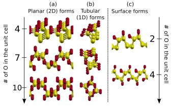
For each oxygen concentration, we use tens of different starting points corresponding to different O atoms arrangements in the pristine phosphorene lattice. After lattice relaxation and geometry optimization, numerous metastable phosphorous oxide (PO) structures are identified. We find that, for each oxygen concentration, the most stable POs can be always divided into two very distinctive classes: planar (2D) and tubular (1D) forms. For convenience, we refer to the planar forms as -P4On and to the tubular forms as -P4On, where is the number of oxygen atoms in the unit cell. We also identify surface forms -P4On. Some representative structures are depicted in Fig.1.
Analysis of the local oxygen atom environments that are common to both -P4On and -P4On forms presented in Fig.1 indicates that all the PO structures identified exhibit two P-O motifs: the dangling and bridging oxygen motifs shown in Fig.2.
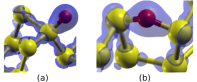
The dangling oxygen motif, represented in Fig.2a, consists of an O atom that forms a bond with only one P atom. The phosphorus lone pair is attracted towards the more electronegative oxygen atom, giving rise to an excess of negative charge localized on the O atom (Fig.2a). The P-O bond is short, strong and polar with bond lengths ranging from 1.44 Å to 1.51 Å depending on phosphorus oxidation number and local environment. 111Oftentimes this bond is classified as a double bondGaleener et al. (1978); Galeener and Jr. (1979); Greenwood and Earnshaw (1997); Brazhkin et al. (2011), although its identification as P=O is not commonly acceptedGilheany (1994); Rai and Symons (1994); Chesnut and Savin (1999). Nevertheless, hereafter we will refer to this bond as P=O for simplicity. Similar to what has been found for dangling oxygens on a phosphorene surfaceA. Ziletti and Neto , in the PO forms identified here, the P=O bonds always point away from the zigzag ridge in which they are chemisorbed, minimizing Coulomb repulsion between the oxygen orbitals.
The P=O bond length decreases monotonically as the number of oxygens linked to the phosphorus involved in the bond increases. Let P(n) be a phosphorus atom that is linked to oxygens, where . With this notation P(1)=O defines a double bond in which a phosphorus is linked to only one oxygen, as shown for example in Fig.2a. The average P=O bond lengths for the sequence of P(n) configurations are given in Table 1 for the planar and tubular forms. The longest P=O is found for (where the P atom establishes a P=O bond and three P-P bonds); conversely, the shortest P=O are found in -P4O10 and -P4O10 (), where the phosphorus atom is bonded to four oxygens. This monotonic decrease in the P=O bond length with increasing oxygen connectivity has an analogue in the molecular series P4Om (=610)Jansen et al. (1981); Walker et al. (1979); Clade et al. (1994).
| planar | tubular | |
|---|---|---|
| P(1)=O | 1.508 | 1.477 |
| P(2)=O | 1.476 | 1.466 |
| P(3)=O | 1.459 | 1.458 |
| P(4)=O | 1.445 | 1.447 |
In the bridge motif, as the name indicates, the oxygen atom bridges two P atoms, occupying a position close to a P-P bond center (Fig.2b). The electron density is mostly distributed on the two P-O bonds, and only a moderate electron density accumulation is observed on the oxygen. Therefore, we expect a much stronger Coulomb repulsion between dangling oxygens than bridging oxygens, especially for high O concentrations. The P-O bondlengths in this case are between 1.61 Å and 1.78 Å, this wide range due to different strain interactions and lattice distortions depending on the oxygen concentration.
Bridging oxygens can either bond with P atoms from the same or from different zigzag ridges, therefore forming intra-ridge (e.g. B or C in Fig. 3b) or inter-ridge bridge (e.g. D in Fig. 3b) structures. In particular, inter-ridge bridges are essential for the formation of the planar forms because they effectively link different ridges that would otherwise separate and form 1D tubular chains. The formation of oxygen bridges increases considerably the lattice parameters, resulting in deformations as large as 90% relative to pristine phosphorene for the maximally oxidized forms (see Table 2).
| System | XC | (Å) | (Å) | a(%) | b(%) |
|---|---|---|---|---|---|
| Phosphorene | PBE | 3.30 | 4.62 | – | – |
| PBEsol | 3.28 | 4.45 | – | – | |
| -P4O10 | PBE | 4.39 | 6.52 | 33 | 41 |
| PBEsol | 4.41 | 6.75 | 34 | 51 | |
| -P4O10 | PBE | 6.12 | 4.53 | 85 | -1.9 |
| PBEsol | 6.28 | 4.58 | 91 | 2.9 | |
| -P4O2 | PBE | 3.44 | 4.52 | 4.2 | -2.2 |
| PBEsol | 3.41 | 4.37 | 3.9 | -1.8 | |
| -P4O4 | PBE | 3.60 | 4.76 | 9.1 | 3.0 |
| PBEsol | 3.57 | 4.85 | 8.2 | 9.0 |
Dangling and bridging oxygens can occupy numerous positions in the lattice, giving rise to a manifold of metastable, nearly degenerate structures. In the following we examine in detail the most stable POs obtained for maximum oxygen concentration: the planar -P4O10 and the tubular -P4O10 forms. These are shown in Fig.3 and Fig.5, respectively.

The planar -P4O10 is a waved structure, like the parent phosphorene. It can be built by placing an oxygen atom near each bond-center and near each lone pair. The space group is (), and the unit cell contains 4 phosphorus and 10 oxygen atoms. There are four inequivalent P-O bonds, three of which connect the phosphorus atom with bridging oxygens (B,C,D in Fig.3b), while the forth links the phosphorus to a dangling oxygen (A in Fig.3b). The respective bond lengths are given in Table 3. To minimize Coulomb repulsion, the bridging oxygens between P atoms in the same zigzag ridge alternate up and down buckling, forming two rows parallel to the -direction and separated by 1.23 Å in the -direction (see brown and grey bonds in Fig.3b), one row being above and the other below the - plane passing through the two P atoms in the zigzag ridge.
Alternatively -P4O10 can be seen as consisting of identical quasi-tetrahedral PO4 units, forming a network of vertex-sharing tetrahedra (Fig.3a). The bond lengths of the PO4 tetrahedra are listed in Table 3. The deviations from the ideal tetrahedral bond angles (109.5∘) can be straightforwardly explained in the framework of valence-shell electron pair repulsion (VSEPR) theoryGillespie and Nyholm (1957); Gillespie (1963, 1992). The P-O(A) bond has in fact a double bond character, hence the repulsion between electron-pair domains in VSEPR is largerGillespie (1992), causing larger bond angles relative to the idealized tetrahedral geometry.
| Planar form: -P4O10 | |||
|---|---|---|---|
| Bondlengths (Å) | Bond angles (∘) | ||
| P=O(A) | 1.444 | O(A)-P-O(B) | 115.1 |
| P-O(B) | 1.585 | O(A)-P-O(C) | 115.7 |
| P-O(C) | 1.613 | O(A)-P-O(D) | 117.8 |
| P-O(D) | 1.606 | ||
| Planar surface form: -P4O2 | |||
| Bondlengths (Å) | Bond angles (∘) | ||
| P=O(A) | 1.477 | O-P(a)-P(b) | 111.8 |
| P=O(B) | 1.516 | O-P(c)-P(d) | 104.1 |
| Planar surface form: -P4O4 | |||
| Bondlengths (Å) | Bond angles (∘) | ||
| P=O(A) | 1.468 | O-P(a)-P(b) | 111.3 |
| P=O(B) | 1.510 | O-P(c)-P(d) | 110.8 |
| Tubular form: -P4O10 | |||
| Bondlengths | Bond angles | ||
| P=O | 1.442 | O-P-O | 115.5 |
| P-O | 1.595 | O-P-O | 115.6 |
| P-O | 1.599 | O-P-O | 118.6 |
| P-O | 1.604 | ||
| P=O | 1.447 | O-P-O | 114.4 |
| P-O | 1.588 | O-P-O | 115.0 |
| P-O | 1.594 | O-P-O | 116.5 |
| P-O | 1.599 | ||
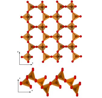
The planar -P4O10 structure is entirely different from the layered o′-P2O5 form previously reported by several experimental studies,Stachel et al. (1995) shown in Fig.4. While both consist of rings of six PO4 tetrahedra, in o′-P2O5 the tetrahedra are organised in alternating rows along the [100]. Further, in profile o′-P2O5 resembles a sawtooth, whereas -P4O10 has the same waved appearence as phosphorene.
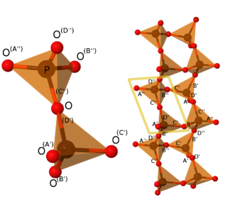
Nearly degenerate in energy with the planar -P4O10 form (Fig.6), we find the tubular -P4O10 structure, shown in Fig.5. The -P4O10 structure is a tubular (1D) form consisting of eight-membered rings, connected to each other at an angle of approximately 100∘ (see for example the angle between the D′ and B′ bonds or D′′ and C′′ bonds in Fig.5), these rings consisting of alternated phosphorus and bridging oxygen atoms. Only inversion symmetry is present, hence the space group is P-1 (C). There are two inequivalent (although very similar) PO4 units, depicted in Fig.5 (left side). The bond lengths and angles are listed in Table 3.
Similar to -P4O10, -P4O10 can be seen as a corner-linked network of PO4 tetrahedra. However, different from polyphosphate chains,Greenwood and Earnshaw (1997) in these tubular forms each tetrahedra shares three vertices, forming a tubular structure rather than a simple chain structure.
For intermediate concentrations, we find two metastable phosphorene oxides in which oxygen is present only on the phosphorene surface, in the form of dangling oxygen atoms. We refer to these as surface forms, and in particular as -P4O2 for the single-surface oxidized, and -P4O4 for the double-surface oxidized; the two structures are shown in Fig.1 panel (c). As a result of oxygen chemisorption, the planes containing the top and the bottom zigzag ridges are not parallel to each other anymore (as in pristine phosphorene). The two P atoms in the same zigzag ridge are shifted from each other by about 0.5 Å in the direction perpendicular to the surface. In contrast with the other planar forms, the lattice deformation in this case is minimal.
III.2 Stability
The stability of the POs can be quantified using the binding energy per oxygen atom, defined as
| (1) |
where is the number of O atoms in the unit cell, , and are the total energies of the PO, the pristine phosphorene, and the O2 (triplet) molecule, respectively. From the definition above, a value of indicates that the oxide formation is energetically favored in the presence of O2. The average binding energies per O atom Eb for the most stable tubular, planar and surface POs calculated with the PBEsol and HSE functionals are shown in Fig.6. The gain in energy due to oxygen chemisorption in phosphorene is very large across the whole concentration range (from 1.8 to 2.9 eV per O atom).
Regardless of the oxygen concentration, we invariably find that the lowest energy structure is a planar (-P4On) phosphorene oxide, with the tubular forms lying slightly higher in energy. In addition, for low and medium oxygen concentrations we find a manifold of states corresponding to different oxygen arrangements that have nearly the same energy. Obviously, for P4O10 the phosphorene lattice is saturated with oxygen, so only two new structures were found (-P4O10 and -P4O10).
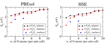
For the lowest concentration considered, =1, the chemisorption of dangling oxygen is favored relative to bridging oxygens; in fact, -P4O1 has a chemisorbed dangling oxygen (Eb=2.1 eV), while on -P4O1 we have a bridging oxygen (Eb=1.4 eV). However, for all O concentrations higher than =1, no clear difference emerges in the energetics of dangling or bridging oxygens. The amount of energy gained after O chemisorption is dictated by the interplay between Coulomb repulsion of localized charges on the phosphorene surface and strain interactions, the former due to dangling oxygens and the latter mainly governed by bridging oxygens. Since bridging oxygens increase the P-P distance by about 0.7 Å (Fig.2b), the insertion of oxygen bridges between neighboring dangling oxygens can substantially reduce their Coulomb repulsion, greatly stabilizing the PO. This explains the counter-intuitive increase in binding energy per oxygen atom with increasing oxygen concentration.
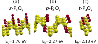
The effect of the reduction of Coulomb repulsion due to the insertion of oxygen bridges is particularly evident for the case =2, shown in Fig.7. Even though for =1 dangling oxygen chemisorption is favored over bridging oxygen chemisorption, when two dangling oxygens are chemisorbed on the same side of the phosphorene surface their strong Coulomb repulsion raises the energy of the compound, resulting in Eb=1.76 eV for the surface form -P4O2 (blue triangle at =2 in Fig.6). This energy can be reduced by forming oxygen bridges instead of dangling oxygen, and therefore there are numerous other -P4O2 and -P4O2 forms containing bridging oxygen that are more stable (up to 1.02 eV and 0.74 eV, respectively) than -P4O2. Similar considerations apply to the double-side surface oxidized form -P4O4, which is 1.8 eV and 1.4 eV higher in energy than the most stable planar and tubular forms, respectively, with the same concentration. From these simple considerations, we can argue that in near-equilibrium conditions the surface forms -P4O2 and -P4O4 will not be the primary outcome of phosphorene oxidation, contrary to what has been recently proposedWang et al. . Nevertheless, these surface oxidized forms can possibly be favored by specific kinetic factors. Still, since the activation energies for dangling oxygen formation and for oxygen insertion (bridges formation) are very similar (0.54 and 0.69 eV, respectively),A. Ziletti and Neto it is unlikely that these surface oxide phases will form even at low temperatures.
We also found that the other surface forms proposed in the literatureDaia and Zenga are not stable at both PBE and PBEsol level, and after lattice relaxation and geometry optimization (with a tight convergence threshold for forces of 10-3 eV/Å) the two forms presented here, -P4O2 and -P4O4, are found.
For the maximum oxygen concentration (=10), the -P4O10 and -P4O10 forms have nearly degenerate energies, with Eb=2.81 eV (2.70 eV) and Eb=2.78 eV (2.70 eV) at the PBEsol (HSE) level, respectively. They are nearly degenerate in energy with the o′ form, which has a binding energy of 2.81 eV with PBEsol (2.68 eV with HSE) in the monolayer form, or 2.88 eV (at the PBEsol level) in the bulk form. They also are the most stable POs among all O concentrations considered in this work. Their structures, which are essentially a homogenous network of three-corner linked PO4 tetrahedra (Fig.3 and Fig.5), minimize both strain interactions due to oxygen bridges and Coulomb repulsion from dangling oxygens.
III.3 Electronic properties
Chemisorption of oxygen atoms and the formation of POs drastically changes the electronic properties of phosphorene. The bandgap Egap of both planar and tubular oxides is plotted as a function of oxygen concentration in Fig.8. Both planar and tubular phosphorene oxides are found to be semiconducting or insulating, depending on the O concentration.
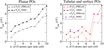
The bandgap of planar POs (black squares) increases monotonically with oxygen concentration, from 2.21 eV () to 7.69 eV () at the HSE level. Note that the bandgaps calculated at the PBEsol level follow almost precisely the same trends, but are simply shifted to smaller gaps compared to HSE by roughly 1.5 eV across the entire O concentration range (see Fig.8).
The increase in bandgap with O concentration in planar POs is due to the increasingly ionic character of the bonds, and the resulting wavefunction localization. The series -P4On (=1,2,3) is a particularly explanatory example of this phenomenon, and it is shown in Fig.9. In -P4O1 one dangling oxygen is chemisorbed, corresponding to a 25% concentration. The band structure is strongly modified compared to pristine phosphorene (shown in Supplemental Material), but the valence band is still delocalized over all the crystal, as shown in Fig.9a, and still exhibits moderate dispersion. The bandgap is now indirect, but its value is still close to pristine phosphorene (2.21 eV vs. 1.77 eV of pristine phosphorene at the HSE level in our calculation). If more oxygen atoms are added, the band structure drastically changes (Fig.9e-9f). In -P4O2, the valence band becomes localized on the top zigzag ridge (Fig.9b). The valence band is now nearly completely flat, and the conduction band generally has less dispersion than that of -P4O1. The bridging oxygen thus effectively creates nanoribbons, and strongly increases the bandgap (3.35 eV with HSE). In -P4O3 (Fig.9c-9f) additional oxygen bridges, intra-ridge and inter-ridge, are formed. The valence band is now localized on P, and both valence and conduction bands have very low dispersion due to their localization. The bandgap is 3.65 eV at the HSE level. For 3, all -PO band structures preserve the low dispersion present in -P4O3, together with an increasing bandgap. Their atomic and electronic band structures are shown in Supplemental Material.
Tubular phosphorene oxides are also insulating, with bandgaps covering a large range of energies from 1.62 eV to 7.78 eV (at HSE level). In contrast to what we have described for planar POs, we did not find that tubular POs exhibit a monotonic increase in bandgap with oxygen concentration (see Fig.8). We can separately consider two regimes, and .
The atomic structure and electronic band structure of some representative tubular POs are shown in Fig.10. The compound -P4O4 (Fig.10a) presents a feature common to all low and medium concentration tubular POs (): both occupied and unoccupied states around the Fermi energy are nearly flat, while the conduction band is dispersive and well-separated from the manifold of occupied (below) and empty states (above). The conduction band in this particular case has 2 eV bandwidth in all directions (-M, M-X and -Y), but it is flat on the X- direction, which is the direction perpendicular to the chain composing this tubular PO; this is the signature of the 1D nature of tubular POs. The strong Coulomb repulsion between dangling oxygens separates the chains, which interact only through (weak) van der Waals forces.
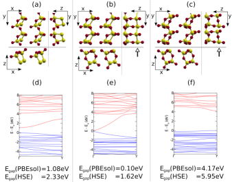
The compound -P4O7 has a similar band structure, but shows increased dispersion of the conduction band (Fig.10b-10e). The conduction band arises from delocalized phosphorus orbitals at the bottom of the PO layer, where no oxygen bridges are present (see right side of Fig.10b). The bandgap is only 1.62 eV (HSE level).
In the high concentration regime (), the scenario is quite different. Chemisorption of a bridging oxygen atom in the phosphorus chain (marked by an arrow in Fig.10b,c) breaks the conjugation, localizing both conduction and valence bands, and greatly increasing Egap. This is particularly evident in -P4O8 (Fig.10c-10f): the conduction band, in contrast to the situation discussed above, is now very close to the manifold of unoccupied states, and presents no dispersion. The bandgap increases by roughly 4 eV at both PBEsol and HSE levels, making the material an insulator. The surface forms -P4O2 and -P4O4 are instead both semiconducting with HSE bandgaps of 2.54 eV and 2.34 eV, respectively.
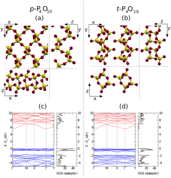
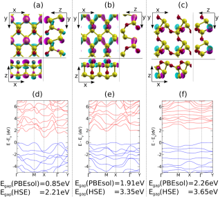
Electronic band structure and density of states (DOS) of the maximally oxidized phosphorene oxides -P4O10 and -P4O10 are shown in Fig.11. Both POs are insulators, and they have remarkably similar band structures. There is a manifold of 8 nearly degenerate dispersion-less occupied states between -1 eV and 0 eV for both species; these states are orbitals localized on dangling oxygens, bridging oxygens or both, and they do not have any appreciable electron density on the P atoms. The conduction band consists instead of orbitals on dangling oxygens and on P-O bridges, and it is slightly dispersive. The bandgap is 5.56 eV for both species at the PBEsol level, enlarged at 7.68 eV for -P4O10 and at 7.78 eV for -P4O10 with the HSE functional. Interestingly, we find that monolayer o′-P2O5 has a similar HSE bandgap (7.45 eV).
For pristine phosphorene and the maximally oxidized phosphorene oxides we calculated the bandgaps also within the GW approximation.Hedin (1965); Hybertsen and Louie (1986) For the pristine phosphorene we obtain 1.70 eV, close to the 1.60 eV of Ref.Rudenko and Katsnelson (2014), but different by 0.3 eV from Tran et al.Tran et al. (2014) We note that all the convergence parameters used in this work are higher than those used in these previous studies (see Supplemental Material for a complete comparison).
For -P4O10 and -P4O10 we obtain GW bandgaps of 8.50 eV and 8.69 eV, respectively, close to the HSE results. The bandgaps for pristine phosphorene and maximal oxidation POs calculated with different levels of theory are summarised in Table 4.
| System | Egap(eV) | ||
|---|---|---|---|
| PBEsol | HSE | GW | |
| Phosphorene | 0.72 | 1.77 | 1.70 |
| -P4O10 | 5.56 | 7.68 | 8.50 |
| -P4O10 | 5.56 | 7.78 | 8.69 |
III.4 Vibrational properties
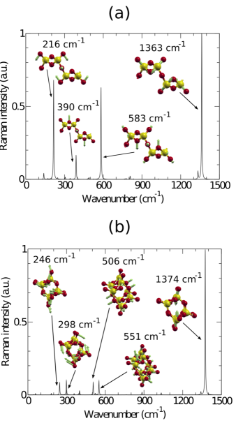
Even though the Raman spectrum of POs changes quite significantly depending on the oxygen concentration, it is possible to identify three distinctive vibrational regions common throughout the whole P4On series. Some of the typical vibrational modes relevant in these different regions are presented in Fig. 12. The low frequency region of the vibrational spectra (300 cm-1) is dominated by bending modes of the P=O groups relative to the rest of the structure, but also includes a small component of P-O-P bending. The presence of P-O-P bending modes increases with frequency, and becomes strongly predominant in the region between 500 and 1000 cm-1. For the surface forms (-P4O2 and -P4O4) in which only dangling oxygens are present, there are indeed no vibrational modes between 500 cm-1 and 1000 cm-1. In the high frequency region, the vibrational spectrum instead only involves P=O stretching modes; these modes are decoupled from the other vibrations because the P=O bonds are much stronger than P-O bridges, as in the case of the molecular (bulk) phosphorus oxidesValentim et al. (1997, 1998). The P=O stretching modes are separated from the other modes by at least 200 cm-1 for all oxygen concentrations, and there are as many as the number of dangling oxygen in the PO. This observation opens the possibility of direct experimental detection of the number of dangling oxygens through Raman spectroscopy. P=O stretching modes start at 1063 cm-1 for =1 (-P4O1) and monotonically blueshift with increasing oxygen concentration till about 1370 cm-1 for maximum oxidation. This is consistent with the shortening of P=O bond found with increasing oxygen linkage, as shown in Table 1 and analogous to the observed blueshift of P=O vibrational frequencies in the series P4Om (=610) measured in solid argonMielke and Andrews (1989).
Raman spectra and Raman active modes of -P4O10 and -P4O10 are shown in Fig.12a and 12b, respectively. In -P4O10, there are three high intensity Raman peaks at 216 cm-1, 583 cm-1 and 1363 cm-1 and a low intensity peak at 390 cm-1. The first peak (216 cm-1) consists of the concerted wag mode of the four P=O dangling oxygens (scissoring), together with an symmetric P-O-P stretch of the two inter-ridge bridges (purple in Fig.3b). The second strong peak at 583 cm-1 is a combination of the P-O-P symmetric stretch of the middle intra-ridge bridges (brown in Fig.3b) and the P=O stretch of all dangling oxygens. The last strong peak (1363 cm-1) involves the stretching of the P=O bonds. The low intensity peak at 390 cm-1 is instead the P-O-P symmetric stretching of the lower and higher intra-ridge bridges (grey in Fig.3b).
In contrast, only one strong peak at 1374 cm-1, corresponding to the P=O stretching of two dangling oxygens, is present in the Raman spectrum of -P4O10, as shown in Fig.12b. Four low intensity peaks, however, appear in the range 250-550 cm-1.
The mode at 246 cm-1 involves P=O bond scissoring with a contribution from an asymmetrical bending of the P-O-P bridges, while the one at 298 cm-1 involves P=O scissoring such that the top and bottom layer dangling oxygen moves towards the tubular oxide center. Finally, at 506 cm-1 we find a ring-breathing mode (P-O-P symmetrical bending of inter-ridge bridges) and at 551 cm-1 another ring breathing mode plus P=O stretching.
Raman and IR spectra for planar and tubular forms for all oxygen concentrations are reported in Supplemental Material.
IV Conclusions
We have described a family of two dimensional phosphorus oxides obtained by oxidation of phosphorene, with oxygen content up to 10 oxygen per phosphorene unit cell (P2O5).
The gain in binding energy due to oxygen chemisorption in phosphorene is very large (from 1.8 to 2.9 eV per O atom), and increases with the oxygen concentration. In parallel with the two-dimensional, planar forms, there are tubular (polymeric) forms with similar formation enthalpy. The planar forms are lower in energy than the tubular forms for all O concentrations; the energy difference is however quite small (typically0.1 eV per O atom), hence both forms are likely to coexist under normal experimental conditions, forming ordered and disordered (amorphous) domains depending on the oxidation process, local impurities and defects. It is also feasible that different forms of POs can be experimentally created by a suitable choice of growth conditions, in much the same way as the (bulk) series P4O6+m (04) can be obtained by varying the atmosphere, temperature and pressure in which P4 is treatedJansen et al. (1981); Greenwood and Earnshaw (1997). Moreover, both planar and tubular forms have very similar formation enthalpy of the thermodynamically most stable known phosphorus pentoxide (o′-P2O5).
The phosphorene oxides can be used as a transparent tunneling materials, namely on black-phosphorus or phosphorene-based devices. The bandgap energy of planar P4On increases with , and is 8.5 eV for . The tubular forms are also insulating, and close to the oxygen saturation limit (P2O5), have nearly the same gap energy, and therefore their coexistence with the planar form does not jeopardize its transparency or electrical insulation properties.
Though the planar and the tubular form of P2O5 give rise to high frequency Raman scattering peaks with very close energies (at 1363 and 1374 cm-1), the planar form can be identified by an intense peak at 583 cm-1, just above the Raman edge of black phosphorus (470 cm-1 at room temperatureSugai and Shirotani (1985)).
Acknowledgments
A.Z. and D.F.C. acknowledge NSF grant CHE-1301157 and also an allocation of computational resources from Boston University’s Office of Information Technology and Scientific Computing and Visualization. A.Z. also acknowledges the support from NSF grant CMMI-1036460, Banco Santander. A.H.C.N., A.C. and P.E.T. acknowledge the National Research Foundation, Prime Minister’s Office, Singapore, under its Medium Sized Centre Programme and CRP award ”Novel 2D materials with tailored properties: beyond graphene” (R-144-000-295-281).
References
- Rodin et al. (2014) S. Rodin, A. A. Carvalho, and H. Castro Neto, A.\̇lx@bibnewblockStrain-induced gap modification in black phosphorus. Phys. Rev. Lett., 112:176801, May 2014. doi: 10.1103/PhysRevLett.112.176801. URL http://link.aps.org/doi/10.1103/PhysRevLett.112.176801.
- Tran et al. (2014) Vy Tran, Ryan Soklaski, Yufeng Liang, and Li Yang. Layer-controlled band gap and anisotropic excitons in few-layer black phosphorus. Phys. Rev. B, 89:235319, Jun 2014. doi: 10.1103/PhysRevB.89.235319. URL http://link.aps.org/doi/10.1103/PhysRevB.89.235319.
- Wei and Peng (2014) Qun Wei and Xihong Peng. Superior mechanical flexibility of phosphorene and few-layer black phosphorus. Applied Physics Letters, 104(25):251915, 2014. doi: http://dx.doi.org/10.1063/1.4885215. URL http://scitation.aip.org/content/aip/journal/apl/104/25/10.1063/1.4885215.
- Koenig et al. (2014) Steven P. Koenig, Rostislav A. Doganov, Hennrik Schmidt, A. H. Castro Neto, and Barbaros Ozyilmaz. Electric field effect in ultrathin black phosphorus. Applied Physics Letters, 104(10):103106, 2014. doi: http://dx.doi.org/10.1063/1.4868132. URL http://scitation.aip.org/content/aip/journal/apl/104/10/10.1063/1.4868132.
- (5) Fengnian Xia, Han Wang, and Yichen Jia. Rediscovering black phosphorus: A unique anisotropic 2d material for optoelectronics and electronics. arXiv:1402.0270.
- Buscema et al. (2014) Michele Buscema, Dirk J. Groenendijk, Sofya I. Blanter, Gary A. Steele, Herre S. J. van der Zant, and Andres Castellanos-Gomez. Fast and broadband photoresponse of few-layer black phosphorus field-effect transistors. Nano Letters, DOI: 10.1021/nl5008085, 2014. doi: 10.1021/nl5008085. URL http://pubs.acs.org/doi/abs/10.1021/nl5008085.
- (7) D. K. Campbell D. F. Coker A. Ziletti, A. Carvalho and A. H. Castro Neto. Oxygen defects in phosphorene. arXiv:1407.5880.
- (8) Rostislav A. Doganov, Eoin C.T. OÕFarrell, Steven P. Koenig, Yuting Yeo, Kenji Watanabe, Takashi Taniguchi, A. Ziletti, A. Carvalho, D.K. Campbell, D. F. Coker, A. H. Castro Neto, and Barbaros Oezyilmaz. unpublished.
- (9) Andres Castellanos-Gomez, Leonardo Vicarelli, Elsa Prada, Joshua O Island, K L Narasimha-Acharya, Sofya I Blanter, Dirk J Groenendijk, Michele Buscema, Gary A Steele, J V Alvarez, Henny W Zandbergen, J J Palacios, and Herre S J van der Zant. Isolation and characterization of few-layer black phosphorus. 2D Materials, 1(2):025001.
- (10) A. Favron, E. Gaufres, F. Fossard, P.L. L’evesque, A-L. Phaneuf-L’Heureux, A. Loiseau N. Y-W. Tang, R. Leonelli, S. Francoeur, and R. Martel. arXiv:1408.0345.
- Yau et al. (1992) Shueh-Lin Yau, Thomas P. Moffat, Allen J. Bard Zhengwei Zhang, and Michael M. Lerner. Chem. Phys. Lett., 198:383, 1992.
- Farnsworth et al. (2014) Tyler W Farnsworth, Rebekah A Wells, Adam H Woomer, Jun Hu, Carrie Donley, and Scott C Warren. Understanding the surface reactivity of 2-d black phosphorus. Informal Phosphore Symposium, 2014.
- Brow (2000) Richard K Brow. Review: the structure of simple phosphate glasses. Journal of Non-Crystalline Solids, 263-264(0):1 – 28, 2000. ISSN 0022-3093. doi: http://dx.doi.org/10.1016/S0022-3093(99)00620-1.
- Jansen (1986) B. Jansen, M.; Luer. Z. Kristallogr., 177(0):149 – 151, 1986.
- Arbib et al. (1996) El Hassan Arbib, Brahim Elouadi, Jean Pierre Chaminade, and Jacques Darriet. {BRIEF} communication: New refinement of the crystal structure of o-p2o5. Journal of Solid State Chemistry, 127(2):350 – 353, 1996. ISSN 0022-4596. doi: http://dx.doi.org/10.1006/jssc.1996.0393. URL http://www.sciencedirect.com/science/article/pii/S002245969690393X.
- Stachel et al. (1995) D. Stachel, I. Svoboda, and H. Fuess. Phosphorus Pentoxide at 233 K. Acta Crystallographica Section C, 51(6):1049–1050, Jun 1995. doi: 10.1107/S0108270194012126. URL http://dx.doi.org/10.1107/S0108270194012126.
- Hulliger (1976) F. Hulliger. Structural Chemistry of Layer-Type Phases. Springer, 1976.
- Galeener and Jr. (1979) F.L. Galeener and J.C. Mikkelsen Jr. The raman spectra and structure of pure vitreous {P2O5}. Solid State Communications, 30(8):505 – 510, 1979. ISSN 0038-1098. doi: http://dx.doi.org/10.1016/0038-1098(79)91227-4. URL http://www.sciencedirect.com/science/article/pii/0038109879912274.
- (19) Gaoxue Wang, Ravindra Pandey, Shashi P. Karna Zhengwei Zhang, and Michael M. Lerner. arXiv:1409.0459.
- Giannozzi et al. (2009) Paolo Giannozzi, Stefano Baroni, Nicola Bonini, Matteo Calandra, Roberto Car, Carlo Cavazzoni, Davide Ceresoli, Guido L Chiarotti, Matteo Cococcioni, Ismaila Dabo, Andrea Dal Corso, Stefano de Gironcoli, Stefano Fabris, Guido Fratesi, Ralph Gebauer, Uwe Gerstmann, Christos Gougoussis, Anton Kokalj, Michele Lazzeri, Layla Martin-Samos, Nicola Marzari, Francesco Mauri, Riccardo Mazzarello, Stefano Paolini, Alfredo Pasquarello, Lorenzo Paulatto, Carlo Sbraccia, Sandro Scandolo, Gabriele Sclauzero, Ari P Seitsonen, Alexander Smogunov, Paolo Umari, and Renata M Wentzcovitch. Quantum espresso: a modular and open-source software project for quantum simulations of materials. Journal of Physics: Condensed Matter, 21(39):395502 (19pp), 2009. URL http://www.quantum-espresso.org.
- Perdew et al. (1996) John P. Perdew, Kieron Burke, and Matthias Ernzerhof. Generalized gradient approximation made simple. Phys. Rev. Lett., 77:3865–3868, Oct 1996. doi: 10.1103/PhysRevLett.77.3865. URL http://link.aps.org/doi/10.1103/PhysRevLett.77.3865.
- Perdew et al. (2008) John P. Perdew, Adrienn Ruzsinszky, Gábor I. Csonka, Oleg A. Vydrov, Gustavo E. Scuseria, Lucian A. Constantin, Xiaolan Zhou, and Kieron Burke. Restoring the density-gradient expansion for exchange in solids and surfaces. Phys. Rev. Lett., 100:136406, Apr 2008. doi: 10.1103/PhysRevLett.100.136406. URL http://link.aps.org/doi/10.1103/PhysRevLett.100.136406.
- Krukau et al. (2006) Aliaksandr V. Krukau, Oleg A. Vydrov, Artur F. Izmaylov, and Gustavo E. Scuseria. Influence of the exchange screening parameter on the performance of screened hybrid functionals. The Journal of Chemical Physics, 125(22):224106, 2006. doi: http://dx.doi.org/10.1063/1.2404663. URL http://scitation.aip.org/content/aip/journal/jcp/125/22/10.1063/1.2404663.
- Heyd et al. (2005) Jochen Heyd, Juan E. Peralta, Gustavo E. Scuseria, and Richard L. Martin. Energy band gaps and lattice parameters evaluated with the heyd-scuseria-ernzerhof screened hybrid functional. The Journal of Chemical Physics, 123(17):174101, 2005. doi: http://dx.doi.org/10.1063/1.2085170. URL http://scitation.aip.org/content/aip/journal/jcp/123/17/10.1063/1.2085170.
- Janesko et al. (2009) Benjamin G. Janesko, Thomas M. Henderson, and Gustavo E. Scuseria. Screened hybrid density functionals for solid-state chemistry and physics. Phys. Chem. Chem. Phys., 11:443–454, 2009. doi: 10.1039/B812838C. URL http://dx.doi.org/10.1039/B812838C.
- Henderson et al. (2011) Thomas M. Henderson, Joachim Paier, and Gustavo E. Scuseria. Accurate treatment of solids with the hse screened hybrid. physica status solidi (b), 248(4):767–774, 2011. ISSN 1521-3951. doi: 10.1002/pssb.201046303. URL http://dx.doi.org/10.1002/pssb.201046303.
- Blöchl (1994) P. E. Blöchl. Projector augmented-wave method. Phys. Rev. B, 50:17953–17979, Dec 1994. doi: 10.1103/PhysRevB.50.17953. URL http://link.aps.org/doi/10.1103/PhysRevB.50.17953.
- Troullier and Martins (1991) N. Troullier and José Luriaas Martins. Efficient pseudopotentials for plane-wave calculations. Phys. Rev. B, 43:1993–2006, Jan 1991. doi: 10.1103/PhysRevB.43.1993. URL http://link.aps.org/doi/10.1103/PhysRevB.43.1993.
- Baroni et al. (2001) Stefano Baroni, Stefano de Gironcoli, Andrea Dal Corso, and Paolo Giannozzi. Phonons and related crystal properties from density-functional perturbation theory. Rev. Mod. Phys., 73:515–562, Jul 2001. doi: 10.1103/RevModPhys.73.515. URL http://link.aps.org/doi/10.1103/RevModPhys.73.515.
- Monkhorst and Pack (1976) Hendrik J. Monkhorst and James D. Pack. Special points for brillouin-zone integrations. Phys. Rev. B, 13:5188–5192, Jun 1976. doi: 10.1103/PhysRevB.13.5188. URL http://link.aps.org/doi/10.1103/PhysRevB.13.5188.
- Blöchl et al. (1994) Peter E. Blöchl, O. Jepsen, and O. K. Andersen. Improved tetrahedron method for brillouin-zone integrations. Phys. Rev. B, 49:16223–16233, Jun 1994. doi: 10.1103/PhysRevB.49.16223. URL http://link.aps.org/doi/10.1103/PhysRevB.49.16223.
- Perdew et al. (1982) John P. Perdew, Robert G. Parr, Mel Levy, and Jose L. Balduz. Density-functional theory for fractional particle number: Derivative discontinuities of the energy. Phys. Rev. Lett., 49:1691–1694, Dec 1982. doi: 10.1103/PhysRevLett.49.1691. URL http://link.aps.org/doi/10.1103/PhysRevLett.49.1691.
- Perdew and Levy (1983) John P. Perdew and Mel Levy. Physical content of the exact kohn-sham orbital energies: Band gaps and derivative discontinuities. Phys. Rev. Lett., 51:1884–1887, Nov 1983. doi: 10.1103/PhysRevLett.51.1884. URL http://link.aps.org/doi/10.1103/PhysRevLett.51.1884.
- Hedin (1965) Lars Hedin. New method for calculating the one-particle green’s function with application to the electron-gas problem. Phys. Rev., 139:A796–A823, Aug 1965. doi: 10.1103/PhysRev.139.A796. URL http://link.aps.org/doi/10.1103/PhysRev.139.A796.
- Hybertsen and Louie (1986) Mark S. Hybertsen and Steven G. Louie. Electron correlation in semiconductors and insulators: Band gaps and quasiparticle energies. Phys. Rev. B, 34:5390–5413, Oct 1986. doi: 10.1103/PhysRevB.34.5390. URL http://link.aps.org/doi/10.1103/PhysRevB.34.5390.
- (36) STEVEN G. LOUIE. FIRST-PRINCIPLES THEORY OF ELECTRON EXCITATION ENERGIES IN SOLIDS, SURFACES, AND DEFECTS, chapter 3, pages 96–142. doi: 10.1142/9789812817006˙0003. URL http://www.worldscientific.com/doi/abs/10.1142/9789812817006_0003.
- Malone and Cohen (2013) Brad D Malone and Marvin L Cohen. Quasiparticle semiconductor band structures including spin-orbit interactions. Journal of Physics: Condensed Matter, 25(10):105503, 2013. URL http://stacks.iop.org/0953-8984/25/i=10/a=105503.
- Ismail-Beigi (2006) Sohrab Ismail-Beigi. Truncation of periodic image interactions for confined systems. Phys. Rev. B, 73:233103, Jun 2006. doi: 10.1103/PhysRevB.73.233103. URL http://link.aps.org/doi/10.1103/PhysRevB.73.233103.
- Gonze et al. (2009) X. Gonze, B. Amadon, P.M. Anglade, J.M. Beuken, F. Bottin, P. Boulanger, F. Bruneval, D. Caliste, R. Caracas, M. Cote, T. Deutsch, L. Genovese, Ph. Ghosez, M. Giantomassi, S. Goedecker, D.R. Hamann, P. Hermet, F. Jollet, G. Jomard, S. Leroux, M. Mancini, S. Mazevet, M.J.T. Oliveira, G. Onida, Y. Pouillon, T. Rangel, G.M. Rignanese, D. Sangalli, R. Shaltaf, M. Torrent, M.J. Verstraete, G. Zerah, and J.W. Zwanziger. Abinit: First-principles approach to material and nanosystem properties. Computer Physics Communications, 180(12):2582 – 2615, 2009. ISSN 0010-4655. doi: http://dx.doi.org/10.1016/j.cpc.2009.07.007. URL http://www.sciencedirect.com/science/article/pii/S0010465509002276. 40 {YEARS} {OF} CPC: A celebratory issue focused on quality software for high performance, grid and novel computing architectures.
- Greenwood and Earnshaw (1997) N N. Greenwood and A Earnshaw. Chemistry of the Elements. Reed Elsevier, 1997.
- Jansen et al. (1981) Martin Jansen, Marlen Voss, and Hans-Jorg Deiseroth. Structural properties of phosphorus oxides in the solid aggregation state. Angewandte Chemie International Edition in English, 20(11):965–965, 1981. ISSN 1521-3773. doi: 10.1002/anie.198109651. URL http://dx.doi.org/10.1002/anie.198109651.
- Walker et al. (1979) Michael L. Walker, Daniel E. Peckenpaugh, and Jerry L. Mills. Mixed phosphorus(iii)-phosphorus(v) oxide chalcogenides. Inorganic Chemistry, 18(10):2792–2796, 1979. doi: 10.1021/ic50200a032. URL http://pubs.acs.org/doi/abs/10.1021/ic50200a032.
- Clade et al. (1994) J. Clade, F. Frick, and M. Jansen. Recent synthetic, structural, spectroscopic, and theoretical studies on molecular phosphorus oxides and oxide sulfides. 41:327 – 388, 1994. ISSN 0898-8838. doi: http://dx.doi.org/10.1016/S0898-8838(08)60175-0. URL http://www.sciencedirect.com/science/article/pii/S0898883808601750.
- Gillespie and Nyholm (1957) R. J. Gillespie and R. S. Nyholm. Inorganic stereochemistry. Q. Rev. Chem. Soc., 11:339–380, 1957. doi: 10.1039/QR9571100339. URL http://dx.doi.org/10.1039/QR9571100339.
- Gillespie (1963) R. J. Gillespie. The valence-shell electron-pair repulsion (vsepr) theory of directed valency. Journal of Chemical Education, 40(6):295, 1963. doi: 10.1021/ed040p295. URL http://pubs.acs.org/doi/abs/10.1021/ed040p295.
- Gillespie (1992) Ronald J. Gillespie. The vsepr model revisited. Chem. Soc. Rev., 21:59–69, 1992. doi: 10.1039/CS9922100059. URL http://dx.doi.org/10.1039/CS9922100059.
- (47) Jun Daia and Xiao Cheng Zenga. Structure and stability of two dimensional phosphorene with =o or =nh functionalization. arXiv:1409.7719.
- Rudenko and Katsnelson (2014) A. N. Rudenko and M. I. Katsnelson. Quasiparticle band structure and tight-binding model for single- and bilayer black phosphorus. Phys. Rev. B, 89:201408, May 2014. doi: 10.1103/PhysRevB.89.201408. URL http://link.aps.org/doi/10.1103/PhysRevB.89.201408.
- Valentim et al. (1997) A. R. S. Valentim, B. Engels, S. Peyerimhoff, J. Clade, and M. Jansen. Study of the p4o7, p4o6s, and p4o6se vibrational spectra. Inorganic Chemistry, 36(11):2451–2457, 1997. doi: 10.1021/ic961202s. PMID: 11669885.
- Valentim et al. (1998) A. R. S. Valentim, B. Engels, S. D. Peyerimhoff, J. Clade, and M. Jansen. A comparative study of the bonding character in the p4on (n = 6?10) series by means of a vibrational analysis. The Journal of Physical Chemistry A, 102(21):3690–3696, 1998. doi: 10.1021/jp9805611.
- Mielke and Andrews (1989) Zofia Mielke and Lester Andrews. Infrared spectra of phosphorus oxides (p4o6, p4o7, p4o8, p4o9 and p4o10) in solid argon. The Journal of Physical Chemistry, 93(8):2971–2976, 1989. doi: 10.1021/j100345a024.
- Sugai and Shirotani (1985) S. Sugai and I. Shirotani. Raman and infrared reflection spectroscopy in black phosphorus. Solid State Communications, 53(9):753 – 755, 1985. ISSN 0038-1098. doi: http://dx.doi.org/10.1016/0038-1098(85)90213-3. URL http://www.sciencedirect.com/science/article/pii/0038109885902133.
- Galeener et al. (1978) F. L. Galeener, J. C. Mikkelsen, R. H. Geils, and W. J. Mosby. The relative raman cross sections of vitreous sio2, geo2, b2o3, and p2o5. Applied Physics Letters, 32(1), 1978.
- Brazhkin et al. (2011) V. V. Brazhkin, J. Akola, Y. Katayama, S. Kohara, M. V. Kondrin, A. G. Lyapin, S. G. Lyapin, G. Tricot, and O. F. Yagafarov. Densified low-hygroscopic form of p2o5 glass. J. Mater. Chem., 21:10442–10447, 2011. doi: 10.1039/C1JM10889A. URL http://dx.doi.org/10.1039/C1JM10889A.
- Gilheany (1994) Declan G. Gilheany. Ylides, phosphoniumno d orbitals but walsh diagrams and maybe banana bonds: Chemical bonding in phosphines, phosphine oxides, and phosphonium ylides. Chemical Reviews, 94(5):1339–1374, 1994. doi: 10.1021/cr00029a008. URL http://pubs.acs.org/doi/abs/10.1021/cr00029a008.
- Rai and Symons (1994) Uma S. Rai and Martyn C. R. Symons. Epr data do not support the p[double bond, length as m-dash]o representation for trialkyl phosphates and phosphine oxides or sulfides. J. Chem. Soc., Faraday Trans., 90:2649–2652, 1994. doi: 10.1039/FT9949002649. URL http://dx.doi.org/10.1039/FT9949002649.
- Chesnut and Savin (1999) D. B. Chesnut and A. Savin. The electron localization function (elf) description of the po bond in phosphine oxide. Journal of the American Chemical Society, 121(10):2335–2336, 1999. doi: 10.1021/ja984314m. URL http://pubs.acs.org/doi/abs/10.1021/ja984314m.