Permanent address: ]Universidad Rey Juan Carlos, Escuela Superior de Ciencias Experimentales y Tecnología, Madrid 28933, Spain
Epitaxial growth of Bi2Pt2O7 pyrochlore
Certain pyrochlore oxidesHorowitz, Longo, and Lewandowski (1981) are among the best oxygen catalysts in alkaline media.Horowitz, Longo, and Horowitz (1983); Tuller (1992); Subramanian, Aravamudan, and Rao (1983); Christensen, Hamnett, and Linares-Moya (2011); Boivin and Mairesse (1998); Wuensch et al. (2000); Kahoul et al. (2001); Konishi et al. (2009) Hence, exploring epitaxial films of these materials is of great fundamental and technological interest. Unfortunately, direct film growth of one of the most promising pyrochlores, Bi2Pt2O7,Beck et al. (2006) has not yet been achieved, owing to the difficulty of oxidizing platinum metal in the precursor material to Pt4+. In this work, in order to induce oxidation of the platinum, we annealed pulsed laser deposited films consisting of epitaxial –Bi2O3 and co–deposited, comparatively disordered platinum. We present synchrotron x–ray diffraction results that show the annealed films are the first epitaxial crystals of Bi2Pt2O7. We also visualized the pyrochlore structure by scanning transmission electron microscopy, and observed ordered cation vacancies in a bismuth–rich film but not in a platinum–rich film. The similarity between the –Bi2O3 and Bi2Pt2O7 structures appears to facilitate the pyrochlore formation. These results constitute a new approach for synthesis of novel pyrochlore thin film oxygen catalysts.
Pyrochlore oxides, A2B2O7,Subramanian, Aravamudan, and Rao (1983) that exhibit catalytic activity are prospective cathode materials for fuel cells. Furthermore, the oxygen ion conductivity of some pyrochlores is comparable to that of oxides with fluorite structure, such as yttria–stabilized zirconia.Tuller (1992); Boivin and Mairesse (1998); Wuensch et al. (2000) In this regard, one of the most promising pyrochlores for catalytic applications is bismuth platinum oxide, Bi2Pt2O7. Previous work on powders of Bi2Pt2O7 found its electrochemical activity essentially identical to pure Pt.Beck et al. (2006) Furthermore, compared to bulk powders, enhanced catalytic activity has been observed in epitaxially grown perovskite oxides and structural derivatives.la O et al. (2010); Liu et al. (2012); Jeen et al. (2013); Kubicek et al. (2013) These results have contributed to growing interest in epitaxial films of pyrochlore oxides as an oxygen catalyst. However, while nanocrystalline powders of Bi2Pt2O7 by thermal oxidation of Bi–Pt nanoparticles has been reported,Dawood, Leonard, and Schaak (2007) epitaxial crystals of Bi2Pt2O7 have not yet been achieved.
Here we report the first, successful formation of epitaxial Bi2Pt2O7 (111) on YSZ(111) single–crystal substrates. Our synthesis strategy consists of a deposition step followed by a post-growth anneal. During growth, epitaxial Bi2O3 is formed in its cubic phase, while co-deposited platinum forms an as-yet undetermined state, exhibiting only minute amounts of metallic platinum in specular x–ray diffraction (XRD). Annealing in air transforms the –Bi2O3 into epitaxial crystals of Bi2Pt2O7 pyrochlore.
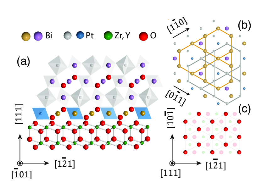
Fig.1 displays the side (a), and top (b, c) views of the structural model of Bi2Pt2O7(111)/YSZ(111). Bi2Pt2O7 has a lattice parameter approximately twice that of YSZ, but consists of eight nearly identical fluorite subunits with intrinsic oxygen vacancies on 1/8 of the oxygen sites. The misfit between these subunits and the YSZ substrate is 0.8% (from our results, =5.147 Å, for YSZ; and, =10.371 Å for Bi2Pt2O7 in bulkBeck and Kemmlersack (1987) form).
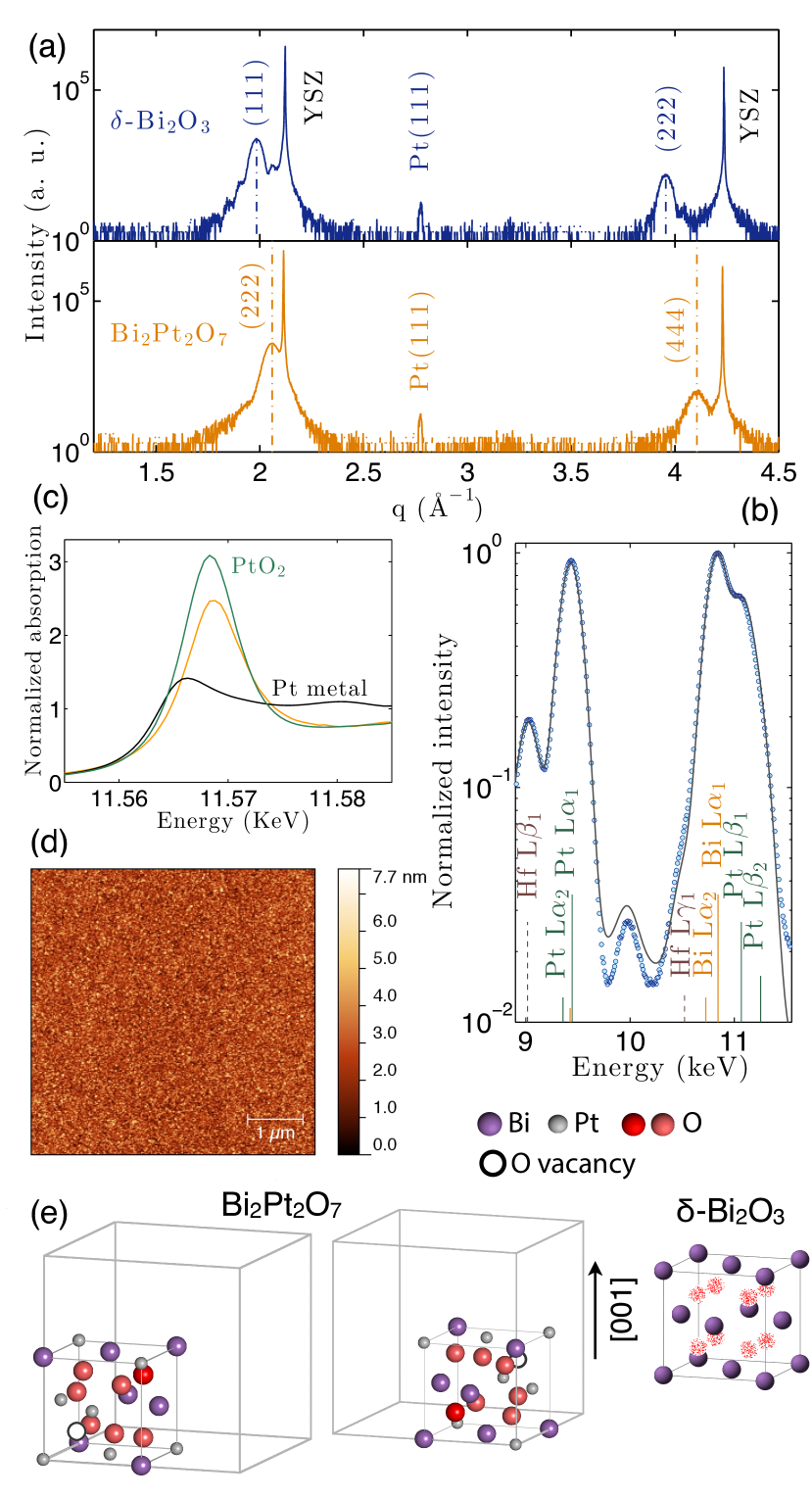
We studied the out–of–plane orientation of the films by XRD. Fig.2 (a) shows – scans of an as–deposited film (blue pattern). The growth was carried out at an oxygen pressure of 10-4 Torr, a substrate temperature of 640∘ C, and a laser fluence of 3 J/cm2. The as–grown pattern reveals intense peaks that can be assigned to (111), (222) and (333) (not shown) cubic –Bi2O3. Distinct thickness fringes are observed surrounding the (111) Bragg peak of –Bi2O3 in the as–grown films (Supplementary Information, Fig. S1) which give evidence of a smooth film surface. Rocking curve measurements for the (111)–Bi2O3 peak (Supplementary Information, Fig. S2) show a FWHM of 0.1000.001∘ in omega (rocking curve on YSZ substrates has a nominal FWHM of 0.014∘ in omega). The measured out–of–plane lattice parameter for –Bi2O3 is 5.519 Å, in good agreement with previous reported values.Switzer, Shumsky, and Bohannan (1999); Proffit et al. (2010)
Although the cubic phase of Bi2O3 in bulk form is stable only from 729∘ C up to its melting point at 825∘ C and transforms to other phases upon cooling, it has previously been stabilized at room temperature on Au substrates by electrodeposition,Switzer, Shumsky, and Bohannan (1999) on polycrystalline YSZ substrates by atmospheric pressure chemical vapour deposition,Takeyama et al. (2006) and as –Bi2O3 nanostructures on perovskite substrates.Proffit et al. (2010)
The weak diffraction peak at =2.77 in Fig.2 (a, blue pattern) is consistent with that of the (111) plane of a trace amount of platinum metal with an fcc structure. Apart from the small amount of (111)–oriented platinum, these data are in agreement with several possibilities, including either amorphous platinum, 1 nm platinum nanocrystals that diffract similarly weakly to amorphous platinum, or platinum that is somehow incorporated into the –Bi2O3 lattice.
The phase segregation of the platinum metal and Bi2O3 is a major difficulty of working with this system. Like the as–deposited film, the PLD target exhibits phase separation of the platinum from the bismuth oxide. The precursor material exhibits the pyrochlore phase (Bi3+, Pt4+) when prepared by solid state reaction at 650∘C from stoichiometric mixtures of Bi2O3 and platinum metal (Supplementary Information, Fig. S3). However, as expected, targets sintered at this temperature are not dense enough for proper ablation. Sintering temperatures higher than 650∘C are required to synthesize high–density PLD targets, but at these temperatures Pt4+ reduces to platinum metal. Thus, PLD targets sintered at 820∘ C (slightly below the melting point of Bi2O3) contain platinum and Bi2O3 in its monoclinic phase (Supplementary Information, Fig. S3). Consequently, platinum metal has to oxidize to Pt4+ so that the pyrochlore phase can form on the substrate. Furthermore, control of the Bi/Pt cation stoichiometry of the film becomes complicated due to the high volatility of the bismuth and the large difference between the melting temperatures of the two components of the target (Bi2O3, 825∘ C; Pt, 1770∘ C). Perovskite Bi–based films with the correct stoichiometry have been grown by PLD from bismuth–rich targetsHavelia et al. (2009); Shelke et al. (2009) as well as from stoichiometric targets.You et al. (2009); Kim et al. (2014) There is no previous work on epitaxial films of Bi2Pt2O7. We used in situ x–ray fluorescence (XRF) at grazing incidence to tune the deposition parameters while keeping track of the Bi/Pt ratio of the films. We chose to use a stoichiometric target, and compensate for the preferential ablation of bismuth that we observed in this system by working at a pressure of 10-4 Torr, lower than the bismuth vapor pressure at 640∘ C (bismuth vapor pressure is 10-4 Torr at 517∘ C; 10-2 Torr at 672∘ C). This leads to the sublimation of a portion of the deposited bismuth. Fig.2 (b) displays the XRF spectrum for the film shown in panel (a) measured in situ once the substrate temperature was cooled down. The quantitative analysis performed by a custom fitting procedure which employs the Elam databaseElam, Ravel, and Sieber (2002) provides an estimate Bi/Pt ratio of 0.88 0.01 within a 95% confidence level.
In order to induce oxidation of the platinum, the film shown in Fig.2 (a, blue scan) was annealed in a tube furnace in air at 640∘ C for 8 h. Fig.2 (a, orange scan) displays the – scan of the film after annealing. This pattern shows two new peaks that can be attributed to (222) and (444) pyrochlore with an out–of–plane lattice parameter of 10.57 0.02 Å. The reflections assigned in the as–grown film to planes of –Bi2O3 have disappeared while the weak peak attributed to some trace of (111) platinum metal is still observable. These results support the hypothesis that the Pt(111) peak in the XRD scan of Fig.2 (a, blue pattern) cannot fully account for all the deposited platinum in the film. Rocking curve measurements for the (222)Bi2Pt2O7 peak (Supplementary Information, Fig. S4) reveal a FWHM of 0.0980.002∘ in omega, comparable to that of the (111)–Bi2O3 peak (Supplementary Information, Fig. S2).
We investigated the oxidation state of platinum in the annealed films. Fig.2(c) shows Pt L3 edge X–ray absorption near edge structure (XANES) spectrum (orange) of a bismuth–rich annealed film (Bi/Pt=1.620.04) with pyrochlore phase (Supplementary Information, Fig. S5 and S6). Fig.2(c) also displays XANES spectra for a platinum metal foil (black), and PtO2 (green) used as reference standards. The higher threshold energy () of the film in relation to that of the platinum reference foil is consistent with a higher oxidation state in the annealed film, most probably Pt4+. The intense peak, referred to as white line, observed in the spectrum of the film (Fig.2(c), orange) is indicative of a metal oxide. It is thus clear that platinum oxidized during the post–growth anneal.
The smoothness of the surface of the annealed film in Fig.2(a),(b) was analyzed by atomic force microscopy (AFM). The AFM image shown in Fig.2(d) reveals a rms roughness of 0.84 nm. It was grown on a YSZ (111) stepped surface with atomically flat terraces (Supplementary Information, Fig. S7).
The structural models shown in Fig.2(e) reveal striking similarity between the cubic –Bi2O3 and the pyrochlore structures. Both are based on an ordered oxygen deficient fluorite structure. Bi3+ cations in –Bi2O3, as well as both Bi3+ and Pt4+ in the pyrochlore structure form an fcc lattice. This strongly suggests the stabilization of the phase of Bi2O3 is essential for the formation of the Bi2Pt2O7 pyrochlore phase during the ex–situ post–growth anneal. In fact, films that contained the monoclinic () phase of Bi2O3 still exhibit –Bi2O3 as the majority phase after annealing, (Supplementary Information, Fig.S8).
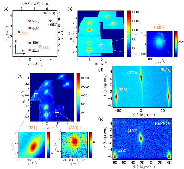
Remarkably, the pyrochlore phase is epitaxial. Along with high intensity peaks characteristic of the parent fluorite structure (, , and are all multiples of four), we observed weak pyrochlore superstructure peaks (their intensity is expected to be three orders of magnitude lower), such as () and ().
The expected positions for the reflections of the epitaxial Bi2Pt2O7/YSZ system are depicted in Fig.3(a). Peaks from the YSZ substrate with a fluorite lattice are also labeled. Area scans at grazing incidence around asymmetrical reflections of the Bi2Pt2O7 phase were transformed into reciprocal space maps (RSM). They are plotted in Fig.3(b) and (c) for a bismuth–rich film and for a slightly platinum–rich film, respectively. –Bi2O3 still present in the bismuth–rich film shows up in the RSM of the Bi2Pt2O7, revealing again the similarity between the fluorite and pyrochlore structures (Supplementary Information, Fig.S9). Superstructure peaks indicative of the pyrochlore phase are highlighted.
In–plane () and out–of–plane () lattice constants of the epitaxial Bi2Pt2O7, along and directions, respectively, determined from the RSMs are = 10.460.03 and =10.4230.006 , for the film in Fig.3(b); and, = 10.530.08 and = 10.530.04 (in accordance, within the error, with the value obtained from the –2 scan in Fig.2(a)) for the film in Fig.3(c). These figures suggest the pyrochlore is relaxed with respect to the substrate lattice.
Azimuthal scans carried out using synchrotron x–ray radiation at grazing incidence on the (200) peak of –Bi2O3 of an as–grown film, Fig.3(d), and on the peak of Bi2Pt2O7 in an annealed film, Fig.3(e), confirm the epitaxial nature of the films with an in–plane orientation which gives rise to peaks with threefold symmetry. The FWHM of (200) peaks of –Bi2O3 is 0.50∘0.01, and that of the peaks separated 120∘ in the scan is 0.57∘0.02. In–plane orientation relationships of either or , rotated 60∘ respect to each other, can be built with the structural model shown in Fig.1(b),(c). However, no twin domains were observed in our films.
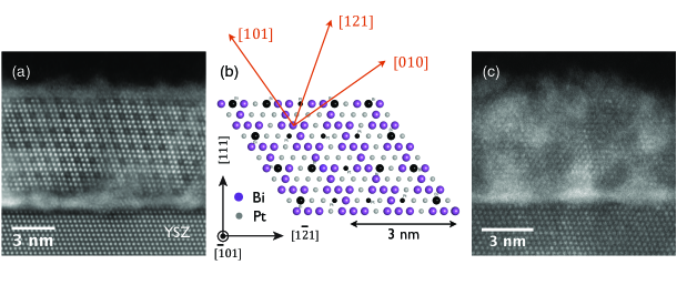
Fig.4 shows cross–sectional high–angle annular dark field (HAADF) scanning TEM images for the films whose RSMs are displayed in Fig.3(b), (c). These studies reveal 100 nm long regions of ordered epitaxial pyrochlore. A wider field of view scanning TEM images showing non–pyrochlore regions are shown in Supplementary Information, Fig.S10. Fig.4(a) exhibits the expected columns of a pyrochlore structure with cation ordered vacancies (no noticeable contrast in the HAADF is expected between Bi and Pt columns). The presence of vacancies in this case can be due to the deviation from the correct stoichiometry of the film composition, Bi/Pt=1.620.04. The platinum–rich film shown in Fig.3(c) with a Bi/Pt ratio of 0.880.01 does not contain cation vacancies in its pyrochlore structure, Fig.4(c).
In conclusion, these results provide a novel route for the formation of epitaxial Bi2Pt2O7 pyrochlore, thought to be one of the most efficient oxide catalysts. Upcoming work includes the investigation of the oxygen reduction activity of the (111) surface of Bi2Pt2O7, and its comparison with powder pellets. However, the difficulty of controlling Bi/Pt stoichiometry suggests that independent control of volatile bismuth and non–volatile platinum sources may be required for routine growth of pristine films.
References
- Horowitz, Longo, and Lewandowski (1981) H. Horowitz, J. Longo, and J. Lewandowski, “New oxide pyrochlores: A2[B2-xAx]O7-y (A : Pb, Bi; B = Ru, Ir),” Materials Research Bulletin 16, 489–496 (1981).
- Horowitz, Longo, and Horowitz (1983) H. S. Horowitz, J. M. Longo, and H. H. Horowitz, “Oxygen electrocatalysis on some oxide pyrochlores,” Journal of the Electrochemical Society 130, 1851–1859 (1983).
- Tuller (1992) H. L. Tuller, “Mixed ionic–electronic conduction in a number of fluorite and pyrochlore compounds,” Solid State Ionics 52, 135–146 (1992).
- Subramanian, Aravamudan, and Rao (1983) M. A. Subramanian, G. Aravamudan, and G. V. S. Rao, “Oxide pyrochlores – a review,” Progress in Solid State Chemistry 15, 55–143 (1983).
- Christensen, Hamnett, and Linares-Moya (2011) P. A. Christensen, A. Hamnett, and D. Linares-Moya, “Oxygen reduction and fuel oxidation in alkaline solution,” Physical Chemistry Chemical Physics 13, 5206–5214 (2011).
- Boivin and Mairesse (1998) J. C. Boivin and G. Mairesse, “Recent material developments in fast oxide ion conductors,” Chemistry of materials 10, 2870–2888 (1998).
- Wuensch et al. (2000) B. J. Wuensch, K. W. Eberman, C. Heremans, E. M. Ku, P. Onnerud, E. M. Yeo, S. M. Haile, J. K. Stalick, and J. D. Jorgensen, “Connection between oxygen-ion conductivity of pyrochlore fuel-cell materials and structural change with composition and temperature,” Solid State Ionics 129, 111–133 (2000).
- Kahoul et al. (2001) A. Kahoul, P. Nkeng, A. Hammouche, F. Nâmoune, and G. Poillerat, “A sol–gel route for the synthesis of Bi2Ru2O7 pyrochlore oxide for oxygen reaction in alkaline medium,” Journal of Solid State Chemistry 161, 379–384 (2001).
- Konishi et al. (2009) T. Konishi, H. Kawai, M. Saito, J. Kuwano, H. Shiroishi, T. Okumura, and Y. Uchimoto, “Electrocatalytic activity of the pyrochlores Ln2M2O7-δ (Ln = Lanthanoids) for oxygen reduction reaction,” Topics in Catalysis 52, 896–902 (2009).
- Beck et al. (2006) N. K. Beck, B. Steiger, G. G. Scherer, and A. Wokaun, “Methanol tolerant oxygen reduction catalysts derived from electrochemically pre–treated Bi2Pt2±yIryO7 pyrochlores,” Fuel Cells 6, 26–30 (2006).
- la O et al. (2010) G. J. la O, S. J. Ahn, E. Crumlin, Y. Orikasa, M. D. Biegalski, H. M. Christen, and Y. Shao-Horn, “Catalytic activity enhancement for oxygen reduction on epitaxial perovskite thin films for solid-oxide fuel cells,” Angewandte Chemie-International Edition 49, 5344–5347 (2010).
- Liu et al. (2012) J. Liu, G. Collins, M. Liu, C. Chen, J. He, J. Jiang, and E. I. Meletis, “Ultrafast oxygen exchange kinetics on highly epitaxial PrBaCo2O5+δ thin films,” Applied Physics Letters 100 (2012).
- Jeen et al. (2013) H. Jeen, Z. Bi, W. S. Choi, M. F. Chisholm, C. A. Bridges, M. P. Paranthaman, and H. N. Lee, “Orienting oxygen vacancies for fast catalytic reaction,” Advanced Materials 25, 6459–6463 (2013).
- Kubicek et al. (2013) M. Kubicek, Z. Cai, W. Ma, B. Yildiz, H. Hutter, and J. Fleig, “Tensile lattice strain accelerates oxygen surface exchange and diffusion in La1-xSrxCoO3-δ thin films,” ACS Nano 7, 3276–3286 (2013).
- Dawood, Leonard, and Schaak (2007) F. Dawood, B. M. Leonard, and R. E. Schaak, “Oxidative transformation of intermetallic nanoparticles: An alternative pathway to metal/oxide nanocomposites, textured ceramics, and nanocrystalline multimetal oxides,” Chemistry of Materials 19, 4545–4550 (2007).
- Beck and Kemmlersack (1987) E. Beck and S. Kemmlersack, “The semiconductor-metal transition in bismuth pyrochlores of the system Bi2Pt2-yIryO7,” Journal of the Less–Common Metals 135, 257–268 (1987).
- Ballabio et al. (2004) G. Ballabio, M. Bernasconi, F. Pietrucci, and S. Serra, “Ab initio study of yttria–stabilized cubic zirconia surfaces,” Physical Review B 70, 075417 (2004).
- Switzer, Shumsky, and Bohannan (1999) J. Switzer, M. G. Shumsky, and E. Bohannan, “Electrodeposited ceramic single crystals,” Science 5412, 293–296 (1999).
- Proffit et al. (2010) D. L. Proffit, G.-R. Bai, D. D. Fong, T. T. Fister, S. O. Hruszkewycz, M. J. Highland, P. M. Baldo, P. H. Fuoss, T. O. Mason, and J. A. Eastman, “Phase stabilization of –Bi2O3 nanostructures by epitaxial growth onto single crystal SrTiO3 or DyScO3 substrates,” Applied Physics Letters 96, 021905 (2010).
- Battle et al. (1986) P. D. Battle, C. R. A. Catlow, J. W. Heap, and L. M. Moroney, “Structural and dynamic studies of –Bi2O3 oxide ion conductors,” Journal of Solid State Chemistry 63, 8–15 (1986).
- Yashima and Ishimura (2003) M. Yashima and D. Ishimura, “Crystal structure and disorder of the fast oxide-ion conductor cubic Bi2O3,” Chemical Physics Letters 378, 395–399 (2003).
- Mohn et al. (2009) C. E. Mohn, S. Stolen, S. T. Norberg, and S. Hull, “Oxide–ion disorder within the high temperature phase of Bi2O3,” Physical Review Letters 102 (2009).
- Takeyama et al. (2006) T. Takeyama, N. Takahashib, T. Nakamurab, and S. Itoh, “–Bi2O3 thin films deposited on dense YSZ substrates by CVD method under atmospheric pressure for intermediate temperature SOFC applications,” Surface & Coatings Technology 200, 4797–4801 (2006).
- Havelia et al. (2009) S. Havelia, S. Wang, M. Skowronski, and P. A. Salvador, “Controlling the Bi content, phase formation, and epitaxial nature of BiMnO3 thin films fabricated using conventional pulsed laser deposition, hybrid pulsed laser deposition, and solid state epitaxy,” Journal of Applied Physics 112 (2009).
- Shelke et al. (2009) V. Shelke, V. N. Harshan, S. Kotru, and A. Gupta, “Effect of kinetic growth parameters on leakage current and ferroelectric behavior of BiFeO3 thin films,” Journal of Applied Physics 106 (2009).
- You et al. (2009) L. You, N. T. Chu, K. Yao, L. Chen, and J. Wang, “Influence of oxygen pressure on the ferroelectric properties of epitaxial BiFeO3 thin films by pulsed laser deposition,” Physical Review B 80 (2009).
- Kim et al. (2014) Y. Kim, A. Morozovska, E. Eliseev, M. P. Oxley, R. Mishra, S. M. Selbach, T. Grande, S. T. Pantelides, S. V. Kalinin, and A. Y. Borisevich, “Direct observation of ferroelectric field effect and vacancy–controlled screening at the BiFeO3/LaxSr1-xMnO3 interface,” Nature Materials 13, 879–883 (2014).
- Elam, Ravel, and Sieber (2002) W. Elam, B. Ravel, and J. Sieber, “A new atomic database for x–ray spectroscopic calculations,” Radiation Physics and Chemistry 63, 121–128 (2002).
- Ravel and Newville (2005) B. Ravel and M. Newville, “ATHENA, ARTEMIS, HEPHAESTUS: data analysis for X–ray absorption spectroscopy using IFEFFIT,” Journal of Synchrotron Radiation 12, 537–541 (2005).
Methods
We sintered the PLD target from a mixture of Bi2O3 (99.999%) and Pt powder (20 m, 99.97%) with controlled cation stoichiometry (Bi/Pt=1) in a solid–state reaction. These mixtures were ground in an agate mortar, pressed into pellets, and heated in a box furnace at 800∘C for 3 h. Then, the pellets were ground and sintered again at 650∘C for 48 h. This annealing at 650∘C for 48 h was repeated five times, with intermediate grinding and pressing. In order to get high-density PLD targets, the powders were reground and repressed, and then fired at 820∘C for further 24 h. X–ray diffraction patterns (Rigaku SmartLab, Cu source) were taken on the intermediate pellets and on the final targets. Their cation stoichiometry was measured by x–ray fluorescence. Polished cross-sections of the targets were studied by high resolution scanning electron microscopy and energy dispersive X–ray spectroscopy (LEO 1550 Field Emission SEM).
Films were grown in the in situ PLD system housed at the G3 hutch of Cornell High Energy Synchrotron Source (CHESS’s) which utilizes a KrF excimer laser (248 nm) focused onto the target. During the growth, oxygen was supplied in the PLD chamber yielding a background pressure up to 10-1 Torr. The substrate temperature, monitored by an optical pyrometer ( = 4.8–5.3 m, = 0.8) and a thermocouple, was set in the range 450 ∘C – 740 ∘C. Single-crystals 8 mol % Y2O3–stabilized ZrO2 ((ZrO2)0.92 (Y2O3)0.08), YSZ) with (111) orientation (MTI Corporation) were used as substrates. Prior to the growth, they were annealed at 1300∘ C for 3 h in air. Atomic Force Microscopy (AFM, Veeco Dimension 3100 system) on the annealed substrates showed steps of height 0.3 nm corresponding to the distance of successive planes (oxygen– metal–oxygen triple layer) separated by terraces.
The Bi and Pt content of the films were monitored in situ by X-ray fluorescence (XRF) at grazing incidence. The excitation energy was 13.64 keV (synchrotron radiation), above the Bi L3 absorption edge (13.42 keV). Lα x–ray emission lines of Bi (L at 10.84 KeV) and Pt (L at 9.44 KeV) were used to quantify the Bi/Pt ratio. The quantitative analysis of the XRF spectrum was performed by a custom fitting procedure implemented in MATLAB which employs the Elam database.Elam, Ravel, and Sieber (2002) The model provides an estimate of the Bi/Pt ratio by fitting a calculated spectrum to the measured one.
Ex situ XRD /2 scans were performed in a four–circle diffractometer (Rigaku SmartLab, Cu source, Ge(220) 2–bounce incident beam monochromator). X–ray diffraction under grazing incidence experiments were conducted at CHESS G2 hutch with an incident energy of 10.04 keV or 13.64 keV. The incidence angle was set to 0.25∘ or 0.275∘. Area scans around the asymmetrical reflections of the film were transformed to reciprocal space maps (RSM) and combined into a single figure. In–plane and out–of–plane lattice parameters were determined from RSMs.
X–ray absorption near edge structure (XANES) measurements at the Pt L3 edge (11.564 keV) were carried out at the F3 beamline of CHESS. A silicon (220) double-crystal monochromator with an energy resolution E/E of 10-4 was used. A Pt reference foil standard, used for energy calibration, was measured in transmission mode downstream of the sample between two ion chambers filled with 100% N2. All XANES data were calibrated and normalized using the Demeter Athena XAS software package.Ravel and Newville (2005)
Cross-sectional TEM specimens were investigated in the 5th–order aberration corrected 100 keV NION UltraSTEM with a probe size of 1 Å for a bismuth-rich sample, and the monochromated 200 keV FEI Tecnai F-20 STEM/TEM with a probe size of 1.6 Å for a platinum-rich sample. The specimens were prepared using the FEI Strata 400 focused ion beam, with a final ion milling with 2 keV gallium ions to minimize surface damage. HAADF-STEM imaging was used in both machines, which offers atomic resolution atomic number contrast, going roughly as the square of the atomic number, with heavier atoms appearing brighter.
Acknowledgements
We acknowledge Hanjong Paik for helpful discussions; Darrrel Schlom for making available the equipment in his laboratory to prepare YSZ substrates; Raymond Burns and Frank DiSalvo for providing us an initial amount of pyrochlore powder to start this work.
This work is based upon research conducted at the Cornell High Energy Synchrotron Source (CHESS) which is supported by the National Science Foundation and the National Institutes of Health/National Institute of General Medical Sciences under NSF awards DMR-0936384 and DMR-1332208. This work also made use of the Cornell Center for Materials Research Shared Facilities that are supported through the NSF MRSEC program (DMR-1120296).
A.G.L acknowledges financial support from the Spanish Ministry of Education, Culture and Sport under research grant PRX12/00405; the CajaMadrid Foundation (Spain) under a research grant, 2012 call; and, the Energy Materials Center at Cornell (emc2), an Energy Frontier Research Center funded by the U.S. Department of Energy, Office of Science, Office of Basic Energy Sciences under award number DE-SC0001086.
Author contributions
A.G.L. designed the experiments and led this research project; synthesized and characterized the targets, prepared and characterized the YSZ substrates, and grew the films; performed the ex situ XRD –2 scans, the in situ XRF, and the AFM measurements; analyzed the XRD (–2), reciprocal space maps and XRF data; prepared the figures and wrote the manuscript. M.C.S., H.J. and A.G.L. tune the PLD system up. A.W. and A.G.L. performed area scans for asymmetrical reflections at G2 (CHESS). A.W. wrote the MATLAB code to transform area scans carried out at G2 (CHESS) into RSMs, and to combine several RSMs into a single figure, and began XRF studies at G3 for this system on silicon substrates. M.E.H. and D.A.M. performed STEM studies. M.C.S. assisted in preliminary growths of films consisting of Bi2O3 ant Pt phases. M.J.W. prepared the PtO2 standard for XANES measurements. M.J.W. and H.J. measured XANES at Pt L3 edge (F3 beamline, CHESS). J.D.B. made available the resources and equipments of his group to develop this research. All the authors discussed the manuscript.
Additional information
Supplementary information is available.
Competing financial interests
The authors declare no competing financial interests.