Present address: ]London Centre for Nanotechnology, University College London, 17-19 Gordon Street, London, WC1H 0AH, United Kingdom
Coherent Control of a Single Silicon-29 Nuclear Spin Qubit
Abstract
Magnetic fluctuations caused by the nuclear spins of a host crystal are often the leading source of decoherence for many types of solid-state spin qubit. In group-IV materials, the spin-bearing nuclei are sufficiently rare that it is possible to identify and control individual host nuclear spins. This work presents the first experimental detection and manipulation of a single 29Si nuclear spin. The quantum non-demolition (QND) single-shot readout of the spin is demonstrated, and a Hahn echo measurement reveals a coherence time of ms – in excellent agreement with bulk experiments. Atomistic modeling combined with extracted experimental parameters provides possible lattice sites for the 29Si atom under investigation. These results demonstrate that single 29Si nuclear spins could serve as a valuable resource in a silicon spin-based quantum computer.
pacs:
03.67.Lx, 71.55.-i, 85.35.Gv, 71.70.Gm, 31.30.GsThe presence of non-zero nuclear spins in a host crystal lattice is known to induce decoherence in a central spin qubit through mechanisms such as spectral diffusion Klauder and Anderson (1962). This “nuclear bath” is the primary source of decoherence for 31P electron and nuclear spin qubits in silicon Pla et al. (2012, 2013), nitrogen-vacancy (NV) centers in diamond Hanson et al. (2008), as well as for GaAs-based quantum dot spin qubits Coish and Loss (2005); Yao et al. (2006). However, for semiconductors composed of majority spin-zero isotopes (such as silicon and carbon), the low abundance of spin-carrying nuclei allows to resolve the hyperfine couplings of individual nuclei with a central electronic spin, permitting the detection and manipulation of single nuclear spins. This has led to the demonstration of a quantum register for the spin of a NV center in diamond, where the electronic spin state can be stored in individual nuclei Dutt et al. (2007) and read out in single shot Robledo et al. (2011). Quantum error correction protocols have been implemented within these nuclear spin registers Waldherr et al. (2014); Taminiau et al. (2014), showing their potential to implement surface-code based quantum computing architectures Nickerson et al. (2013). Natural silicon contains a 4.7% abundance of the spin-carrying () 29Si isotope which, in combination with a localized electron spin, could in principle be used as quantum register or ancilla qubit equivalently to 13C in NV-diamond. In addition, the 29Si nuclear spin has itself been championed as a quantum bit in an “all-silicon” quantum computer Ladd et al. (2002); Itoh (2005).
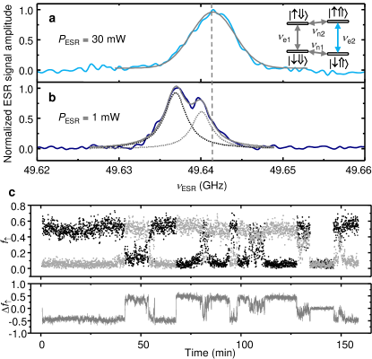
Here we present the first experimental demonstration of single-shot readout, coherent control, and measurement of the coherence properties of an individual 29Si nuclear spin in natural Si. All measurements were performed with a magnetic field T, in a dilution refrigerator with electron temperature mK. This work follows from previous experiments where the electron Pla et al. (2012) and nuclear Pla et al. (2013) spins of a single 31P donor were detected using a compact nano-scale device Morello et al. (2009) consisting of ion-implanted phosphorus donors Jamieson et al. (2005), tunnel-coupled to a silicon MOS single-electron transistor (SET) Angus et al. (2007). Spin control was achieved through microwave and RF excitations generated by an on-chip broadband transmission line Dehollain et al. (2013). The 31P donor in silicon represents a two-qubit system, with an electron spin () bound at cryogenic temperatures to a nuclear spin (). The eigenstates of this system are displayed as an inset to Fig. 1a – with thin arrows representing the spin state of the electron () and thick the nucleus (). There are two electron spin resonance (ESR) frequencies , and two 31P nuclear magnetic resonance (NMR) frequencies .
The detection of a single 29Si spin was achieved by first performing an ESR experiment about one of the 31P hyperfine peaks. We chose the transition corresponding to the state, i.e. , since the nuclear spin is predominantly polarized here as a result of the differing and nuclear spin relaxation mechanisms Pla et al. (2013). The ESR experiment involves using the SET to monitor the induced electron spin-up fraction in response to a microwave excitation with varying frequency , resulting in the spectrum of Fig. 1a. The line-shape is well described by a Gaussian with full-width-at-half-maximum (FWHM) MHz (or T) at the largest applied ESR power mW. This figure corresponds to the bulk value for the inhomogeneous broadening caused by the 29Si nuclear spin bath Tyryshkin et al. (2003). From the measured Rabi frequency at this power Pla et al. (2012) we extract T, confirming that power broadening does not occur here. However, by further reducing the excitation power to 1 mW ( T) the ESR line splits in two, and shifts to lower frequency (Fig. 1b). A double-Lorentzian fit best captures the shape of the line and yields a FWHM MHz for both peaks, with the center frequency decreasing by 3 MHz with respect to Fig. 1a. Overall, the observed low-power behavior indicates a polarization and a narrowing of the 29Si nuclear bath. The behavior is reproducible over several measurements, and does not depend on the direction of the frequency sweep. The microscopic origin of this phenomenon is currently not understood. It is not consistent with the standard Overhauser effect, where excitation of the electron spin to the state, in combination with a fast electron-nuclear spin-conserving relaxation channel results in a predominant bath polarization. The line shift to lower frequencies indicates instead a polarization, since 29Si has a negative gyromagnetic ratio MHz/T (Ref. 19). Several papers have discussed nuclear polarization with anomalous direction, but under conditions that do not apply to our experiment Laird et al. (2007); Rudner and Levitov (2007); Rudner et al. (2011); Urbaszek et al. (2013); Yang and Sham (2013). The line shift and narrowing occurs at low power, when , and the resonance is measured through counting single-shot electron spin readout events. Therefore the experiment effectively constitutes a projective measurement of the nuclear bath state, which can result in a narrowed bath distribution Cappellaro (2012). However, the shift to lower frequencies remains unexplained.
The splitting of the line indicates the presence of a single 29Si nuclear spin, strongly hyperfine-coupled to the donor-bound electron. This allows us to read the 29Si spin state in the same way as the 31P spin Pla et al. (2013). Here we apply adiabatic frequency sweeps Laucht et al. (2014) over the first half of the resonance, i.e. from far-detuned to a point mid-way between the two peaks. After each passage we acquire a single-shot measurement of the electron spin to obtain . The process is then repeated on the second half of the hyperfine-split peak. We observe clear “quantum jumps” (Fig. 1c), providing strong evidence that the splitting does indeed originate from a single spin coupled to the electron. Occasionally, both sides of the split peak produces no resonance, indicating that the 31P nuclear spin has flipped to . We therefore periodically measure the state of the donor nuclear spin and initialize it in the state if it has flipped sup .
Next we perform an NMR experiment on the single 29Si nucleus. The whole system is described by the spin Hamiltonian Feher (1959); Levitt (2008):
| (1) | |||||
where are the electron, 31P and 29Si spin operators and GHz/T, MHz/T, MHz/T (Ref. 19) are their respective gyromagnetic ratios. We assume that the electron-29Si interaction is dominated by a contact hyperfine term, i.e. we omit the dipolar coupling between 29Si and the electron. This omission is justified by the fact that we observe an extremely small probability to flip the 29Si spin by ionizing/neutralizing the donor ( flip every 100,000 readout events), which indicates that the secular approximation for the electron-nuclear interaction is almost exact, and non-diagonal interaction terms are negligible. For this reason, the nuclear spin measurement is almost exactly quantum-nondemolition (QND) Braginsky and Khalili (1996).
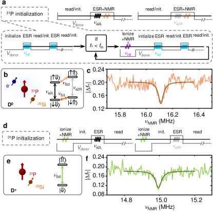
Calling the 29Si NMR frequency for a electron, and for (Fig. 2b), one has . Since the 29Si hyperfine splitting observed in Fig. 1b is MHz at T, we extract MHz and MHz. We then perform a NMR experiment where we first initialize the electron spin, for example , and apply a long NMR pulse at a frequency before attempting to adiabatically invert and read the electron spin. The electron spin-up fraction is then recorded, where and are the 29Si spin-dependent ESR transition frequencies defined as .
For each we calculate and plot the result for the transition in Fig. 2c. Off-resonance we find . At resonance, a randomization of the 29Si spin state produces an almost equal probability of having an “active” or transition. The trough observed at MHz is remarkably close to the estimated value for .
The tunnel-coupled SET used for readout can also be utilized to ionize the 31P donor and perform NMR on the isolated 29Si nuclear spin (Fig. 2e). Here the NMR frequency is simply . The pulse sequence for such a measurement is shown in Fig. 2d with the resulting resonance plot in Fig. 2f. The trough at MHz, together with the external magnetic field T – calibrated using the measured 31P NMR frequencies Pla et al. (2013) – implies a gyromagnetic ratio of MHz/T, very close to the bulk value of 8.458 MHz/T (Ref. 19). These experiments also yield an accurate value for the hyperfine coupling MHz.
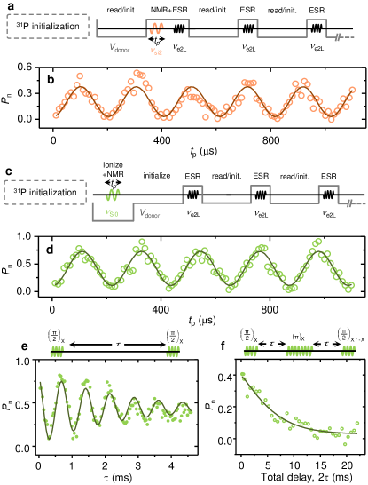
We demonstrate the ability to coherently manipulate the 29Si nuclear spin – with both a neutral (D0) and ionized (D+) donor – by observing Rabi oscillations. The protocols for such measurements are illustrated in Figs. 3a and c, and the 29Si nuclear spin flip probabilities as a function of the pulse duration are shown in Figs. 3b and 3d for the donor in the D0 and D+ charge states, respectively. The D+ data displays higher visibility oscillations than the D0 case, due to its immunity to electron spin state initialization errors.
Next we probe the coherence of the isolated (ionized donor) 29Si nuclear spin by performing Ramsey fringe and Hahn echo experiments (Fig. 3). Fitting the Ramsey data in Fig. 3e with a damped cosine function of the form yields a dephasing time of ms. Also from this fit we get , the average detuning from resonance, which enables us to provide a more accurate estimate of the gyromagnetic ratio MHz/T. The echo decay curve of Fig. 3f, fitted with an exponential function , reveals a coherence time of ms and an exponent . The coherence time is in excellent agreement with Hahn echo measurements in bulk Dementyev et al. (2003), where decoherence is caused by the dipole interactions with other 29Si nuclear spins.
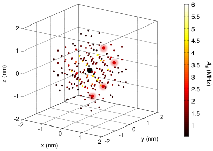
The individual hyperfine couplings between 29Si nuclei and a donor-bound electron are known from early work in bulk samples Hale and Mieher (1969a, b, 1971); Ivey and Mieher (1972, 1975a, 1975b). By adapting metrology techniques demonstrated for 31P Mohiyaddin et al. (2013), we can narrow down the possible locations of the 29Si atom measured here. A device-specific wavefunction was obtained by first calculating, with a finite-elements Poisson equation solver, the electrostatic potential profile surrounding the donor, then solving the full atomistic tight-binding Hamiltonian with the tool Nano Electronic MOdeling 3D (NEMO 3D) Klimeck et al. (2007). Calculating the shift from the bulk value in the probability density of the electron wavefunction at each lattice site, allows us to appropriately scale the 29Si Fermi contact hyperfine splittings from bulk data. Figure 4 shows a 3D plot of the 31P donor (large black circle) and the surrounding 29Si nuclei with known hyperfine constants. The 29Si nuclei with couplings in the range MHz are plot as enlarged circles. They all belong to a shell at 1.11 nm distance from the 31P nucleus sup . We have thus been able to narrow down the location of our 29Si atom to 4 out of a known possible sites.
In conclusion, we have performed electrical single-shot QND readout on a single 29Si nuclear spin, and demonstrated its coherent control though Rabi, Ramsey and Hahn echo experiments, which yield coherence values similar to those observed in bulk samples. While the isotopic purification of 28Si is an exciting avenue to achieve the best possible coherence and fidelity benchmarks Muhonen et al. (2014), the present work shows that isolated 29Si nuclear spins can be utilized as an additional resource Robledo et al. (2011) for quantum information processing in silicon.
We thank R.P. Starrett, D. Barber, C.Y. Yang and R. Szymanski for technical assistance, and W.A. Coish for discussions. This research was funded by the Australian Research Council Centre of Excellence for Quantum Computation and Communication Technology (project number CE110001027) and the US Army Research Office (W911NF-13-1-0024). We acknowledge support from the Australian National Fabrication Facility. Computational resources on nanoHUB.org, funded by the NSF grant EEC-0228390, were used in this work.
References
- Klauder and Anderson (1962) J. R. Klauder and P. W. Anderson, Phys. Rev. 125, 912 (1962).
- Pla et al. (2012) J. J. Pla, K. Y. Tan, J. P. Dehollain, W. H. Lim, J. J. Morton, D. N. Jamieson, A. S. Dzurak, and A. Morello, Nature 489, 541 (2012).
- Pla et al. (2013) J. J. Pla, K. Y. Tan, J. P. Dehollain, W. H. Lim, J. J. Morton, F. A. Zwanenburg, D. N. Jamieson, A. S. Dzurak, and A. Morello, Nature 496, 334 (2013).
- Hanson et al. (2008) R. Hanson, V. Dobrovitski, A. Feiguin, O. Gywat, and D. Awschalom, Science 320, 352 (2008).
- Coish and Loss (2005) W. A. Coish and D. Loss, Phys. Rev. B 72, 125337 (2005).
- Yao et al. (2006) W. Yao, R.-B. Liu, and L. Sham, Physical Review B 74, 195301 (2006).
- Dutt et al. (2007) M. V. G. Dutt, L. Childress, L. Jiang, E. Togan, J. Maze, F. Jelezko, A. S. Zibrov, P. R. Hemmer, and M. D. Lukin, Science 316, 1312 (2007).
- Robledo et al. (2011) L. Robledo, L. Childress, H. Bernien, B. Hensen, P. F. Alkemade, and R. Hanson, Nature 477, 574 (2011).
- Waldherr et al. (2014) G. Waldherr, Y. Wang, S. Zaiser, M. Jamali, T. Schulte-Herbrüggen, H. Abe, T. Ohshima, J. Isoya, J. Du, P. Neumann, et al., Nature 506, 204 (2014).
- Taminiau et al. (2014) T. H. Taminiau, J. Cramer, T. van der Sar, V. V. Dobrovitski, and R. Hanson, Nature nanotechnology 9, 171 (2014).
- Nickerson et al. (2013) N. H. Nickerson, Y. Li, and S. C. Benjamin, Nature communications 4, 1756 (2013).
- Ladd et al. (2002) T. D. Ladd, J. R. Goldman, F. Yamaguchi, Y. Yamamoto, E. Abe, and K. M. Itoh, Phys. Rev. Lett. 89, 017901 (2002).
- Itoh (2005) K. M. Itoh, Solid State Communications 133, 747 (2005).
- Morello et al. (2009) A. Morello, C. C. Escott, H. Huebl, L. H. Willems van Beveren, L. C. L. Hollenberg, D. N. Jamieson, A. S. Dzurak, and R. G. Clark, Phys. Rev. B 80, 081307 (2009).
- Jamieson et al. (2005) D. N. Jamieson, C. Yang, T. Hopf, S. M. Hearne, C. I. Pakes, S. Prawer, M. Mitic, E. Gauja, S. E. Andresen, F. E. Hudson, A. S. Dzurak, and R. G. Clark, Applied Physics Letters 86, 202101 (2005).
- Angus et al. (2007) S. J. Angus, A. J. Ferguson, A. S. Dzurak, and R. G. Clark, Nano Letters 7, 2051 (2007).
- Dehollain et al. (2013) J. P. Dehollain, J. J. Pla, E. Siew, K. Y. Tan, A. S. Dzurak, and A. Morello, Nanotechnology 24, 015202 (2013).
- Tyryshkin et al. (2003) A. M. Tyryshkin, S. A. Lyon, A. V. Astashkin, and A. M. Raitsimring, Phys. Rev. B 68, 193207 (2003).
- Hale and Mieher (1969a) E. B. Hale and R. L. Mieher, Phys. Rev. 184, 739 (1969a).
- Laird et al. (2007) E. Laird, C. Barthel, E. Rashba, C. Marcus, M. Hanson, and A. Gossard, Physical review letters 99, 246601 (2007).
- Rudner and Levitov (2007) M. Rudner and L. Levitov, Physical review letters 99, 036602 (2007).
- Rudner et al. (2011) M. Rudner, F. Koppens, J. Folk, L. Vandersypen, and L. Levitov, Physical Review B 84, 075339 (2011).
- Urbaszek et al. (2013) B. Urbaszek, X. Marie, T. Amand, O. Krebs, P. Voisin, P. Maletinsky, A. Högele, and A. Imamoglu, Reviews of Modern Physics 85, 79 (2013).
- Yang and Sham (2013) W. Yang and L. Sham, Physical Review B 88, 235304 (2013).
- Cappellaro (2012) P. Cappellaro, Phys. Rev. A 85, 030301 (2012).
- Laucht et al. (2014) A. Laucht, R. Kalra, J. T. Muhonen, J. P. Dehollain, F. A. Mohiyaddin, F. Hudson, J. C. McCallum, D. N. Jamieson, A. S. Dzurak, and A. Morello, Applied Physics Letters 104, 092115 (2014).
- (27) See Supplemental Material for extended figure captions.
- Feher (1959) G. Feher, Phys. Rev. 114, 1219 (1959).
- Levitt (2008) M. H. Levitt, Spin dynamics: basics of nuclear magnetic resonance (Wiley, 2008).
- Braginsky and Khalili (1996) V. B. Braginsky and F. Y. Khalili, Rev. Mod. Phys. 68, 1 (1996).
- Dementyev et al. (2003) A. E. Dementyev, D. Li, K. MacLean, and S. E. Barrett, Phys. Rev. B 68, 153302 (2003).
- Hale and Mieher (1969b) E. B. Hale and R. L. Mieher, Phys. Rev. 184, 751 (1969b).
- Hale and Mieher (1971) E. B. Hale and R. L. Mieher, Phys. Rev. B 3, 1955 (1971).
- Ivey and Mieher (1972) J. L. Ivey and R. L. Mieher, Phys. Rev. Lett. 29, 176 (1972).
- Ivey and Mieher (1975a) J. L. Ivey and R. L. Mieher, Phys. Rev. B 11, 822 (1975a).
- Ivey and Mieher (1975b) J. L. Ivey and R. L. Mieher, Phys. Rev. B 11, 849 (1975b).
- Mohiyaddin et al. (2013) F. A. Mohiyaddin, R. Rahman, R. Kalra, G. Klimeck, L. C. L. Hollenberg, J. J. Pla, A. S. Dzurak, and A. Morello, Nano Letters 13, 1903 (2013).
- Klimeck et al. (2007) G. Klimeck, S. Ahmed, H. Bae, N. Kharche, S. Clark, B. Haley, S. Lee, M. Naumov, H. Ryu, F. Saied, M. Prada, M. Korkusinski, T. Boykin, and R. Rahman, Electron Devices, IEEE Transactions on 54, 2079 (2007).
- Muhonen et al. (2014) J. T. Muhonen, J. P. Dehollain, A. Laucht, F. E. Hudson, T. Sekiguchi, K. M. Itoh, D. N. Jamieson, J. C. McCallum, A. S. Dzurak, and A. Morello, arXiv:1402.7140 (2014).