Spectromicroscopy of electronic phase separation in KxFe2-ySe2 superconductor
Abstract
Structural phase separation in AxFe2-ySe2 system has been studied by different experimental techniques, however, it should be important to know how the electronic uniformity is influenced, on which length scale the electronic phases coexist, and what is their spatial distribution. Here, we have used novel scanning photoelectron microscopy (SPEM) to study the electronic phase separation in KxFe2-ySe2, providing a direct measurement of the topological spatial distribution of the different electronic phases. The SPEM results reveal a peculiar interconnected conducting filamentary phase that is embedded in the insulating texture. The filamentary structure with a particular topological geometry could be important for the high Tc superconductivity in the presence of a phase with a large magnetic moment in AxFe2-ySe2 materials.
The discovery of superconductivity in Fe-based materials Kamihara08 gave a new boost to the research on this intriguing quantum phenomenon. Among others, the FeSe was found to show superconductivity at a T 8 K Hsu08 , that could be increased to over 30 K by physical pressure Medvedev_Nat_09 . Furthermore, it can be increased to similar values by intercalating alkali atoms A, yielding in a new series of AxFe2-ySe2 materials Guo10 ; Mizuguchi11 . One of the interesting features of Fe2-ySe2 system is the intrinsic phase separation in coexisting crystallographic phases Liu11a ; Wang11 ; Li11b ; Ricci11b ; Shermadini12 ; Chen_PRX ; Yuan12 ; Wen12 . The minority phase has a stoichiometry close to Fe2Se2 (122) Fang11 , embedded into a majority phase that manifests iron-vacancy ordering. This majority phase has a stoichiometry of Fe1.6Se2 (245) and shows a Fe superstructure vacancy ordering Fang11 ; Bao11 ; Zhao12 . It exhibits a particular block antiferromagnetic state with a large magnetic moment per Fe atom of 3.3 Bao11 . It has been argued that the minority 122-phase is metallic and becomes superconducting while the sample is cooled through the transition temperature Tc. In contrary, the majority 245-phase with iron vacancy order remains magnetic, and hence the superconductivity occurs in the presence of the magnetic order.
Although, this phase separation scenario is established from the crystallographic point of view, the electronic nature of the different phases and their topological distribution is not yet clear. Scanning photoelectron microscopy (SPEM) allows to spatially resolve the normally averaged spectroscopic information spectromicrosc . In this work, for the first time, we have used SPEM technique to investigate the spatial distribution of the coexisting electronic phases in KxFe2-ySe2.
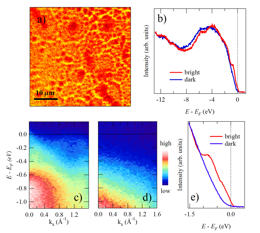
Figure 1a shows a typical SPEM image for the angle-integrated photoelectron yield off the KxFe2-ySe2 at K around the -point. The photoemission intensity is integrated over a wide binding energy interval of to 0.5 eV around the Fermi level , with a spatial resolution of m2 that reveals a clear inhomogeneous spectral distribution. The valence band spectra, measured around the -point, on bright and dark regions are shown as Fig. 1b. The bright region in the spectral image (Fig. 1a) corresponds to the minority metallic phase showing a significant electronic density of states (DOS) at (Fig. 1b). On the other hand, dark region has hardly any DOS near typical of a semiconducting/insulating phase. Interestingly, the bright region is organized into interconnected thin filamentary stripes.
In order to make a further characterization of the observed electronic phases, we have performed angle-resolved photoemission spectroscopy (ARPES) measurements of the bright and dark regions to observe the band structure in the -M direction of the Brillouin zone. The band dispersions for the bright and dark regions, using a photon energy of 27 eV, are shown in Figs. 1c and 1d respectively. Despite limited energy and momentum resolutions ( 50 meV and 0.05 ), the energy dispersive structures in the bright regions of SPEM looks similar to the band structure measured by space integrated ARPES Zhang11 ; Qian11 ; Mou11 ; Liu12b ; Chen12 . On the other hand, no dispersing structures are seen close to in the dark region, neither around (zone center) nor around M (zone corner), suggesting the dark region being due to an insulating/semiconducting phase. The difference between the two regions near the can be better seen in Fig. 1e, showing energy distribution of the spectral weight in the vicinity of the obtained by integrating the band dispersions for the two regions (Figs. 1c and 1d). Therefore, we can make a clear distinction between the different electronic phases and their spatial distribution in the target material. Indeed, the bright filamentary stripes should represent the metallic and superconducting 122 phase, while the dark texture is the Fe vacancy ordered antiferromagnetic and semiconducting/insulating 245 phase.
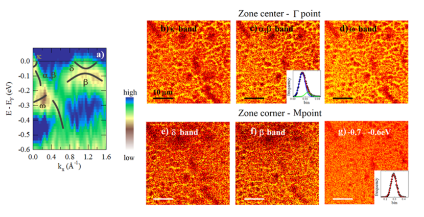
The second derivative of the measured band dispersions for the bright filamentary stripes is shown in Fig. 2a. Following earlier high resolution space integrated ARPES, the spectral weight around the zone center (-point) can be assigned to weak electron-like band crossing . This weak band is known to be mainly due to the Fe orbitals with admixed Se orbitals Liu12b ; Chen12 . At slightly higher binding energies, around 80 meV below , spectral weight due to and bands with hole-like character can be seen. These bands are mainly derived from a combination of the Fe and Fe orbitals. At around 0.35 eV below , one can see the band, originating mainly from the Fe orbital. Around the zone corner (M point), spectral weight due to electron-like band can be identified that crosses having a complicated orbital character. In addition, the band can be visualized also at the M point, around 0.2 eV below . The overall structures appear consistent with the band dispersions reported in the earlier space integrated ARPES Zhang11 ; Chen_PRX .
To investigate energy dependence of the spatial distribution of the filamentary phase, we have measured SPEM images with different binding energy windows identified from the band dispersions. Figs. 2b to 2g show the spectral intensity distributions for the different bands. Figure 2b represents spectral intensity distribution of the electron-like band close to ( to 0 eV below ). From the band-selected SPEM images (in particular, those for the and / bands around the -point), it is clearly seen that the spatial phase separation in KxFe2-ySe2 occurs with interconnected thin filamentary stripes of the superconducting 122 phase organized in a peculiar geometry. The filamentary stripes have a width of less than 1 m, and are embedded into the dark region (i.e., the semiconducting/insulating 245 phase).
From the spectral intensity map, the ratio of the 245 phase with lower DOS to the 122 phase with higher DOS can be determined. A detailed analysis of the distribution histogram of the / bands shown in the inset of Fig. 2c reveals an asymmetric intensity distribution which can be fitted to a sum of two Gaussians. This analysis indicates that % of the sample can be attributed to the insulating/semiconducting 245 and the remaining % to the metallic 122 phase. This is consistent with the earlier estimations of the volume fractions of the majority and minority phases determined by different experiments Shermadini12 ; Ricci11b ; Wen12 . The SPEM images underline that instead of granular superconductivity a scenario of filamentary superconductivity is more plausible for KxFe2-ySe2. In addition, the SPEM image of the / bands around shows very similar features as the band around (see Fig. 2c). Also the band shows (Fig. 2d) network of filaments of the 122 phase. As expected a very similar spatial distribution of the spectral weight of the bands around the M point, where the band crossing , is observed (Fig. 2e). The same is observed for the band at M (see Fig. 2f). In comparison to the energy integrated SPEM image presented in Fig. 1b, the SPEM images with energy and momentum selected windows show significantly higher contrast resolution in the distribution of the filamentary metallic 122 phase.
At higher binding energies, the SPEM images appear quite uniform (Fig. 2g), however, still some inhomogeneities can be seen. The corresponding intensity histogram is symmetric and can be fitted by a single Gaussian (see the inset to Fig. 2g). Nevertheless, the SPEM images permit to conclude unambiguously that the peculiar electronic phase separation in KxFe2-ySe2 is solely due to the bands close to with Fe 3 character.
Here, it is worth mentioning that a structural phase separation was observed, with uniform areas of the 245 phase enclosed by a network of the 122 phase having a width of about 1 m Charnukha12 ; Liu12 ; Wen12 . Also a combination of scanning and transmission electron microscopy has shown a structural phase separation in this scale Wang12 . The peculiar topological distribution of the metallic phase in KxFe2-ySe2 is characterstic of the system. The distribution of the filamentary phase may be different depending on the sample preparation procedure and thermal history. Indeed, the contrast resolution and topological distribution of coexisting phases appear to depend on the sample heat treatment, also underlined by transmission electron microscopy Wang12 , measureing different structural phases unlike the present work in which the electronic structure of the coexisting phases is determined.
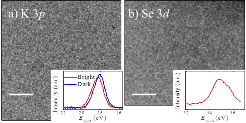
Clearly, the electronic phase separation is characterized by Fe electronic states. In order to have a further insight to the role of other elements, core level SPEM of K and Se were performed. Figure 3a shows the spatial distribution of the integrated photoemission yield measured using a photon energy of 74 eV around the K 3 core level. No clear inhomogeneities can be observed in the SPEM image, indicating an overall homogeneous distribution of K. However, a more detailed investigation leads to the observation of the binding energy shift in the K 3 peak. The inset to Fig. 3a shows the K 3 photoemission peaks of the metallic 122 (bright) and the insulating/semiconducting 245 (dark) phases. The K 3 binding energy for the 245 phase is lower than the one for the 122 phase, indicating stronger screening. It appears that the valence of K is different for the two phases, perhaps due to different interactions with the neighbouring atoms (e.g., degree of covalency). Also, the K 3 core level appears wider for the 122 phase, which might be due to mixing of the 245 phase in the 122 phase due to the fact that the filaments of the latter are less than 1m wide. Similarly, the spatial distribution of Se 3 core level photoemission intensity was measured. Although the Se 3 peak is overlapping with the Fe 3 core level, the lower cross section of Fe 3 compared to the Se 3 at the photon energy used allowed to measure the desired SPEM image. The resulting map of the Se shows no evident inhomogeneities (see Figs. 3b). Thus, it appears that, similar to K, Se is also distributed uniformly in the sample. Within the experimental resolution, no evidence of Se 3 core level shift was found, suggesting almost similar oxidation state for Se in the two phases.
From the SPEM imaging, the electronic phase separation is clearly observed in KxFe2-ySe2. The sample consists predominantly of the insulating 245 phase that is interrupted by filamentary interconnected stripes with less than 1 m size of 122 phase. It seems that K and Se are distributed homogeneously in the sample leading to the conclusion that mainly the Fe content is varying due to the phase separation into the stoichiometric 122 and the Fe deficient 245 phase. That could indicate that and are strongly correlated and solely the Fe content is responsible for the electronic phase separation. Consequently, Fe might posses different spin states in the corresponding electronic phases. Such a scenario was already observed in X-ray emission studies with a majority phase having a high spin Fe2+ and a minority phase having an intermediate Fe2+ state Simonelli12 . This appears consistent with the observation of different covalance of K depending on the corresponding phase as observed in the shift of the K 3 core level SPEM. The present results may have implications on the recent indications of superconductivity in a single layer of FeSe grown on SrTiO3 substrate WangFeSe12 , where electronic structure studies have shown absence of hole bands LiuFeSe12 ; TanFeSe13 as the case of KxFe2-ySe2. Indeed, a particular filamentary network can give rise to high Tc superconductivity as argued in the oxide interfaces Caprara13 . From the SPEM results, it can be hypothesized that a particular electronic structure topology of the conducting phase (FeSe) embedded in an insulating texture (SrTiO3 substrate) is a key to the higher Tc superconductivity in the single layer FeSe.
Methods
Sample growth The KxFe2-ySe2 single crystals were prepared using the Bridgman method
Mizuguchi11 . After the growth the single crystals were sealed into a quartz tube and annealed for 12 hours in 600∘C. Some of the crystals were annealed in vacuum at 400∘C for 1 hour followed by fast cooling to room temperature. The crystals were characterized by X-ray diffraction, resistivity in a PPMS (Quantum Design), and magnetization measurements in a SQUID (Quantum Design) magnetometer. The samples exhibit a sharp superconducting transition with K.
Spectromicroscopy. The SPEM measurements were performed on the spectromicroscopy beamline at the ’Elettra synchrotron facility’ at Trieste spectromicrosc . Photons at 27 eV and 74 eV were focused through a Schwarzschild objective, to obtain a submicron size spot. This allows to sample the electronic structure at the Fermi level as well as several core levels along with the valence level in order to visualize the normally averaged spectroscopic information in the electronic structure. For the present measurements the total energy resolution was about 50 meV while the angle resolution was limited to 1∘. The measurements were performed in ultra-high vacuum (p 2 10-10 mbar) on an in-situ prepared (cleaved) surfaces. The SPEM images contain 150x144 points with a step of 0.3 and the measurement time for each point is 100 milliseconds. A standard photoemission microscopy procedure was used to remove topographic features from the images presented surface .
References
- (1) Kamihara, Y., et. al. Iron-based layered superconductor La[O1-xFx]FeAs () with K. J. Am. Chem. Soc. 130, 3296 (2008).
- (2) Hsu, F. C., et. al. Superconductivity in the PbO-type structure -FeSe. Proc. Natl. Acad. Sci. USA 105, 14262 (2008).
- (3) Medvedev, S.,et. al. Electronic and magnetic phase diagram of -Fe1.01Se with superconductivity at 36.7 K under pressure. Nature Mater. 8, 630 (2009).
- (4) Guo, J., et. al. Superconductivity in the iron selenide KxFe2Se2 (). Phys. Rev. B 82, 180520(R) (2010).
- (5) Mizuguchi, Y., et. al. Transport properties of the new Fe-based superconductor KxFe2Se2 ( K). Appl. Phys. Lett. 98, 042511 (2011).
- (6) Liu, R. H., et. al. Coexistence of superconductivity and antiferromagnetism in single crystals A0.8Fe2-ySe2 (A = K, Rb, Cs, Tl/K and Tl/Rb): Evidence from magnetization and resistivity. Europhys. Lett. 94, 27008 (2011).
- (7) Wang, Z., et. al. Microstructure and ordering of iron vacancies in the superconductor system KyFexSe2 as seen via transmission electron microscopy. Phys. Rev. B 83, 140505(R) (2011).
- (8) Li, W., et. al. Phase separation and magnetic order in K-doped iron selenide superconductor, Nature Phys. 8, 126 (2011).
- (9) Ricci, A., et. al. Intrinsic phase separation in superconducting K0.8Fe1.6Se2 ( K) single crystals. Supercond. Sci. Technol. 24, 082002 (2011); Ricci, A. et. al.. Nanoscale phase separation in the iron chalcogenide superconductor K0.8Fe1.6Se2 as seen via scanning nanofocused x-ray diffraction Phys. Rev. B 84, 060511 (2011).
- (10) Shermadini, Z., et. al. Superconducting properties of single-crystalline Fe2-ySe2 ( Rb, K) studied using muon spin spectroscopy Phys. Rev. B 85, 100501(R) (2012).
- (11) Chen, F., et. al. Electronic Identification of the Parental Phases and Mesoscopic Phase Separation of KxFe2-ySe2 Superconductors. Phys. Rev. X 1, 021020 (2011).
- (12) Yuan, R. H., et. al. Nanoscale phase separation of antiferromagetic order and superconductivity in K0.75Fe1.75Se2. Sci. Rep. 2, 221 (2012).
- (13) Wen, H.-W., Overview on the physics and materials of the new superconductor KxFe2-ySe2, Rep. Prog. Phys. 75, 112501 (2012).
- (14) Fang, M.-H., et. al. Fe-based superconductivity with K bordering an antiferromagnetic insulator in (Tl,K)FexSe2. Europhys. Lett. 94, 27009 (2011).
- (15) Bao, W., et. al. A Novel Large Moment Antiferromagnetic Order in K0.8Fe1.6Se2 Superconductor. Chin. Phys. Lett. 28, 086104 (2011).
- (16) Zhao, J., et. al. Neutron-Diffraction Measurements of an Antiferromagnetic Semiconducting Phase in the Vicinity of the High-Temperature Superconducting State of KxFe2-ySe2. Phys. Rev. Lett. 109, 267003 (2012).
- (17) Dudin, P., et. al. Angle-resolved photoemission spectroscopy and imaging with a submicrometric probe at the SPECTROMICROSCOPY-3.2 beamline at Elettra. J. Synchr. Rad. 17, 445 E50 (2010).
- (18) Zhang, Y., et. al. Nodeless superconducting gap in AxFe2Se2 (A = K, Cs) revealed by angle-resolved photoemission spectroscopy Nature Mater. 10, 273 (2011).
- (19) Qian, T., et. al. Absence of a holelike Fermi surface for the iron-based K0.8Fe1.7Se2 superconductor revealed by angle-resolved photoemission spectroscopy. Phys. Rev. Lett. 106, 187001 (2011).
- (20) Mou, D., et. al. Distinct Fermi Surface Topology and Nodeless Superconducting Gap in a (Tl0.58Rb0.42)Fe1.72Se2 Superconductor. Phys. Rev. Lett. 106, 107001 (2011).
- (21) Liu, Z.-H., et. al. Three-dimensionality and orbital characters of Fermi surface in (Tl,Rb)yFe2-xSe2. Phys. Rev. Lett. 109, 037003 (2012).
- (22) Fei, C.,, et. al. The orbital characters of low-energy electronic structure in iron-chalcogenide superconductor KxFe2-ySe2. Chin Sci Bull 57, 3829 (2012).
- (23) Charnukha, A., et. al. Nanoscale Layering of Antiferromagnetic and Superconducting Phases in Rb2Fe4Se5 Single Crystals. Phys. Rev. Lett. 109, 017003 (2012).
- (24) Liu, Y., et. al. Evolution of precipitate morphology during heat treatment and its implications for the superconductivity in KxFe1.6+ySe2 single crystals. Phys. Rev. B 86, 144507 (2012).
- (25) Wang, Z.-W., et. al. Structural Phase Separation in K0.8Fe1.6+xSe2 Superconductors. J. Phys. Chem. C 116, 17847 (2012).
- (26) Simonelli, L., et. al. Coexistence of different electronic phases in the K0.8Fe1.6Se2 superconductor: a bulk-sensitive hard x-rays spectroscopy study. Phys. Rev. B 85, 224510 (2012).
- (27) Wang Q.-Y., et. al. Interface-Induced High-Temperature Superconductivity in Single Unit-Cell FeSe Films on SrTiO3. Chin. Phys. Lett. 29, 037402 (2012).
- (28) Liu, D., et. al. Phase Diagram and Electronic indication of High Temperature Superconductivity at 65 K in Single-Layer FeSe Films. Nature Mat. 12, 605 (2013).
- (29) Tan, S.Y, et. al. Interface-induced superconductivity and strain-dependent spin density wave in FeSe/SrTiO3 thin films. Nature Mat. 12, 634 (2013).
- (30) Caprara, S., et. al. Multiband superconductivity and nanoscale inhomogeneity at oxide interfaces. Phys. Rev. B 88, 020504(R) (2013);Bianconi, A. Quantum materials: Shape resonances in superstripes. Nature Physics 9, 536 (2013).
- (31) Marsi, M., et. al. ESCA microscopy at ELETTRA: what it is like to perform spectromicroscopy experiments on a third generation synchrotron radiation source. J. Electron Spectrosc. Relat. Phenom. 84, 73 (1997).
Acknowledgements
The authors would like to thank Elettra staff for the experimental help during the beamtime. This work is a part of the on-going collaboration between Sapienza University of Rome and Japanese institutes including University of Tokyo, NIMS and Tohoku University. D.I. and A. Bi. acknowledge support from superstripes – onlus. M. B. acknowledges support from the Swiss National Science Foundation (grant number PBZHP2_143495).
Author contributions
N.L.S.,T.M., M.B., A.F., Y.T. Y.K, A.Ba. and A.Bi programmed and coordinated the study. H. T., Y. M., Y. T., T. N., T. H. and Y. K. have synthesized the single crystals used for the study. M.B., N.L.S., A.Ba., M.H., T.M., A.I., D.I. and D.O. performed the experiments at the Spectromicroscopy beamline of Elettra synchrotron facility and contributed in the data analysis. M.B., B.J. and N.L.S wrote the paper with contributions from A.Ba. and T.M. All authors discussed the results and commented on the manuscript.
Additional information
The authors declare no competing financial interests. Reprints and permissions information is available online at http://www.nature.com/reprints. Correspondence and requests for materials should be addressed to N.L.S. (Naurang.saini@roma1.infn.it)