Composition variation and underdamped mechanics
near membrane proteins and coats
Abstract
We study the effect of transmembrane proteins on the shape, composition and thermodynamic stability of the surrounding membrane. When the coupling between membrane composition and curvature is strong enough the nearby membrane composition and shape both undergo a transition from over-damped to under-damped spatial variation, well before the membrane becomes unstable in the bulk. This transition is associated with a change in the sign of the thermodynamic energy and hence favors the early stages of coat assembly necessary for vesiculation (budding) and may suppress the activity of mechanosensitive membrane channels and transporters. Our results suggest an approach to obtain physical parameters of the membrane that are otherwise difficult to measure.
pacs:
87.14.ep 87.15.kt 87.16.D-Biological membranes are crucial to the structure and function of living cells Alberts et al. (2008). Transmembrane proteins essential for transport, adhesion and signalling are embedded in membranes Singer and Nicolson (1972); Engelman (2005) consisting of a mixture of lipids and other amphipathic components. The interaction with the adjacent lipid molecules is known to regulate the function of membrane proteins Suchyna et al. (2004); Moe and Blount (2005); Krepkiy et al. (2009); Milescu et al. (2009). Here, we are primarily interested in the non-specific lipid-protein interactions that arise from the coupling of their hydrophobic regions Lee (2004); Mitra et al. (2004); Dowhan et al. (2004); Nyholm et al. (2007); Andersen and Koeppe (2007); Phillips et al. (2009); Lundbaek et al. (2010), although we can also allow for selective enrichment of membrane component(s) near the protein. We employ a continuum theory in which small deformations of the lipid environment near a rigid inclusion can be described by a number of local field variables, such as the profile of the mid-plane of the bilayer, its composition and membrane thickness Canham (1970); Huang (1986); Aranda-Espinoza et al. (1996); Nielsen et al. (1998); Mondal et al. (2014); Sens and Safran (2000); Fournier (1999); Weikl et al. (1998); Wiggins and Phillips (2004, 2005); Dan et al. (1993); Goulian et al. (1993); Kim et al. (1998); Turner and Sens (1999); Markin and Sachs (2004); Ursell et al. (2007); Haselwandter and Phillips (2013a, b); Leibler (1986); Leibler and Andelman (1987); Andelman et al. (1992); Helfrich (1973). Furthermore, the free-energy cost associated with thickness deformation is completely decoupled at lowest order Fournier (1999), and it can be independently analyzed although we do not do so here.
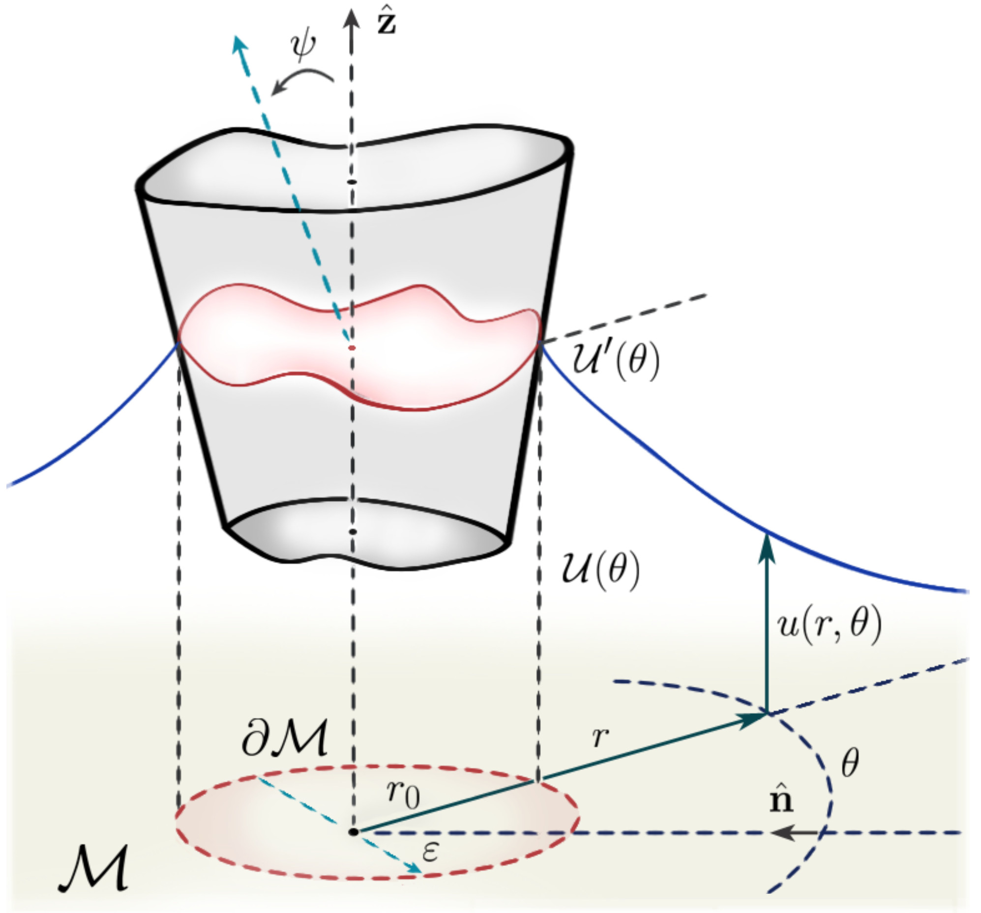
We allow for selective enrichment/depletion of curvature sensitive inclusions in the vicinity of a membrane protein or, equivalently, lipid asymmetry between leaflets that is characterized by a local spontaneous curvature, the preferred mean curvature in the absence of any mechanical stresses on the membrane Leibler (1986); Leibler and Andelman (1987); Andelman et al. (1992); Helfrich (1973); Seifert (1997); Israelachvili et al. (1976); Gruner (1989); McMahon and Gallop (2005); Callan-Jones et al. (2011). This local variation may be relatively large near a membrane protein if its geometry is such that it bends or deforms the surrounding membrane (see Fig 1). Our approach leads to a real-space description of the membrane around an inclusion of arbitrary symmetry.
We consider a two-component membrane in which the local compositional asymmetry between the different layers and/or the density of curvature-sensitive inclusions is phenomenologically coupled to the local mean curvature of the membrane Leibler (1986); Leibler and Andelman (1987). When the compositional variation is weak and the membrane displacement is small, the free-energy can be written as a Landau-Ginzburg expansion Leibler (1986); Leibler and Andelman (1987); Andelman et al. (1992); Sunil Kumar et al. (1999); Schick (2012); Seifert (1993),
| (1) |
where only the lowest-order terms are retained and , and are phenomenological constants. The scalar fields and are the local composition difference (as an area fraction) and bilayer mid-plane height, respectively, see Fig 1. Both deformation fields are described within a Monge representation, which allows us to write the free-energy associated with mid-plane deformation as
| (2) |
where and are the surface tension and bending rigidity of the membrane, respectively Nelson et al. (2004).
We now seek the ground state of the membrane and neglect fluctuations throughout. The membrane shape and its compositional field can then be computed exactly by minimizing the free-energy functional, , leading to the Euler-Lagrange equations:
| (3) | |||
| (4) |
where and the coefficients , and represent the relevant inverse length scales of the model 111The sign choice of is simply a convention. The fields and are invariant under a sign change in , itself related to the convention of a direction for “up” and whether one refers to the enrichment of a component that couples to positive curvature (depending on one’s choice for “up”) or the depletion of one that couples to negative curvature.. By combining (3) and (4), a single equation for can be obtained 222See Supplemental Material, which includes Refs. Riley et al. (2006); Byron and Fuller (1992); Gradshteyn et al. (2000).:
| (5) |
where is given by
| (6) |
By separation of variables, a solution to equation (5) that vanishes in the far-field limit can be found to be
| (7) |
where and are the usual polar coordinates, as illustrated in Fig 1, and is defined by
| (8) |
where are the modified Bessel functions of the second kind of order , and , with and arbitrary constants. From this we obtain the membrane shape through Eq (3), which yields
| (9) |
where the solutions that diverge at infinity are excluded. Here, is the homogeneous solution of (3), namely
| (10) |
where , with and some constants. The remaining two terms in (9) are the inhomogeneous solutions, which are found to be
| (11) |
The angular functions and are determined by the boundary conditions at the interface , located at a distance from the symmetry axis. These are specified by the height and contact angle at which the mid-plane of the bilayer meets the inclusion (see Fig 1). This choice is motivated by assuming a strong coupling between the transmembrane domain of the inclusion and the membrane hydrophobic core. Also, the normal derivative of is chosen to vanish on , which is used to obtain an unique solution 333Although Dirichlet boundary conditions could be used as well, our choice gives the ground state solution in the absence of any constraints on the composition asymmetry field at the boundary; see Ref. Note (2) for more details.
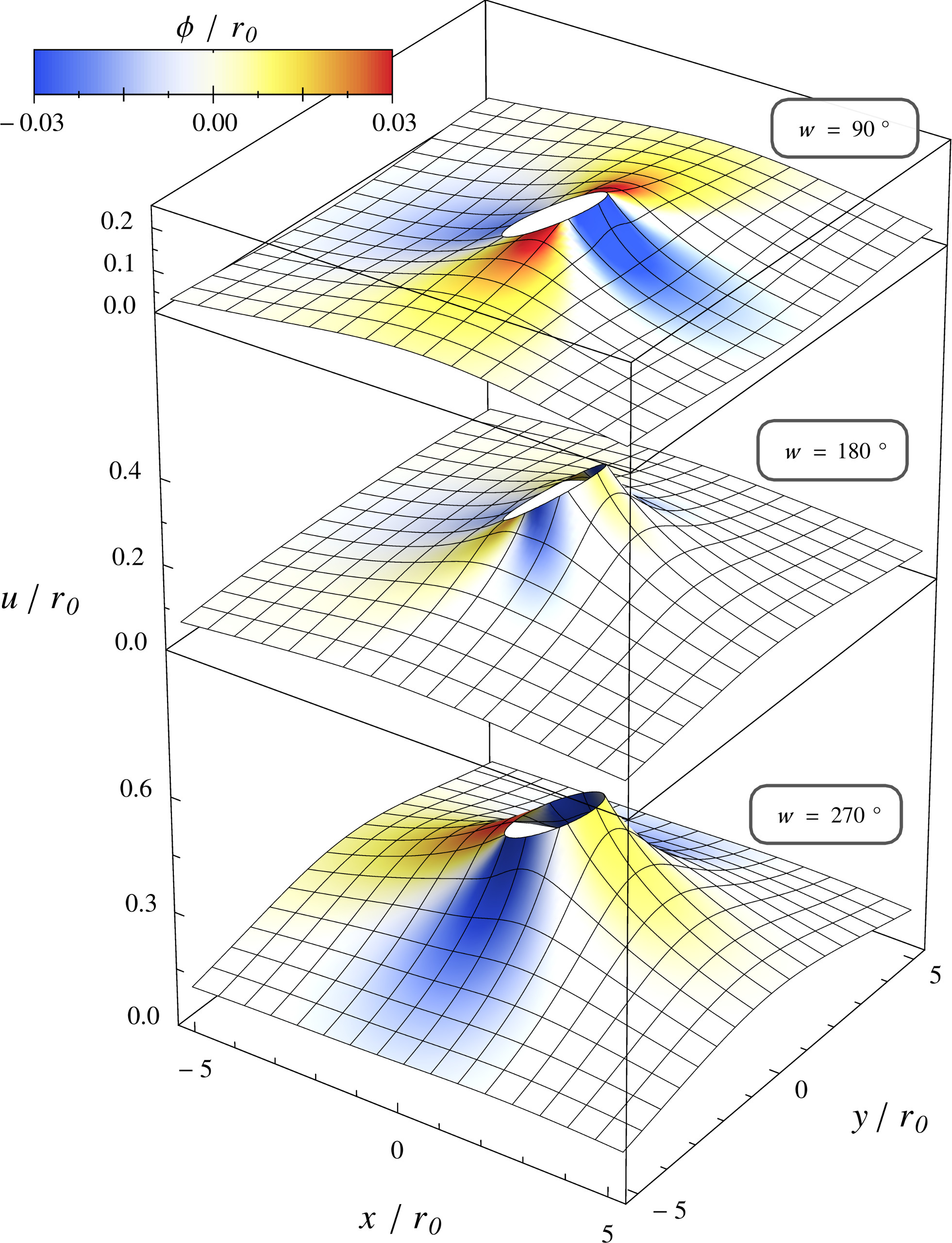
This methodology allows us to compute exactly the lowest order estimates to the membrane profile, its local phase behavior, and the total deformation energy, given an arbitrary model for the shape of the inclusion, through and , i.e. a general solution to the problem. This makes contact with experiments that might measure membrane shape (cryo TEM Shimada et al. (2007) or perhaps TIRF microscopy) and composition (NMR Judge and Watts (2011) or FRET Loura and Prieto (2011)). First, we consider a simple illustrative example in which the height, , is chosen to be a constant , while the contact angle has a non-zero value only within an angular interval , see Fig 2. This corresponds to a rigid inclusion that induces a local mid-plane deformation only within a specific region along its hydrophobic belt, with the remaining part preferring a flat membrane. The Connolly surface of a leucine transporter, LeuT, exhibits similar features Yamashita et al. (2005); Singh et al. (2008). The height is not entirely arbitrary, being set by the overall balance of normal forces. Similarly, the condition of torque balance leads to a tilt of the inclusion (see Note (2) for details), as illustrated in Fig 1. Typical solutions due to such an asymmetrically-shaped inclusion that exerts no net torque are shown in Fig 2 for physiologically reasonable values of , , and 444Typically the membrane correlation length might be Sheetz and Dai (1996). Eq. (1) can be associated with a typical interfacial width for a strongly segregated system of a few nm, say , and a line tension of about a pN ( pN) Tian et al. (2007), which combine to give . Sorting of strongly curvature-coupling components into membrane tubes gives an upper bound of and hence a value for Aimon et al. (2014), indicating that the all regimes discussed in the main text are accessible. While the use of a phase separated system to estimate the parameters and is questionable, given that they are motivated within a model in which the system remains essentially one phase, with very small, we are reassured that the primary limit on the accessibility of the underdamped regime is that is not too large. Given that spontaneous phase separation is often seen on biological membranes there would seem to be no lower limit on a reasonable magnitude for and hence .. The induced shows a rich variation as the angle is varied between and , which correspond to inclusions with a cylindrical and a conical shape, respectively.
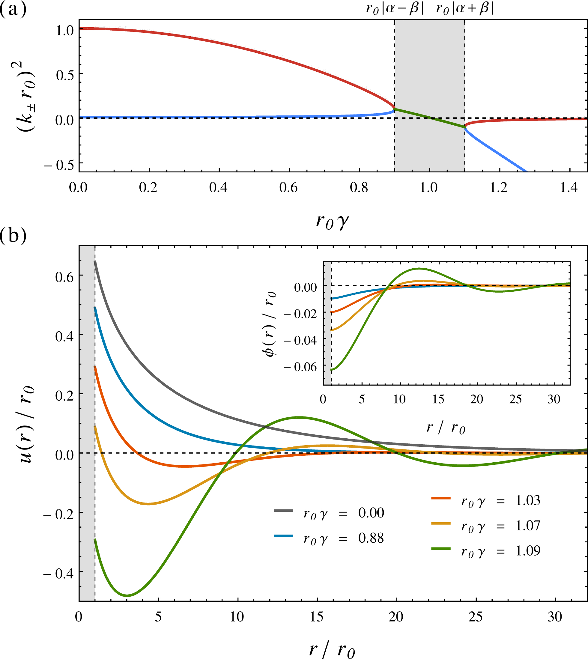
To better understand the role of the coupling constant , we consider symmetric conical inclusions () in what follows, noting that the transition from over- to under-damped variation also appears for rigid inclusions with other (or no) symmetry. For values of less than , the solutions are found to be monotonically decaying, see Fig 3(b). However, as is increased above this point, the solutions show an underdamped behavior, with the membrane displacement decaying to zero for large distances. The magnitude of this amplitude becomes large as approaches , suggesting the presence of an instability. In fact , where as shown in Fig 3(a), corresponds to Leibler’s criterion for curvature-induced instabilities in (bulk) membranes Leibler (1986); Leibler and Andelman (1987). The point instead corresponds to a critically damped system, separating the real and complex domain of . The solutions are thermodynamically stable on either side of this boundary. When , the decay rate of the membrane undulations and its wave number can be determined by approximating for in Eq (9) Abramowitz and Stegun (1965), i.e.
| (12) |
where is a phase angle that only depends on , and . Here, and are given by the real and imaginary parts of (6), respectively. Thus, we find that the wavelength of the pre-critical undulations diverges as we approach , and the decay length diverges for , which signals the presence of a bulk membrane instability. Physically and mathematically distinct underdamped solutions have also been found in studies of membrane thickness mismatch without any compositional field that couples to mid-plane curvature Aranda-Espinoza et al. (1996).
While can be measured through various experimental techniques Evans and Rawicz (1990); Bo and Waugh (1989); Kummrow and Helfrich (1991); Pécréaux et al. (2004), the parameters and are more elusive Note (3). Our analysis suggests a possible method to measure them, e.g. by tuning the system to lie near the instability threshold . Here, the amplitude of the undulations are large and long-ranged, and and can be inferred by comparison with (9) or (12). This tuning might be achieved by controlling the surface tension, e.g. using a micropipette aspiration technique Evans and Rawicz (1990), so as to approach the critical tension , although the presence of thermal fluctuations may mean that some averaging will be required to resolve the ground state, particularly far away from the membrane inclusion. This illustrates the predictive power of our model.
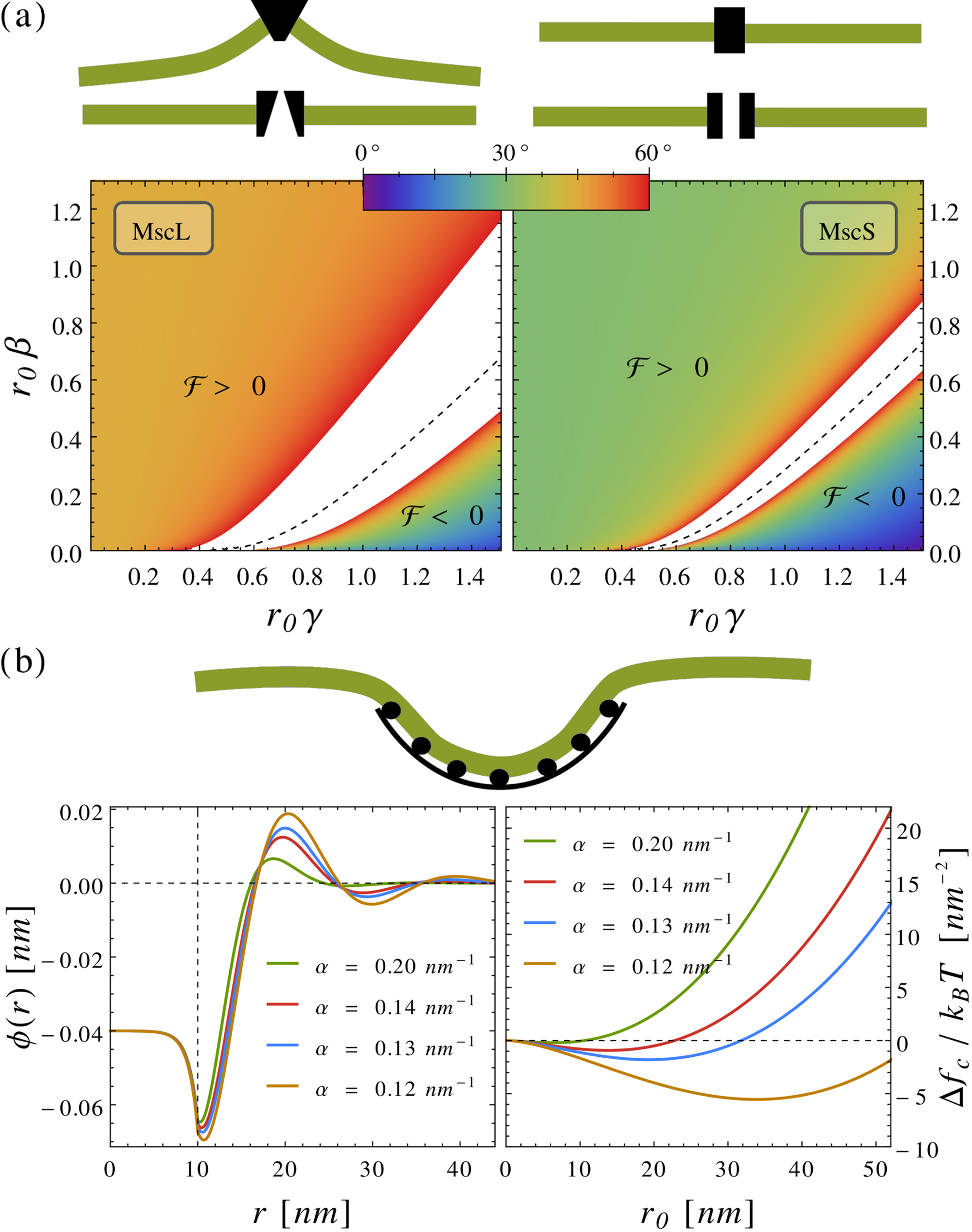
Mechanosensitive membrane channels have been widely studied and reveal the interplay between the biological function of transmembrane proteins and the adjacent membrane structure and composition. Through conformational changes from a closed to an open state that allows the passage of solvent through the membrane, they can equilibrate an osmotic imbalance between the interior and exterior of cells Perozo (2006); Booth et al. (2007); Haswell et al. (2011). Although many examples of these channels are found in nature, the bacterial mechanosensitive channels of large (MscL) and small conductance (MscS) are prototypes of such proteins. Experimental studies have have shown that the channel opening probability is related to the membrane tension and the size of the open pore Sukharev et al. (1994); Perozo (2006); Booth et al. (2007); Haswell et al. (2011); Kung et al. (2010); Chiang et al. (2004); Sukharev (2002); Perozo et al. (2002). One possibility is that the channel simply dilates open at high tension but the transition between the closed and open states might also involve, e.g. a change in slope at the protein-membrane interface (here, ) Turner and Sens (2004). In a two-component membrane such a change in boundary conditions between the closed and open states couples to both the shape and asymmetry field in the nearby membrane and hence contributes to a change in the free energy of the channel-membrane system. Here, for simplicity, we consider the angle at the channel wall to be non-zero in a conical closed state and in the open state. We explore the thermodynamic effect of this gating-by-tilt by comparing the deformation energy of the membrane to the experimental estimate of the energy required to open the channels at zero tension, inferred by assuming purely dilational opening. Fig 4(a) shows that the even modest changes in the boundary angle at the face of the channel could give rise to a significant thermodynamic energy under gating-by-tilt. Moreover, a regime is identified in which the membrane can act to close, rather than open, the channel, characterized by a negative total energy relative to the open state, although a similar result was previously identified in a model that neglects spatial variation of coupling to curvature Wiggins and Phillips (2004, 2005). Our results indicate that lipid composition variation, and its coupling to mean curvature, could play a role in regulating the function of mechanosensitive membrane channels.
Finally, the presence of a negative deformation energy in the underdamped regime motivated us to study the thermodynamics of protein coat formation on a membrane. Such coats are important in regulation, e.g. membrane trafficking using clathrin coats, or in infection, where viral coats assemble at the plasma membrane Alberts et al. (2008). Fig 4(b) shows both the compositional field around a protein coat of size and the variation with of , i.e. the change in membrane energy due to coupling to only, scaled by the coat area. Thus, renormalizes the chemical potential for binding of early coat monomers to the membrane, and it is computed by adding both the contribution from the membrane inside (under) and outside the coat Note (2). Two striking features are observed in the underdamped regime. Firstly, this free-energy change can support an initial decrease with coat size. In this case the deformation of the membrane (with its associated composition) is energetically favorable. This is true (even) for membranes that remain thermodynamically stable, i.e. in the absence of bulk instability. This may represent a new mechanism for driving (controlling) coat formation in cells. This might be tested by tracking coat assembly at different (tension), e.g. controlled by micropipette aspiration Evans and Rawicz (1990): We predict a dramatic increase in the rate of assembly near the critical tension . Secondly, the existence of a minimum in corresponds to a characteristic coat size with metastable character; we note that partially formed coats are often observed Heuser (1980).
Acknowledgements.
We acknowledge the stimulating discussions with Dr P. Sens (Paris) and Profs M. Freissmuth and H. Sitte (Vienna) and funding from EPSRC under grant EP/E501311/1 (a Leadership Fellowship to MST).References
- Alberts et al. (2008) B. Alberts, A. Johnson, J. Lewis, M. Raff, K. Roberts, and P. Walter, Molecular Biology of the Cell, 5th ed. (Garland Science, 2008).
- Singer and Nicolson (1972) S. J. Singer and G. L. Nicolson, Science 175, 720 (1972).
- Engelman (2005) D. M. Engelman, Nature 438, 578 (2005).
- Suchyna et al. (2004) T. M. Suchyna, S. E. Tape, R. E. Koeppe, O. S. Andersen, F. Sachs, and P. A. Gottlieb, Nature 430, 235 (2004).
- Moe and Blount (2005) P. Moe and P. Blount, Biochemistry 44, 12239 (2005).
- Krepkiy et al. (2009) D. Krepkiy, M. Mihailescu, J. A. Freites, E. V. Schow, D. L. Worcester, K. Gawrisch, D. J. Tobias, S. H. White, and K. J. Swartz, Nature 462, 473 (2009).
- Milescu et al. (2009) M. Milescu, F. Bosmans, S. Lee, A. A. Alabi, J. I. Kim, and K. J. Swartz, Nature Struct. Biol. 16, 1080 (2009).
- Lee (2004) A. G. Lee, Biochim. Biophys. Acta 1666, 62 (2004).
- Mitra et al. (2004) K. Mitra, I. Ubarretxena-Belandia, T. Taguchi, G. Warren, and D. M. Engelman, Proc. Natl. Acad. Sci. USA 101, 4083 (2004).
- Dowhan et al. (2004) W. Dowhan, E. Mileykovskaya, and M. Bogdanov, Biochim. Biophys. Acta 1666, 19 (2004).
- Nyholm et al. (2007) T. K. M. Nyholm, S. Ozdirekcan, and J. A. Killian, Biochemistry 46, 1457 (2007).
- Andersen and Koeppe (2007) O. S. Andersen and R. E. Koeppe, Annu. Rev. Biophys. Biomol. Struct. 36, 107 (2007).
- Phillips et al. (2009) R. Phillips, T. Ursell, P. Wiggins, and P. Sens, Nature 459, 379 (2009).
- Lundbaek et al. (2010) J. A. Lundbaek, S. A. Collingwood, H. I. Ingólfsson, R. Kapoor, and O. S. Andersen, J. R. Soc. Interface 7, 373 (2010).
- Canham (1970) P. B. Canham, J. Theor. Biol. 26, 61 (1970).
- Huang (1986) H. W. Huang, Biophys. J. 50, 1061 (1986).
- Aranda-Espinoza et al. (1996) H. Aranda-Espinoza, A. Berman, N. Dan, P. Pincus, and S. A. Safran, Biophys. J. 71, 648 (1996).
- Nielsen et al. (1998) C. Nielsen, M. Goulian, and O. S. Andersen, Biophys. J. 74, 1966 (1998).
- Mondal et al. (2014) S. Mondal, G. Khelashvili, and H. Weinstein, Biophys. J. 106, 2305 (2014).
- Sens and Safran (2000) P. Sens and S. Safran, Eur. Phys. J. E 1, 237 (2000).
- Fournier (1999) J.-B. Fournier, Eur. Phys. J. B 11, 261 (1999).
- Weikl et al. (1998) T. Weikl, M. Kozlov, and W. Helfrich, Phys. Rev. E 57, 6988 (1998).
- Wiggins and Phillips (2004) P. Wiggins and R. Phillips, Proc. Natl. Acad. Sci. USA 101, 4071 (2004).
- Wiggins and Phillips (2005) P. Wiggins and R. Phillips, Biophys. J. 88, 880 (2005).
- Dan et al. (1993) N. Dan, P. Pincus, and S. A. Safran, Langmuir 9, 2768 (1993).
- Goulian et al. (1993) M. Goulian, R. Bruinsma, and P. Pincus, Europhys. Lett. 22, 145 (1993).
- Kim et al. (1998) K. S. Kim, J. Neu, and G. Oster, Biophys. J. 75, 2274 (1998).
- Turner and Sens (1999) M. S. Turner and P. Sens, Biophys. J. 76, 564 (1999).
- Markin and Sachs (2004) V. S. Markin and F. Sachs, Phys. Biol. 1, 110 (2004).
- Ursell et al. (2007) T. Ursell, K. C. Huang, E. Peterson, and R. Phillips, PLoS Comput. Biol. 3, e81 (2007).
- Haselwandter and Phillips (2013a) C. A. Haselwandter and R. Phillips, Europhys. Lett. 101, 68002 (2013a).
- Haselwandter and Phillips (2013b) C. A. Haselwandter and R. Phillips, PLoS Comput. Biol. 9, e1003055 (2013b).
- Leibler (1986) S. Leibler, J. Phys. France 47, 507 (1986).
- Leibler and Andelman (1987) S. Leibler and D. Andelman, J. Phys. France 48, 2013 (1987).
- Andelman et al. (1992) D. Andelman, T. Kawakatsu, and K. Kawasaki, Europhys. Lett. 19, 57 (1992).
- Helfrich (1973) W. Helfrich, Z. Naturforsch C. 28, 693 (1973).
- Seifert (1997) U. Seifert, Adv. Phys. 46, 13 (1997).
- Israelachvili et al. (1976) J. N. Israelachvili, D. J. Mitchell, and B. W. Ninham, J. Chem. Soc., Faraday Trans. 72, 1525 (1976).
- Gruner (1989) S. M. Gruner, J. Phys. Chem. 93, 7562 (1989).
- McMahon and Gallop (2005) H. T. McMahon and J. L. Gallop, Nature 438, 590 (2005).
- Callan-Jones et al. (2011) A. Callan-Jones, B. Sorre, and P. Bassereau, Cold Spring Harb Perspect Biol. 3 (2011).
- Sunil Kumar et al. (1999) P. B. Sunil Kumar, G. Gompper, and R. Lipowsky, Phys. Rev. E 60, 4610 (1999).
- Schick (2012) M. Schick, Phys. Rev. E 85, 1 (2012).
- Seifert (1993) U. Seifert, Phys. Rev. Lett. 70, 1335 (1993).
- Nelson et al. (2004) D. Nelson, T. Piran, and S. Weinberg, Statistical Mechanics of Membranes and Surfaces, 2nd ed. (World Scientific Publishing Company, 2004).
- Note (1) The sign choice of is simply a convention. The fields and are invariant under a sign change in , itself related to the convention of a direction for “up” and whether one refers to the enrichment of a component that couples to positive curvature (depending on one’s choice for “up”) or the depletion of one that couples to negative curvature.
- Note (2) See Supplemental Material, which includes Refs. Riley et al. (2006); Byron and Fuller (1992); Gradshteyn et al. (2000).
- Note (3) Although Dirichlet boundary conditions could be used as well, our choice gives the ground state solution in the absence of any constraints on the composition asymmetry field at the boundary; see Ref. Note (2) for more details.
- Shimada et al. (2007) A. Shimada, H. Niwa, K. Tsujita, S. Suetsugu, K. Nitta, K. Hanawa-Suetsugu, R. Akasaka, Y. Nishino, M. Toyama, L. Chen, Z.-J. Liu, B.-C. Wang, M. Yamamoto, T. Terada, A. Miyazawa, A. Tanaka, S. Sugano, M. Shirouzu, K. Nagayama, T. Takenawa, and S. Yokoyama, Cell 129, 761 (2007).
- Judge and Watts (2011) P. J. Judge and A. Watts, Curr. Opin. Chem. Biol. 15, 690 (2011).
- Loura and Prieto (2011) L. M. S. Loura and M. Prieto, Front. Psychol. 2, 82 (2011).
- Yamashita et al. (2005) A. Yamashita, S. K. Singh, T. Kawate, Y. Jin, and E. Gouaux, Nature 437, 215 (2005).
- Singh et al. (2008) S. K. Singh, C. L. Piscitelli, A. Yamashita, and E. Gouaux, Science 322, 1655 (2008).
- Note (4) Typically the membrane correlation length might be Sheetz and Dai (1996). Eq. (1) can be associated with a typical interfacial width for a strongly segregated system of a few nm, say , and a line tension of about a pN ( pN) Tian et al. (2007), which combine to give . Sorting of strongly curvature-coupling components into membrane tubes gives an upper bound of and hence a value for Aimon et al. (2014), indicating that the all regimes discussed in the main text are accessible. While the use of a phase separated system to estimate the parameters and is questionable, given that they are motivated within a model in which the system remains essentially one phase, with very small, we are reassured that the primary limit on the accessibility of the underdamped regime is that is not too large. Given that spontaneous phase separation is often seen on biological membranes there would seem to be no lower limit on a reasonable magnitude for and hence .
- Abramowitz and Stegun (1965) M. Abramowitz and I. A. Stegun, Handbook of Mathematical Functions (Dover Publications Inc., 1965).
- Evans and Rawicz (1990) E. Evans and W. Rawicz, Phys. Rev. Lett. 64, 2094 (1990).
- Bo and Waugh (1989) L. Bo and R. Waugh, Biophys. J. 55, 509 (1989).
- Kummrow and Helfrich (1991) M. Kummrow and W. Helfrich, Phys. Rev. A 44, 8356 (1991).
- Pécréaux et al. (2004) J. Pécréaux, H.-G. Döbereiner, J. Prost, J.-F. Joanny, and P. Bassereau, Eur. Phys. J. E 13, 277 (2004).
- Chiang et al. (2004) C.-S. Chiang, A. Anishkin, and S. Sukharev, Biophys. J. 86, 2846 (2004).
- Sukharev (2002) S. Sukharev, Biophys. J. 83, 290 (2002).
- Perozo (2006) E. Perozo, Nat. Rev. Mol. Cell Biol. 7, 109 (2006).
- Booth et al. (2007) I. R. Booth, M. D. Edwards, S. Black, U. Schumann, and S. Miller, Nature Rev. Microbiol. 5, 431 (2007).
- Haswell et al. (2011) E. S. Haswell, R. Phillips, and D. C. Rees, Structure 19, 1356 (2011).
- Sukharev et al. (1994) S. I. Sukharev, P. Blount, B. Martinac, F. R. Blattner, and C. Kung, Nature 368, 265 (1994).
- Kung et al. (2010) C. Kung, B. Martinac, and S. Sukharev, Annu. Rev. Microbiol. 64, 313 (2010).
- Perozo et al. (2002) E. Perozo, A. Kloda, D. M. Cortes, and B. Martinac, Nature Struct. Biol. 9, 696 (2002).
- Turner and Sens (2004) M. S. Turner and P. Sens, Phys. Rev. Lett. 93, 118103 (2004).
- Heuser (1980) J. Heuser, J. Cell. Biol. 84, 560 (1980).
- Riley et al. (2006) K. F. Riley, M. P. Hobson, and S. J. Bence, Mathematical Methods for Physics and Engineering, 3rd ed. (Cambridge University Press, 2006).
- Byron and Fuller (1992) F. W. Byron and R. W. Fuller, The Mathematics of Classical and Quantum Physics (Dover Publications Inc., 1992).
- Gradshteyn et al. (2000) I. S. Gradshteyn, I. M. Ryzhik, A. Jeffrey, and D. Zwillinger, Table of Integrals, Series, and Products, 6th ed. (Academic Press, 2000).
- Sheetz and Dai (1996) M. P. Sheetz and J. Dai, Trends Cell Biol. 6, 85 (1996).
- Tian et al. (2007) A. Tian, C. Johnson, W. Wang, and T. Baumgart, Phys. Rev. Lett. 98, 208102 (2007).
- Aimon et al. (2014) S. Aimon, A. Callan-Jones, A. Berthaud, M. Pinot, G. E. S. Toombes, and P. Bassereau, Dev. Cell 28, 212 (2014).