Generation of magnonic spin wave traps
Abstract
Spatially resolved measurements of the magnetization dynamics induced by an intense laser pump-pulse reveal that the frequencies of resulting spin wave modes depend strongly on the distance to the pump center. This can be attributed to a laser generated temperature profile. On a CoFeB thin film magnonic crystal, Damon-Eshbach modes are expected to propagate away from the point of excitation. The experiments show that this propagation is frustrated by the strong temperature gradient.
pacs:
75.78.-n, 75.30.Ds, 75.70.Ak, 75.40.Gb,The manipulation of spin wave frequency and propagation characteristics are of great interest for the design of switching devices such as logic gates in the field of spintronics, and the number of studies in this field grows rapidly Kruglyak et al. (2010); Lenk et al. (2011). The most promising techniques include (i) current-injected magnetic solitons in thin films with perpendicular anisotropy, which could transmit information directly or alternatively be used to selectively influence another spin wave’s propagation Mohseni et al. (2013), and (ii) a change in the ferromagnet’s temperature and therewith its saturation magnetization. The latter can either be brought about by direct contact with e.g. a peltier element, which has been demonstrated by Brillouin-Light-Scattering (BLS) experiments on YIG waveguides Obry et al. (2012), or it can be optically induced: The authors of a recent study Kolokoltsev et al. (2012) were able to show that by punctually heating up the signal conducting stripline in their network analyzer configuration by up to using a focused cw laser, the magnetostatic surface spin waves propagating along the stripline could be trapped in the resulting potential well. In this letter, we address the generation of a spin wave trap on a magnonic crystal by means of a temperature gradient induced by intense laser pulses.
In contrast to the experiments mentioned above, rich magnetization dynamics can be produced without any need for direct contact with the sample by using short optical pulses. One approach is using the inverse Faraday effect, which in combination with a spatially shaped pump spot can create propagating droplets of backward volume magnetostatic waves Satoh et al. (2012). On the other hand, the technique applied in this work relies on a thermally induced anisotropy field pulse in the sample to induce magnetization oscillations. A common method to access these dynamics, described by the Landau-Lifshitz model of magnetization precession, makes use of the magneto-optical Kerr effect (MOKE) Lenk et al. (2010). Both temporal and spatial information can be obtained by applying time resolved scanning Kerr microscopy (TRSKM). Using this technique, propagating spin wave modes have been observed by focusing pump pulses with a full width half maximum (FWHM) of only on a thin Permalloy film Au et al. (2013). The spin wave spectrum originating from such optical excitation is usually quite broad: Ultrafast demagnetization leads to a dense population of high energy excitations which then gradually decays into lower energy spin wave modes on a timescale of a few picoseconds. Energy transfer from high frequency to low frequency spin waves after excitation by short microwave pumping pulses has been systematically studied by Brillouin-Light-Scattering and it was shown that this mechanism leads to the formation of Bose-Einstein condensates if the pumping is strong enough Demidov et al. (2008). The result is an overpopulation of the lowest energy states which on a continuous film are given by the uniform precession or Kittel mode and by a series of perpendicular standing spin waves. Using microstructured magnetic films, so-called magnonic crystals, energy is also transferred into a Damon-Eshbach type mode whose frequency can be tuned in a wide range by choosing appropriate lattice parameters Ulrichs et al. (2010).
In this work, we use CoFeB as the sample material due to its low Gilbert damping and high saturation magnetization. Ultrashort laser pulses from a regeneratively amplified Ti:Sapphire system are used to (i) excite the magnetization dynamics, (ii) probe the magnetic response of the magnonic crystal, and (iii) create a spin wave trap / resonator.
The software package COMSOL has been used to calculate the thermal response of a thin a metallic film to ultrafast laser excitation. The sample system for these calculations consisted of of ruthenium capping a cobalt-iron-boron (Co20Fe60B20) magnetic film on a Si(100) substrate. The heat diffusion equation,
is solved in rotational symmetry for isolating sample edges and a fixed temperature at the bottom of the substrate using the material parameters listed in table 1.
| Material | (kg m-3) | (J kgK-1) | (W mK-1) | |
|---|---|---|---|---|
| Ru | 12370 Walter et al. (2011) | 238 Walter et al. (2011) | 117 Walter et al. (2011) | 0.70 Hass and Hunter (1981) |
| Co20Fe60B20 | 7700 O’Handley et al. (1976) | 440 Walter et al. (2011) | 87 Walter et al. (2011) | 0.72 Johnson and Christy (1974) |
| Si | 2330 Enghag (2004) | 712 Enghag (2004) | 153 Enghag (2004) | - |
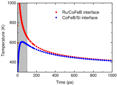
Starting from equilibrium at room temperature, energy is deposited by an ultrashort laser pulse with a duration of . The optical penetration depth is in accordance with the value for ruthenium Palik (1985) as well as with the average value of cobalt and iron, respectively Johnson and Christy (1974). In the film plane, a Gaussian intensity profile is assumed with a FWHM of . The energy carried by each pulse amounts to a total of , as will be the case in the experiments presented below. The results of the simulation are shown in Fig. 1: In the beginning, the laser pulse produces a sudden rise in temperature. After thermalization of the optically excited electrons and equilibration of the spin and phonon subsystems, known to take place on timescales of fs and ps respectively, the modeling yields an effective sample temperature, i.e the temperature of the magnetic system. During the first ps the spatial as well as the temporal heat gradient are rather large, whereas at later times the temperature remains at a high mean value and only a negligible depth profile remains for most part of the CoFeB film.
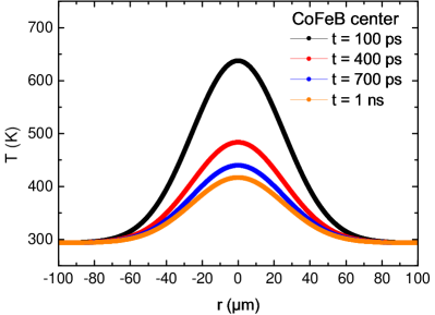
While the temperature is mainly homogeneous throughout the sample depth, it changes significantly across its plane, as shown in Fig. 1 (bottom). The Gaussian distribution of laser intensity in the pump spot produces a temperature profile that persists longer than the lifetime of the observed coherent spin wave modes. During this time (up to 1 ns), no significant heat transport takes place on a micrometer scale and the FWHM of the lateral temperature distribution remains unchanged. In accordance with the Curie-Weiss law, the temperature increase quenches sample’s saturation magnetization so that a potential well is formed which effectively prevents the escape of spin waves from this region.
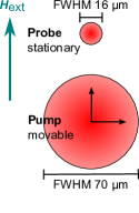
Magnetization dynamics experiments were conducted on amorphous -thick Co40Fe40B20 films magnetron-sputtered onto a Si(100) substrate and capped with a Ru layer to prevent oxidation. Ultrashort laser pulses (central wavelength , pulse duration ) amplified by a Coherent RegA 8040 regenerative amplifier were used to excite and detect the magnetization dynamics in a pump-probe experiment as described in ref. Djordjevic et al. (2006). The experimental parameters are analogous to those in the presented COMSOL simulation. Additionally, an external magnetic field is applied at with respect to sample plane. The experiments were performed separating the pump and probe spots on the sample and measuring the magnetization dynamics as a function of pump-probe distance, thus allowing us to determine the shift in magnetization oscillation frequency along the temperature gradient (i.e the spin wave well).
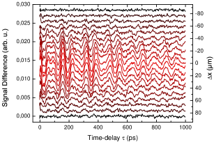
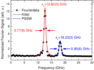
Using variable time delay between pump and probe pulses, the time-resolved magneto-optical Kerr effect (TRMOKE) reveals magnetization precession on timescales of up to , which changes phase by between positive and negative (i.e. reversed) field directions. For the quantitative analysis, the difference between both field directions is calculated. An incoherent background remains which originates from high frequency and high- magnons excited by the intense pump beam Lenk et al. (2010). After respective subtraction, a fast Fourier transform is performed and the resulting peaks in frequency domain can be analyzed (see figure 2).
The dataset presented in Fig. 2 has been obtained on a continuous CoFeB reference film of thickness . Two modes of magnetic precession are observed. Based on earlier results, these can be identified as the in-phase precession of all spins (uniform Kittel mode) at and a first order (i.e. ) standing spin wave with wave vector perpendicular to the sample plane (PSSW) at Ulrichs et al. (2010); Lenk et al. (2010). Both Kittel and PSSW modes have no wave vector components in the lateral direction. In other words, they do not propagate on the sample but have a rather localized character at the spot of (optical) excitation. Consequently, spatially resolved measurements should show no significant precession outside of the pump laser spot.
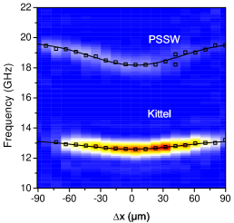
Fig. 3 (top) shows the color-coded Fourier power of magnetization oscillation as a function of spatial separation between the centers of pump and probe spot. In this dataset, pump displacement was performed parallel to the external field direction. For better comparison of different measurements, the Fourier power is normalized to the sum of all transformed data points, allowing to see how much the signal stands out against background noise. On the one hand, the precessional amplitude (represented by the color code) depends on the distance to the center of the pump pulse. This is due to the laser intensity profile and the localized character of the observed modes. On the other hand, also the frequency is strongly position dependent. The latter effect can be explained as a consequence of the increased disorder caused by the intense heating, which leads to a decrease in saturation magnetization and therefore to a change of the spin wave spectrum.
Using the theoretical dispersion of Kittel mode in the Landau-Lifshitz formalism,
| (1) |
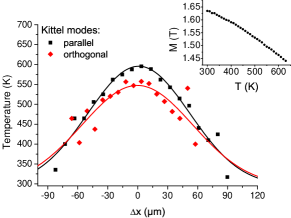
the profile in observed frequency can be used to calculate the laser induced temperature increase: In Eq. (1) the saturation magnetization is regarded as the only free parameter, such that . Comparing this with the experimentally observed frequency profile (open squares in Fig. 3(top)), a corresponding profile in magnetization is calculated. The magnetization profile is then compared to the magnetization curve obtained for a CoFeB sample of equal thickness and composition using a Vibrating Sample Magnetometer (VSM) (inset in Fig. 3). The resulting position dependent temperature profile is shown in figure 3 (bottom).
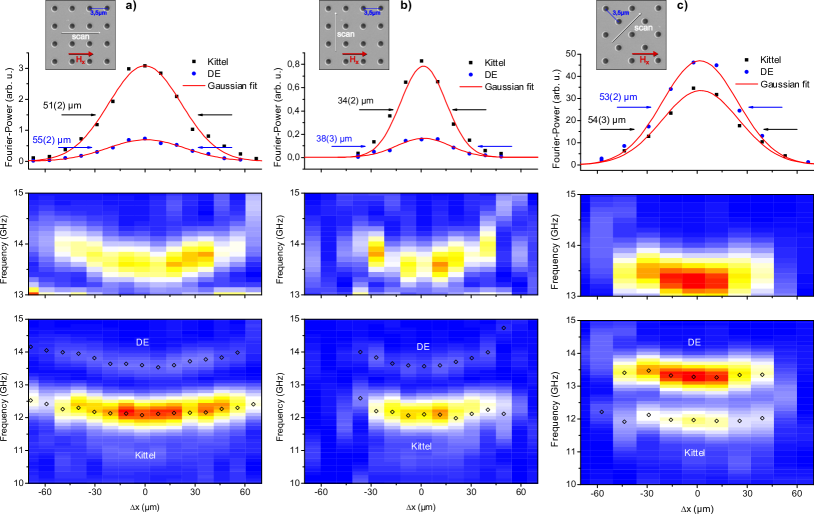
While we expect that the Kittel and PSSW do not propagate across the sample plane, in magnonic crystals composed of periodically arranged antidots the excitation of dipolar Damon-Eshbach surface waves (DE) of selective wave vector has been shown Ulrichs et al. (2010). This provides a possibility for information transport via local excitation of propagating spin waves followed by a spatially separated detection. Magnetization dynamics measurement on a magnonic crystal and its analysis are presented in Fig. 4. In these measurements, pump and probe beam were separated (a) parallel, (b) orthogonal and (c) at an angle of 45∘ to the direction of the external magnetic field. In contrast to the measurements on the continuous film, an additional (magnonic) Damon-Eshbach mode is visible (bottom images of Fig. 4). As has already been observed above for the Kittel and PSSW modes, the precession frequency of DE mode shows a Gaussian dependence on position with a minimal frequency at the position of maximal pump intensity, which is caused by the increased temperature. The measurements should reveal a second effect, though: Damon-Eshbach surface waves are known to propagate mainly orthogonal to the direction of the in-plane magnetic field. In magnonic antidot crystals, only a DE mode parallel to the direction of the smallest hole-to-hole distance is detected Ulrichs et al. (2010). Since the propagation direction of a dipolar surface wave is rotated by 180∘ when the external field is reversed, and because the data shows the difference between the signals measured for positive and negative field direction, a spatial widening of the DE-mode is expected. From the damping time in the TRMOKE data of , the propagation length of spin waves in CoFeB can be estimated to be at least so that detection of the DE-mode should be possible well outside of the pump spot.
The magnetization oscillation’s Fourier power for each measurement is plotted in the top row of Fig. 4. Solid lines represent Gaussian fits to the experimental points, the fitted widths amount to around . By comparison of the (localized) Kittel and the (propagating) DE mode, the surface mode’s propagation characteristics can be analyzed. Since both modes show the same FWHM, it can be concluded that no propagation occurs into the direction of the external field (Fig. 4(a)), as could be presumed. Peculiarly, for the orthogonal and 45∘ configuration there is no significant broadening of the DE mode either, meaning that there is no propagation out of the excitation spot.
Two damping mechanisms can be considered to explain the observed behavior: On the one hand, the pump pulse creates a magnon population of very high density, rendering the picture of ballistic spin wave propagation invalid. Instead, intense scattering takes place that results in a strong overall damping. On the other hand, a spin wave travelling away from the spot of excitation would propagate towards an increasing effective saturation magnetization due to the heat gradient imposed by the pump laser. As we have shown, this change in saturation magnetization drastically impacts the supported frequency, and consequently must result in repeated scattering of the DE-magnons.
The presented experiments and their analysis carry two important points: Firstly, an effective spin-wave well is formed by the local absorption of the optical pump pulse. In analogy to Kolokoltsev et al. (2012) we observe a magnetization profile that follows the intensity profile of optical excitation and strongly influences the observed spin wave spectrum. Despite the ultrashort character of the excitation, the temperature profile remains in effect over the complete range of observed time delays, namely up to . In view of magnonics and their applications, a possible scenario is an optical lattice on a continuous magnetic film. Effectively, a dynamic magnonic crystal can be created in this way without limitations by lithography.
Secondly, we observed the absence of spin wave propagation away from the excitation spot, which is mainly caused by two distinct mechanisms: As discussed in references Djordjevic and Münzenberg (2007); Lenk et al. (2010, 2011), optical spin wave excitation is highly non-equilibrium. The resulting spin wave density is far above the ballistic limit, thus leading to a high probability for scattering between spin waves and a drastically reduced mean free path. A temperature gradient imposes additional scattering as spin waves are continuously reflected when entering a colder region with higher saturation magnetization. This effect might be used to trap spin waves or selectively block their propagation and must certainly be considered when optically exciting propagating surface waves.
Acknowledgements.
Maria Mansurova thanks Soham Manni for assistance and discussion during VSM measurements. We thank the German Research Foundation (DFG) for funding through MU 1780/ 6-1 Photo-Magnonics, SPP 1538 SpinCaT and SFB 1073.References
- Kruglyak et al. (2010) V. V. Kruglyak, S. O. Demokritov, and D. Grundler, J. Phys. D: Appl. Phys. 43, 264001 (2010), URL http://iopscience.iop.org/0022-3727/43/26/260301.
- Lenk et al. (2011) B. Lenk, H. Ulrichs, F. Garbs, and M. Münzenberg, Phys. Rep. 507, 107 (2011), ISSN 0370-1573, URL http://www.sciencedirect.com/science/article/pii/S0370157311001694.
- Mohseni et al. (2013) S. M. Mohseni, S. R. Sani, J. Persson, T. N. A. Nguyen, S. Chung, Y. Pogoryelov, P. K. Muduli, E. Iacocca, A. Eklund, R. K. Dumas, et al., Science 339, 1295 (2013), ISSN 1095-9203, URL https://www.sciencemag.org/content/339/6125/1295.
- Obry et al. (2012) B. Obry, V. I. Vasyuchka, A. V. Chumak, A. A. Serga, and B. Hillebrands, Appl. Phys. Lett. 101 (2012), URL http://scitation.aip.org/content/aip/journal/apl/101/19/10.1063/1.4767137.
- Kolokoltsev et al. (2012) O. Kolokoltsev, N. Qureshi, E. Mejía-Uriarte, and C. L. Ordóñez Romero, Journal of Applied Physics 112, 013902 (2012), URL http://scitation.aip.org/content/aip/journal/jap/112/1/10.1063/1.4730927.
- Satoh et al. (2012) T. Satoh, Y. Terui, R. Moriya, B. A. Ivanov, K. Ando, E. Saitoh, T. Shimura, and K. Kuroda, Nature Photon. 6, 662 (2012), ISSN 1749-4885, URL http://www.nature.com/nphoton/journal/v6/n10/full/nphoton.2012.218.html.
- Lenk et al. (2010) B. Lenk, G. Eilers, J. Hamrle, and M. Münzenberg, Phys. Rev. B 82, 134443 (2010), URL http://link.aps.org/doi/10.1103/PhysRevB.82.134443.
- Au et al. (2013) Y. Au, M. Dvornik, T. Davison, E. Ahmad, P. S. Keatley, A. Vansteenkiste, B. Van Waeyenberge, and V. V. Kruglyak, Phys. Rev. Lett. 110, 097201 (2013), URL http://link.aps.org/doi/10.1103/PhysRevLett.110.097201.
- Demidov et al. (2008) V. E. Demidov, O. Dzyapko, S. O. Demokritov, G. A. Melkov, and A. N. Slavin, Phys. Rev. Lett. 100, 047205 (2008), URL http://link.aps.org/doi/10.1103/PhysRevLett.100.047205.
- Ulrichs et al. (2010) H. Ulrichs, B. Lenk, and M. Münzenberg, Applied Physics Letters 97, 092506 (2010), URL http://scitation.aip.org/content/aip/journal/apl/97/9/10.1063/1.3483136.
- Walter et al. (2011) M. Walter, J. Walowski, V. Zbarsky, M. Münzenberg, M. Schäfers, D. Ebke, G. Reiss, A. Thomas, P. Peretzki, M. Seibt, et al., Nat. Mater. 10, 742 (2011), ISSN 1476-1122, URL http://www.nature.com/nmat/journal/v10/n10/abs/nmat3076.html#supplementary-information.
- Hass and Hunter (1981) G. Hass and W. R. Hunter, Appl. Opt. 20, 2334_1 (1981), URL http://ao.osa.org/abstract.cfm?URI=ao-20-14-2334_1.
- O’Handley et al. (1976) R. C. O’Handley, R. Hasegawa, R. Ray, and C.-P. Chou, Appl. Phys. Lett. 29, 330 (1976), URL http://scitation.aip.org/content/aip/journal/apl/29/6/10.1063/1.89085.
- Johnson and Christy (1974) P. B. Johnson and R. W. Christy, Phys. Rev. B 9, 5056 (1974), URL http://link.aps.org/doi/10.1103/PhysRevB.9.5056.
- Enghag (2004) P. Enghag, Encyclopedia of the Elements - data, history, processing, applications (Wiley VCH, 2004).
- Palik (1985) E. D. Palik, ed., Handbook of Optical Constants of Solids (Academic Press, Boston, 1985).
- Djordjevic et al. (2006) M. Djordjevic, G. Eilers, A. Parge, M. Münzenberg, and J. S. Moodera, J. Appl. Phys. 99 (2006), URL http://scitation.aip.org/content/aip/journal/jap/99/8/10.1063/1.2177141.
- Djordjevic and Münzenberg (2007) M. Djordjevic and M. Münzenberg, Phys. Rev. B 75, 012404 (2007), URL http://link.aps.org/doi/10.1103/PhysRevB.75.012404.