Nuclear and electronic resonance spectroscopy of single molecules by radio-frequency scanning tunnelling microscopy
The ongoing miniaturization in nanoscience and -technology challenges the sensitivity and selectivity of experimental analysis methods to the ultimate level of single atoms and molecules. A promising new approach, addressed here, focuses on the combination of two well-established complementary techniques that have proven to be very successful in their own fields: (i) low-temperature scanning tunneling microscopy (STM), offering high spatial resolution for imaging and spectroscopy together with the capability of manipulating single atoms and molecules in a well-controlled manner Chen (2008); (ii) radio-frequency (rf) magnetic resonance techniques, providing paramount analytical power based on a high energy resolution combined with the versatility of being sensitive to a great variety of different properties of matter Abragam and Bleaney (1970); Slichter (1996); Levitt (2001). Here, we demonstrate the successful resonant excitation and detection of nuclear and electronic magnetic transitions of a single quantum spin in a single molecule by rf tunneling of electrons applied through the tip of a modified STM instrument operated at 5 K. The presented rf-STM approach allows the unrivalled spectroscopic investigation of electronic hyperfine levels in single molecules with simultaneous sub-molecular spatial resolution. The achieved single-spin sensitivity represents a ten orders of magnitude improvement compared to existing methods of magnetic resonance – offering, atom-by-atom, unprecedented analytical power and spin control with impact to physics, chemistry, biology, medicine, nanoscience and -technology.
While classical electron- and nuclear magnetic resonance methods utilize electromagnetic fields to excite electron and nuclear spin transitions, electric currents in a magnetic system can efficiently excite its magnetic moments, as well. For instance, dc electron tunneling has been demonstrated to excite magnons Balashov et al. (2006), flip the spin of a single atom Heinrich et al. (2004); Loth et al. (2010), or switch on and off molecular spins Komeda et al. (2011) by inelastic processes. Rf currents through nanoscale magnetic bars have been shown to excite ferromagnetic resonance Fang et al. (2011). Compared to these earlier approaches, the resonant spin excitation in single molecules, described in the present work, is based on simultaneous dc tunneling through molecular electronic levels while superposing an additional rf tunneling current. Tuning the rf component in resonance with different spin transitions is found to affect the molecular electron tunneling conductance. Thereby, conductance measurements during rf excitation enable a new type of “magnetic resonance” spectroscopy at low magnetic fields of only a few mT and excitation frequencies ranging presently between MHz and 4 GHz.
As model system for rf-STM based spin resonance, we have chosen the well-known “terbium double decker” molecule bis(phthalocyaninato)terbium(III) (, Fig. 1a), consisting of a Tb3+ ion sandwiched between two organic phthalocyanine ligands Ishikawa et al. (2003a, b). The Tb3+ ion has an electronic configuration of [Xe] resulting in a total spin of and a total orbital angular momentum of according to Hund’s rules. A strong spin-orbit coupling yields a total electronic angular momentum of . Due to the exceptionally high ligand field induced by the two phthalocyanine ligands (negative axial zero-field splitting) the electron-spin ground state doublet of is well-separated by more than 50 meV from the next higher doublet of Ishikawa et al. (2003a, b, 2004, 2005). This property makes an exceptional single-molecule magnet Gatteschi et al. (2006) that behaves like an Ising spin at temperatures up to Ishikawa et al. (2003b). Since each phthalocyanine ligand carries a nominal charge of , the charge state of molecules is singly negative in solution and bulk phase Ishikawa et al. (2003b, 2005), however, also neutral when adsorbed on a Au(111) surface Katoh et al. (2009); Komeda et al. (2011).
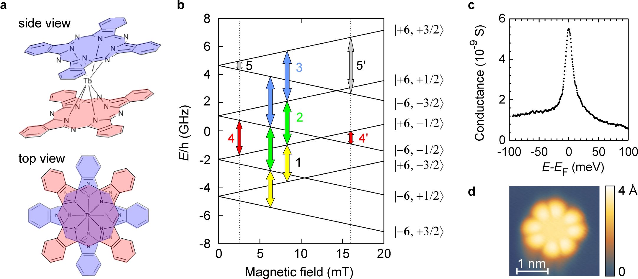
In addition to the electronic spin, the Tb3+ ion carries a nuclear spin of . A large hyperfine interaction (dipole and quadrupole) between the nuclear spin and the electronic angular momentum causes a fourfold splitting of each electronic spin level. The nuclear quadrupole term results in a non-equidistant spacing of the electronic hyperfine levels as illustrated in the Zeeman diagram of Fig. 1b, showing the lowest sub-states of the ground-state doublet of the Tb3+ ion in []0. The labels on the right side mark the eight different hyperfine levels with and nuclear spin and , respectively. The yellow, green and blue arrows mark “purely nuclear” hyperfine transitions, labeled 1–3, with and the electron angular momentum being conserved (). The transitions 1–3 depend only weakly on the magnetic field (nuclear Zeeman effect) due to the large proton-to-electron mass ratio of . In comparison, the red arrow marks a “purely electronic” hyperfine transition with and , labeled 4, which is considerably Zeeman shifted between the two experimental field values of and mT marked by dotted lines. In the following, we demonstrate that the different transitions between electronic hyperfine levels of Tb3+ shown in Fig. 1b can be resonantly excited and detected by rf-STM.
In order to establish well-defined experimental conditions, throughout the present work, we have studied single neutral []0 molecules on Au(111), which are well-known to adsorb non-dissociatively on Au(111) with the phthalocyanine planes aligned parallel to the substrate surface Katoh et al. (2009). Furthermore, neutral []0 molecules exhibit two electronic spin systems: (i) the Tb3+ ion with and (ii) an open-shell system on the phthalocyanine ligands with an electronic spin of . Komeda et al. Komeda et al. (2011) have demonstrated that neutral []0 molecules on Au(111) are clearly identified by a characteristic Kondo signature observed by dc tunneling spectroscopy (Fig. 1c) – as compared to []- anions. Neutral []0 molecules adopt an achiral geometric configuration Ishikawa et al. (2003b); Komeda et al. (2011); Fu et al. (2012) characterized by a 45∘ twist between the lower and upper phthalocyanine ligand (Fig. 1a), showing a characteristic STM topographic appearance Komeda et al. (2011); Fu et al. (2012) with a symmetric 8-lobe structure (Fig. 1d).
The experimental setup is based on a modified commercial STM instrument (Createc) described earlier by our group for the successful detection of rf tunneling current signals in the 100-MHz regime Müllegger et al. (2014a, b). The electronic hyperfine transitions of shown in Fig. 1b correspond to transition frequencies, , between about 100 MHz and 15 GHz. Compared to our previous experiments reported in Refs. Müllegger et al. (2014a, b), in the present work, we increased the experimental bandwidth to GHz utilizing improved rf elements. By operating our rf-STM at two different static magnetic field values at the sample (marked by dotted lines in Fig. 1b), we have been able to resonantly excite as much as 9 different hyperfine transitions of Tb3+ within our experimental bandwidth, including purely nuclear, purely electronic as well as mixed nuclear/electronic spin transitions (see below).
For resonant spin excitation, the STM tip is placed at a fixed position over single molecules at a constant tunneling current between 0.5 and 2 nA. The sample voltage was set at V, facilitating electron tunneling from the STM tip into an unoccupied molecular orbital of the phthalocyanine ligand of /Au(111) Komeda et al. (2011). The -type electronic structure of phthalocyanine permits coupling between the Tb3+ ion and the environment Lodi Rizzini et al. (2011); Komeda et al. (2011). The phthalocyanine provides a transport channel, where the conductance of the ligand is coupled to the spin state of the Tb3+ ion as shown in recent transport experiments on single molecules in a molecular spin transistor Vincent et al. (2012) as well as in a supramolecular spin-valve geometry Urdampilleta et al. (2013). In our rf-STM experiments, the dc tunneling conductance, , is measured while simultaneously modulating the sample bias at a variable radio frequency. The conductance of the phthalocyanine ligand is affected by the excitation of hyperfine-split electronic states of Tb3+ by rf tunneling electrons – a process not restricted by the selection rules of photon-induced magnetic dipole transitions.
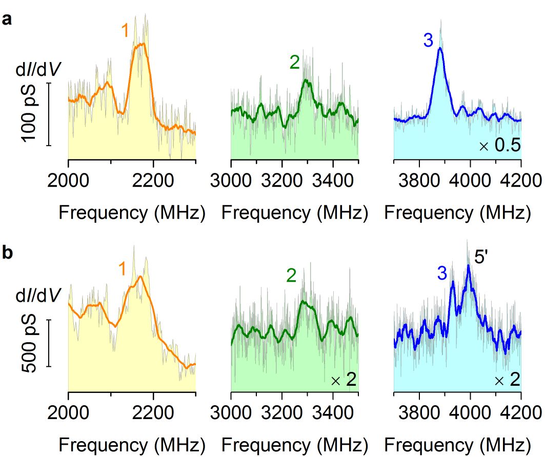
Intriguingly, with the STM tip over single []0 molecules we observe distinct peaks (increased ) at certain rf values, compared to a constant signal over bare Au(111). Figure 2a shows the signal obtained over single molecules while simultaneously ramping the external rf modulation over different frequency ranges in the presence of a static magnetic field of mT at the sample. The experimental peak positions are in very good agreement with the calculated frequency values of the “purely nuclear” hyperfine transitions 1–3 in Fig. 1b within the measurement error (discussed below). The electronic hyperfine levels of Tb3+ in []0 (Fig. 1b) are obtained by numerical diagonalization of the spin Hamiltonian analogous to the calculations by Ishikawa et al. Ishikawa et al. (2003b, 2005) on negatively charged []-. Increasing the static magnetic field to mT leaves the peak positions almost unchanged (Fig. 2b), which is consistent with the expected weak B-field dependence of the transitions 1–3. Notice, that transition 3 corresponds to the left peak of Fig. 3b, right; peak 5’ is a mixed electronic-nuclear transition subject to a strong electronic Zeeman shift (compare Fig. 1b). Both peaks 3 and 5’ in Fig. 2b lie closely adjacent, but are still discernible as separate peaks; thereof, we derive an approximate spectroscopic resolution of our rf-STM method of , which is at least one order of magnitude higher compared to conventional dc-STM spin spectroscopy Heinrich et al. (2004); Fu et al. (2009). Repeating the experiment with different STM tips and different molecules adsorbed over different sites of the Au(111) lattice reproduces the peaks within an experimental uncertainty of MHz.
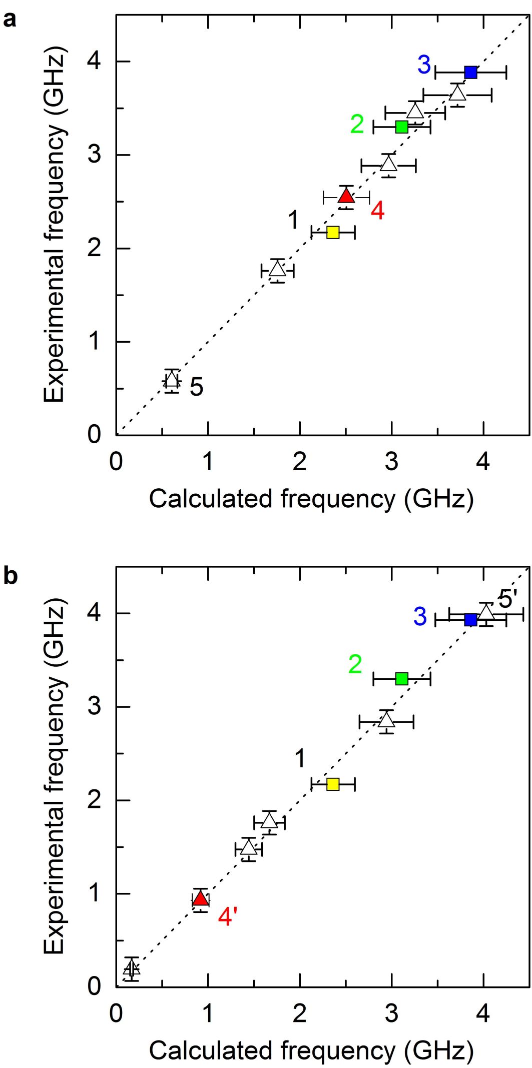
Indeed, we observe peaks that lie very close to the predicted frequencies of all the possible hyperfine transitions of the lowest substates of Tb3+ in that lie within our experimental bandwidth. We performed measurements at two different static magnetic fields of and mT at the sample. The observed transitions include purely nuclear, purely electronic as well as mixed nuclear-electronic hyperfine transitions. Figure 3 juxtaposes the experimental frequencies obtained over single molecules on Au(111) with the calculated hyperfine transition frequencies of Tb3+ for a static magnetic field of 2.5 mT (Fig. 3a) and 16.1 mT (Fig. 3b). Within the error bars of experiment and calculation, all data points lie on the dotted line (guide to the eye), thus revealing a stunningly good agreement for all observed frequencies with the calculated ones. The above results strongly evidence that the frequency values of the resonant peaks observed by rf-STM, indeed, relate to the transition frequencies of different hyperfine levels of the Tb3+ ion in adsorbed on Au(111). Compared to dc-STM based spin spectroscopy performed Heinrich et al. (2004); Fu et al. (2009), the magnetic excitation energies of only a few micro-electronvolts, detected by our rf-STM, are three orders of magnitudes lower than the mean thermal energy (few milli-electronvolt) of the sample at 5 K. Whenever a new method under test, such as our rf-STM, is capable of correctly and unambiguously detecting the manifold of different transition frequencies simultaneously, this can be taken as strong evidence confirming proper functioning. In comparison, no signatures of the magnetic moment of the Tb3+ ion have been detected by conventional dc-STM tunneling spectroscopy Komeda et al. (2011).
In Fig. 3 the experimental error bars (vertical) of GHz for hyperfine transitions with electronic spin flips , plotted as triangles (), are dominated by the uncertainty of the static magnetic field of our experimental setup ( mT). For all transitions, the different adsorption sites of different molecules investigated by rf-STM cause a measurement uncertainty of about GHz; this is the dominant contribution to the experimental error bar of the “purely nuclear” transitions plotted as boxes (). In addition, we consider the uncertainty of the calculated values (horizontal in Fig. 3), which are based on numerical parameters obtained from negatively charged []- in bulk phase Ishikawa et al. (2005). In comparison, single neutral []0 molecules adsorbed on Au(111), studied in the present work, are expected to exhibit a different ligand field and hyperfine parameters due to the different charge state and molecule-substrate interaction – as recently pointed out by Urdampilleta et al. Urdampilleta et al. (2013). The error bars of % represent the approximate deviations of the ligand field and hyperfine parameters of the adsorbed neutral molecule compared to bulk phase.
In conclusion, our rf-STM results evidence the well-controlled resonant excitation and detection of electronic and nuclear spin transitions with single-spin sensitivity and submolecular spatial resolution combined with fast data acquisition. Compared to classical magnetic resonance methods, the presented approach utilizes rf tunneling electrons and is, thus, not restricted by the selection rules of magnetic dipole transitions. We successfully combined STM with magnetic resonance principles into a new analysis technique easily adaptable to a variety of nanoscale systems in the fields of nanoscience and -technology, molecular electronics/spintronics, quantum computation/information, radical chemistry, chemical and biological catalysis, medical therapeutics. This promises unprecedented possibilities for characterizing and controlling physical properties at the scale of single atoms and molecules – including spin excitation and resonance spectroscopy, magnetic writing/reading, chemical analysis, and characterization of oxidation state.
Methods
molecules were synthesized by the procedure reported in Refs. De Cian et al., 1985; Gonidec et al., 2012 followed by purification in a column chromatograph with silicagel and toluene as an eluent. The single-crystal Au(111) substrate was prepared by repeated cycles of Ar+ ion bombardement and annealing at 700 K. was thermally evaporated at ultra-high vacuum conditions from a quartz crucible at 700 K after thorough degassing for h at 363 K and h at 473 K. The static magnetic field perpendicular to the sample was obtained by mounting a SmCo permanent magnet close to the sample and measured ex situ. STM experiments were performed after in situ transfer of the sample, employing electrochemically etched W tips deoxidized by annealing in vacuum. Impurity- and tip effects were minimized by multiple tip formings between the experiments, resulting in Au-coated tips. The signal was obtained from the first-harmonic tunneling current signal detected by lock-in technique with sinusoidal modulation of the sample voltage at 704 Hz and 10–20 mV peak-to-peak. Reliable tip performance was established by accurately reproducing the characteristic conductance signature of the Au(111) surface state well-known in the literature Chen et al. (1998). For the rf resonance experiments, a sinusoidal ac tunneling voltage from an rf signal generator is coupled in via a bias tee to the sample (further details see Ref. Müllegger et al. (2014b)). The modulation frequency was swept between 150 MHz and 4 GHz, respectively, at a rate of 10 MHz/s. The resulting rf current does not affect the tunnel distance, because the radio frequency is several orders of magnitude larger than, both, the cutoff frequency of the STM feedback loop and the bandwidth of the high-gain current amplifier of the STM. The maximum rf power level at the sample was dBm.
References
- Chen (2008) C. J. Chen, Introduction to Scanning Tunneling Microscopy, 2nd ed. (Oxford University Press, 2008).
- Abragam and Bleaney (1970) A. Abragam and B. Bleaney, Electron Paramagnetic Resonance of Transition Ions (Oxford University Press, Oxford, 1970).
- Slichter (1996) C. P. Slichter, Principles of Magnetic Resonance, 3rd ed. (Springer-Verlag, Berlin, 1996).
- Levitt (2001) M. Levitt, Spin Dynamics: Basics of Nuclear Magnetic Resonance (Wiley, 2001).
- Balashov et al. (2006) T. Balashov, A. F. Takacs, W. Wulfhekel, and J. Kirschner, Phys. Rev. Lett. 97, 187201 (2006).
- Heinrich et al. (2004) A. J. Heinrich, J. A. Gupta, C. P. Lutz, and D. M. Eigler, Science 306, 466 (2004).
- Loth et al. (2010) S. Loth, M. Etzkorn, C. P. Lutz, D. M. Eigler, and A. J. Heinrich, Science 329, 1628 (2010).
- Komeda et al. (2011) T. Komeda, H. Isshiki, J. Liu, Y. F. Zhang, N. Lorente, K. Kato, B. K. Breedlove, and M. Yamashita, Nature Commun. 2, 217 (2011).
- Fang et al. (2011) D. Fang, H. Kurebayashi, J. Wunderlich, K. Vyborny, L. P. Zarbo, R. P. Campion, A. Casiraghi, B. L. Gallagher, T. Jungwirth, and A. J. Ferguson, Nat. Nano. 6, 413 (2011).
- Ishikawa et al. (2003a) N. Ishikawa, M. Sugita, T. Ishikawa, S. Koshihara, and Y. Kaizu, J. Am. Chem. Soc. 125, 8694 (2003a).
- Ishikawa et al. (2003b) N. Ishikawa, M. Sugita, T. Okubo, N. Tanaka, T. Iino, and Y. Kaizu, Inorg. Chem. 42, 2440 (2003b).
- Ishikawa et al. (2004) N. Ishikawa, M. Sugita, T. Ishikawa, S. Koshihara, and Y. Kaizu, The Journal of Physical Chemistry B, J. Phys. Chem. B 108, 11265 (2004).
- Ishikawa et al. (2005) N. Ishikawa, M. Sugita, and W. Wernsdorfer, Angew. Chem. Int. Edit. 44, 2931 (2005).
- Gatteschi et al. (2006) D. Gatteschi, R. Sessoli, and J. Villain, Molecular Nanomagnets (Oxford University Press, 2006).
- Katoh et al. (2009) K. Katoh, Y. Yoshida, M. Yamashita, H. Miyasaka, B. K. Breedlove, T. Kajiwara, S. Takaishi, N. Ishikawa, H. Isshiki, Y. F. Zhang, T. Komeda, M. Yamagishi, and J. Takeya, Journal of the American Chemical Society, J. Am. Chem. Soc. 131, 9967 (2009).
- Fu et al. (2012) Y.-S. Fu, J. Schwöbel, S.-W. Hla, A. Dilullo, G. Hoffmann, S. Klyatskaya, M. Ruben, and R. Wiesendanger, Nano Letters, Nano Lett. 12, 3931 (2012).
- Müllegger et al. (2014a) S. Müllegger, M. Rashidi, K. Mayr, M. Fattinger, A. Ney, and R. Koch, Phys. Rev. Lett. 112, 117201 (2014a).
- Müllegger et al. (2014b) S. Müllegger, A. K. Das, K. Mayr, and R. Koch, Nanotechnology 25, 135705 (2014b).
- Lodi Rizzini et al. (2011) A. Lodi Rizzini, C. Krull, T. Balashov, J. J. Kavich, A. Mugarza, P. S. Miedema, P. K. Thakur, V. Sessi, S. Klyatskaya, M. Ruben, S. Stepanow, and P. Gambardella, Phys. Rev. Lett. 107, 177205 (2011).
- Vincent et al. (2012) R. Vincent, S. Klyatskaya, M. Ruben, W. Wernsdorfer, and F. Balestro, Nature 488, 357 (2012).
- Urdampilleta et al. (2013) M. Urdampilleta, S. Klyatskaya, M. Ruben, and W. Wernsdorfer, Phys. Rev. B 87, 195412 (2013).
- Fu et al. (2009) Y.-S. Fu, T. Zhang, S.-H. Ji, X. Chen, X.-C. Ma, J.-F. Jia, and Q.-K. Xue, Phys. Rev. Lett. 103, 257202 (2009).
- De Cian et al. (1985) A. De Cian, M. Moussavi, J. Fischer, and R. Weiss, Inorganic Chemistry, Inorg. Chem. 24, 3162 (1985).
- Gonidec et al. (2012) M. Gonidec, D. B. Amabilino, and J. Veciana, Dalton Trans. 41, 13632 (2012).
- Chen et al. (1998) W. Chen, V. Madhavan, T. Jamneala, and M. Crommie, Phys. Rev. Lett. 80, 1469 (1998).