Förster-induced energy transfer in functionalized graphene
Abstract
Carbon nanostructures are ideal substrates for functionalization with molecules, since they consist of a single atomic layer giving rise to an extraordinary sensitivity to changes in their surrounding. The functionalization opens a new research field of hybrid nanostructures with tailored properties. Here, we present a microscopic view on the substrate-molecule interaction in the exemplary hybrid material consisting of graphene functionalized with perylene molecules. First experiments on similar systems have been recently realized illustrating an extremely efficient transfer of excitation energy from adsorbed molecules to the carbon substrate - a process with a large application potential for high-efficiency photovoltaic devices and biomedical imaging and sensing. So far, there has been no microscopically founded explanation for the observed energy transfer. Based on first-principle calculations, we have explicitly investigated the different transfer mechanisms revealing the crucial importance of Förster coupling. Due to the efficient Coulomb interaction in graphene, we obtain strong Förster rates in the range of 1/fs. We investigate its dependence on the substrate-molecule distance and describe the impact of the momentum transfer for an efficient energy transfer. Furthermore, we find that the Dexter transfer mechanism is negligibly small due to the vanishing overlap between the involved strongly localized orbital functions. The gained insights are applicable to a variety of carbon-based hybrid nanostructures.
keywords:
American Chemical Society, LaTeXInstitut für Theoretische Physik, Technische Universität Berlin, 10623 Berlin, Germany \alsoaffiliationFritz-Haber-Institut der Max-Planck-Gesellschaft, Faradayweg 4-6, D-14195 Berlin, Germany \abbreviationsIR,NMR,UV
The continuing trend to miniaturization of devices in modern technology leads to fundamental physical limits of applied materials.1, 2 The search for new materials and new functionalities brings hybrid systems into the focus of current research.3, 4 They consist of low-dimensional nanostructures functionalized with single molecules combining the remarkable properties of both subsystems. In particular, carbon nanostructures are excellent substrates, since they offer a variety of metallic and semiconducting systems showing a large sensitivity to changes in their surrounding.5, 6, 7, 8 Non-covalent functionalization based on stacking preserves the intrinsic properties of the substrate to a large extent.9 At the same time, the interaction with the attached molecules induces additional properties desired for specific technological applications.10, 11, 12, 13, 14, 15, 16
First experiments have been realized illustrating the successful functionalization of carbon nanotubes with photoactive
molecules suggesting the design of efficient carbon-based molecular switching.10, 17, 12, 18, 19 Recently, a strong excitation energy transfer has been observed in
perylene- and porphyrin-functionalized carbon nanotubes suggesting efficient photo-detection and light harvesting.20, 21, 22 First studies
on functionalized graphene also reveal high energy transfer rates between the attached molecules and the graphene layer.23
The combination of unique transport properties of graphene including ballistic transport and strong light absorption of organic molecules results in new hybrid nanostructures with large application potential for
high-efficiency photodetectors, biomedical sensors, and photovoltaic devices.23
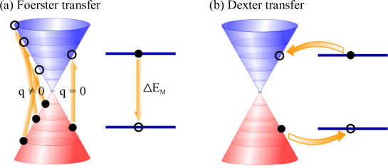
The observed energy transfer could be explained by two major non-radiative energy transfer mechanisms (as depicted in Fig. 1):26 (i) Förster coupling24 describes a direct transfer of energy from the optically excited molecule to graphene. This leads to a quenching of the molecular emission, since the energy is non-radiatively transferred to the electrons in graphene, cf. Fig. 1(a). The Förster transfer rate strongly depends on the molecular transition dipole moment and it exhibits a dependence for hybrid nanostructures on top of a spatially extended two-dimensional substrate27, 23 (in contrast to the well-known scaling for dipolar Förster coupling in molecule-molecule complexes). (ii) Dexter coupling25 is based on a charge transfer between the molecule and graphene states, cf. Fig. 1(b). After the process, the molecule is brought into its ground state and graphene becomes excited and can emit light through carrier recombination. It is a short-range transfer mechanism that directly depends on the spatial overlap of involved molecule and graphene orbital functions resulting in an exponential decay with the substrate-molecule distance .
Recent studies indicate that the observed energy transfer in carbon-based hybrid nanostructures can probably be traced back to a Förster-like transfer process.23, 28
In these studies, the molecule-substrate distance is clearly larger than 10 Å due to the presence of long non-conducting linker molecules. However, for functionalization procedures without such additional molecules, the distance is in the range of just a few Å corresponding to the Van der Waals radius of the involved atoms.20 Here, the Dexter transfer mechanism is expected to be a competing energy transfer mechanism.
In this Letter, we present a systematic first-principle study on the substrate-molecule interaction in the exemplary hybrid system consisting of graphene functionalized with perylene molecules. The obtained insights should be applicable to other carbon-based hybrid nanostructures. We study the molecule-induced changes in the electronic bandstructure and the optical properties of graphene as well as the charge rearrangements within the two sub-systems. We explicitly calculate the Förster transfer rate and investigate its importance as a function of the substrate-molecule distance . Combining first-principle calculations with the tight-binding approximation, we obtain an analytic expression for the transfer rate. Furthermore, we discuss the competing Dexter transfer mechanism by estimating the spatial overlap of the involved substrate and molecule orbitals.
The investigations are based on density
functional theory (DFT) calculations performed within the FHI-aims code package29. It is an all-electron full-potential electronic structure code including numerical atom-centered orbitals, which are very efficient
allowing the investigation of structures containing hundreds of atoms. All calculations are done within the tight settings including a tier 2 basis set for the carbon and hydrogen atoms.29. Calculations with increased accuracy in the basis functions revealed that the chosen settings already lead to converged results with respect to the total energy. We focus on graphene functionalized with perylene molecules (), cf. Figs. 2(a)-(b) illustrating the top and side view of the studied structure. For graphene, we choose a supercell covering unit cells corresponding to 98 carbon atoms with a lattice constant of 1.42 Å. The investigated situation corresponds to a moderate functionalization degree with a molecule-molecule distance of approximately 7 Å. The electron interactions are described within the PBE exchange-correlation functional30 including the recently implemented Van der Waals
correction31 to account for the long-rang van der Waals interaction. The latter plays a fundamental role in describing the weak molecule-nanostructure coupling that is of paramount importance to quantitative estimate the relative contribution of the Dexter transfer mechanism. We also performed additional calculations with the hybrid functional PBE032 and HSE0633, 34
to investigate the alignment of molecular levels.
We found that the molecular HOMO (LUMO) level is located below (above) the Fermi energy in graphene and thus, initial spurious charge transfer does not occur.
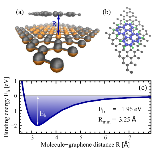
The initial perylene-functionalized graphene structure is fully relaxed using the Broyden-Fletcher-Goldfarb-Shanno method minimizing all force components to values smaller than eV/Å. Figure 2(a) illustrates the hybrid nanostructure after geometric relaxation. The comparison with the perfectly flat graphene layer (orange color) reveals a slight dent of carbon atoms of less than 0.1 Å close to the molecule. This geometric pillow effect is a direct consequence of the presence of the perylene molecule and can be traced back to the Pauli pushback35, 36. It also gives rise to a charge rearrangement, which will be discussed below.
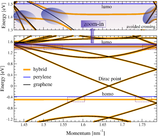
We find an optimal substrate-molecule distance of Å, which is slightly smaller than the initial value of the Van der Waals diameter of the carbon atom, cf. Fig. 2(c). The optimal binding energy at is eV corresponding to a binding energy of meV per atom in the perylene molecule. This is in the expected range for a Van der Waals-induced non-covalent adsorption of the molecule to the graphene surface. The -electronic system of the perylene molecule is linked to the graphene surface via stacking, which is much less invasive compared to the covalent adsorption.3 This can be well observed on the only minor changes in the electronic structure of the substrate, cf. Fig. 3. The unique bandstructure of graphene including the Dirac point and the linear bands is entirely preserved after the functionalization with perylene molecules. The observable changes appear at the points where the molecular HOMO and LUMO levels cross the graphene electronic states, as illustrated in the inset of Fig. 3. Here, the resulting states of the hybrid nanostructure exhibit avoided crossings. This well-known behavior in quantum chemistry is further illustrated within the zoomed-in region around the molecular LUMO level, which anti-crosses the graphene electronic states several times.
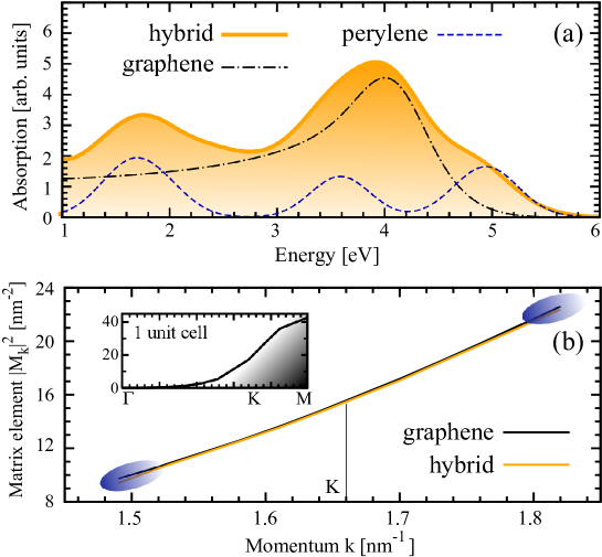
As a direct consequence of the almost completely preserved electronic bandstructure, the optical properties of graphene remain unchanged to a large extent, cf. Fig. 4. The optical absorption of the hybrid nanostructure corresponds to an overlap of the absorption peaks of the pristine graphene and isolated perylene molecule, as shown in Fig. 4(a). The optical matrix element corresponding to the expectation value of the momentum operator7 exhibits only slight changes in the region, where avoided crossing takes place, cf. the blue-shaded circles in Fig. 4(b). Note, however, that the energy transfer within the hybrid nanostructure is not directly included within the DFT treatment and will be further discussed below.
The absorption spectrum of graphene is characterized by the well pronounced peak at approximately 4 eV corresponding to the transition at the saddle point (M point) in the Brillouin zone.37, 38, 9 The widely delocalized electronic system in the perylene molecule gives rise to strong absorption peaks at 1.7 eV, 3.6 eV, and 4.9 eV. The obtained transition energies are lower than in experiment due to the shortcoming of the applied exchange-correlation functional. Calculations based on hybrid functionals give a much better agreement with the experiment. Since in this work, we focus on the Förster and Dexter energy transfer mechanisms between the perylene molecule and graphene, the energetic deviations within PBE exchange-correlation function do not play a qualitative role. Due to the linear gap-less bandstructure of graphene in the relevant energy region, there are always electronic states that are in resonance with the energetically lowest HOMO-LUMO transition of the perylene molecule.
Furthermore, we have investigated the charge rearrangement within the hybrid nanostructure. As already seen in Fig. 2, the adsorbed molecule leads to a spatial pillow-like effect39 pushing the graphene’s carbon atoms further away and giving rise to a small dent of < 0.1 Å . This also affects the mobility of charge carriers within the graphene layer resulting in charge rearrangements. Figure 5(a) shows an surface plot illustrating the molecule-induced charge density difference for the exemplary iso-value of Å3. One can clearly see the accumulation of negative (blue) and positive (red) charges. According to the pillow effect, the electrons are pushed away from the region directly below the molecule. As a result, this region is characterized by a positive charge, i.e. the lack of electrons (red areas). At the same time, electrons accumulate further away at the graphene surface at the graphene-facing side of the molecule (blue areas). To further illustrate the charge distribution along the z-direction (perpendicular to the graphene surface), we show the plane-averaged charge density difference and the charge difference , cf. Figs. 5(b) and (c), respectively. The charge distribution around the graphene layer qualitatively reflects the spatial shape of the most relevant carbon orbitals reaching above and below the graphene sheet. A similar charge distribution can also be observed around the position of the perylene molecule illustrating a positive (negative) charge accumulation slightly below (above) the molecule. Note, however, that the quantitative effect of charge rearrangements is relatively small. The predicted small charge difference of up to is in agreement with what one would expect for a non-covalent functionalization.
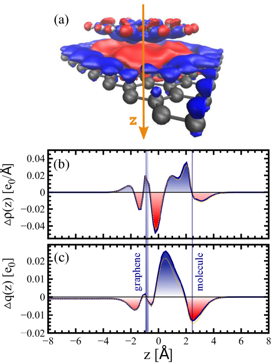
The dashed lines in Fig. 5 reflect the charge distribution obtained within constrained DFT calculations,40 i.e. we imposed particular initial occupations of molecular HOMO and LUMO levels while solving the Kohn-Sham equations. The aim was to investigate the change of the substrate-molecule interaction once the molecule is optically excited. Therefore, we promoted one electron from the HOMO to the LUMO level. The calculations show only marginal changes in the charge distribution (cf. Fig. 5) or in the electronic bandstructure (not shown). This insight is important for the discussion of the excitation energy transfer in the investigated hybrid structure. Time-dependent DFT calculations41 are beyond the scope of this study and will be performed in future work.
After having characterized the perylene-functionalized graphene including its electronic and optical properties, we now focus on the investigation of the possible energy transfer mechanisms in such a hybrid structure, cf. Fig. 1. The Förster and Dexter energy transfer rates can be analytically expressed via the Fermi golden rule
| (1) |
with the momentum-dependent initial and final states of the graphene substrate and the HOMO and LUMO states of the molecule . The delta function makes sure that only energy-conserving processes contribute. The Förster rate is determined by the direct contribution of the Coulomb interaction24
| (2) |
where denotes the elementary charge and the vacuum permitivity. The exchange Coulomb contribution gives the Dexter rate with25
| (3) |
For Dexter coupling, a large spatial overlap between graphene and molecular orbitals ( and ) is of key importance.25, 26 As a result, shows an exponential dependence on the substrate-molecule distance and occurs only for small distances (typically, smaller than 10 Å).26 In contrast, the Förster coupling is dominated by the factor in Eq. (2).
Considering the conventional energy transfer between donor and acceptor molecules, the Förster coupling is based on the dipole-dipole interaction and is characterized by a dependence.24, 42, 26 In the case of functionalized graphene, the substrate is not a spatially localized molecule, but a periodically extended two-dimensional nanostructure. Following the approach of Swathi et al.27, the Förster coupling can be considered as an interaction of the molecular transition dipole located in the electrostatic potential arising from the transition charge density of graphene Then, the Förster energy transfer rate can be written as
| (4) |
where the electrostatic potential is evaluated at the fixed position of the molecule.
Combining DFT calculations on the molecular transition dipole moment with the tight-binding approximation of the graphene wave functions allows us to obtain an analytic expression for . For the molecular transition dipole moment, we obtain e0Å with e0Å. As expected for the flat perylene molecule lying in the x-y plane, is nearly zero. The dipole moment is obtained for the perylene molecule that has been fully geometrically relaxed in the presence of the graphene substrate. Furthermore, we have also performed constrained DFT calculations40 modeling an initially excited molecule (one electron promoted from the HOMO into the LUMO level) to account for the changes of the molecular states due to the optical excitation taking place before the actual energy transfer process, as illustrated in Fig. 1. Our calculations reveal only negligibly small changes of the dipole components in agreement with the marginal changes observed for the charge distribution in Fig. 5.
Within the tight-binding approximation, the transition charge density of graphene can be obtained analytically. Taking into account only the strongest overlaps one obtains for the Förster transfer rate27
| (5) |
with the HOMO-LUMO gap and the slope in the electronic bandstructure of graphene . The Förster coupling explicitly depends on the square of the parallel (in the x-y plane) and the perpendicular component (z-axis) of the molecular transition dipole moment . For the investigated perylene-functionalized graphene, can be neglected, as shown above.
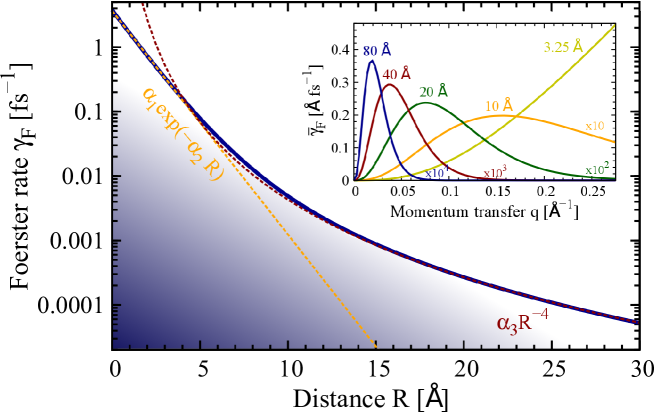
Figure 6 illustrates the Förster rate as a function of the substrate-molecule distance . Generally, the integral over all processes involving the momentum transfer cannot be analytically solved. We find that within the simplest tight-binding approximation taking into account only the strongest overlaps, the direct transitions with do not contribute to the energy transfer. The inset of Fig. 6 shows the integrand of Eq. (5) (denoted as ) as a function of for different fixed distances . For R<10 Å, quickly increases with and the Förster rate shows an exponential decay with , i.e. with fs-1 and Å-1, cf. Fig. 6. For large distances, the behavior drastically changes: is characterized by a maximum centered at , i.e. only processes involving a certain momentum transfer significantly contribute to the energy transfer rate. In the limit of large substrate-molecule distances ( Å), the Förster coupling exhibits a clear dependence, i.e. with fs-1, cf. Fig. 6. This is in excellent agreement with the observations in a recent experiment varying the distance between graphene and attached molecular emitters by depositing additional layers.23
Inserting the molecular transition dipole moment for the investigated exemplary perylene-functionalized graphene, we obtain a very efficient Förster energy transfer rate of fs-1. This can be traced back to the strong Coulomb interaction in the graphene substrate and the short substrate-molecule distance of Å obtained within a full geometric relaxation of the entire hybrid nanostructure. At such a short distance, transitions involving different momentum transfers crucially contribute to the Förster rate, cf. the inset of Fig. 6.
Our result is in line with experimental time-resolved investigations of the energy transfer in functionalized carbon nanotubes suggesting
that the transfer process occurs on an ultrafast femtosecond timescale.43
Often, it is necessary to include additional linker molecules to experimentally achieve the functionalization19 resulting in much larger substrate-molecule distances. For example, Å and 50 Å result in a Förster rate of ps-1 and 6.88 ps-1, respectively. The drastic decrease in efficiency is in agreement with the experimental findings of L. Gaudreau and co-workers.23
In spite of the short distance between the graphene layer and the perylene molecule, our calculations reveal that the Dexter energy transfer rate is negligibly small compared to the discussed Förster transfer mechanism. The Dexter rate is determined by the spatial overlap between the strongly localized graphene and perylene orbitals. To estimate , we calculate the ratio between the overlaps and appearing in the Dexter and the Förster rate, respectively, cf. Eqs. (3) and (2). We obtain . Since in the rates the square of the product of two such overlaps appears, the Dexter rate is
expected to be approximately four orders of magnitude smaller than the Förster rate .
In conclusion, we have investigated the energy transfer in perylene-functionalized graphene. Having characterized the hybrid material within DFT calculations including a fully geometric relaxation of the structure, its electronic bandstructure, optical properties, and charge rearrangements, we focus on the energy transfer that has been measured in recent experiments. Combining DFT-based calculation of the molecular transition dipole moment and tight-binding-based consideration of graphene wave functions allows us to obtain an analytic expression for the Förster energy transfer rate. Our calculations reveal strongly efficient Förster coupling with rates in the range of fs-1. In contrast, the Dexter energy transfer mechanism is found to be negligibly small due to small overlap between the involved strongly localized substrate and molecule orbital functions. The obtained results can be applied to other carbon-based hybrid nanostructures and in general to the description of energy transfer processes in molecular functionalised nanostructures, once the molecular dipole moment and the substrate-molecule separation are known.
We thank the Einstein Foundation Berlin and the Deutsche Forschungsgemeinschaft (within the collaborative research center SFB 658) for financial support. O. T. H. acknowledges the support from FWF (Project: J 3285-N20). A.R acknowledge financial support from the European Research Council (ERC-2010-AdG-267374), Spanish Grant (FIS2010-21282-C02-01), Grupos Consolidados UPV/EHU del Gobierno Vasco (IT578-13), and the EU project (280879-2 CRONOS CP-FP7) .
References
- Avouris et al. 2007 Avouris, P.; Chen, Z.; Perebeinos, V. Nature Nanotechnology 2007, 2, 605
- Bonaccorso et al. 2010 Bonaccorso, F.; Sun, Z.; Hasan, T.; Ferrari, A. C. Nature Photonics 2010, 4, 611
- Hirsch and Vostrowsky 2005 Hirsch, A.; Vostrowsky, O. 2005, 245, 193
- Burghard 2005 Burghard, M. Surf. Sci. Rep. 2005, 58, 1–109
- Reich et al. 2004 Reich, S.; Thomsen, C.; Maultzsch, J. Carbon Nanotubes: Basic Concepts and Physical Properties; Wiley-VCH: Berlin, 2004
- Jorio et al. 2008 Jorio, A.; Dresselhaus, M.; Dresselhaus, G. Carbon nanotubes: advanced topics in the synthesis, structure, properties and applications; Springer: Berlin, 2008
- Malić and Knorr 2003 Malić, E.; Knorr, A. Graphene and Carbon Nanotubes: Ultrafast Optics and Relaxation Dynamics; WILEY-VCH: Berlin, 2003
- Avouris et al. 2008 Avouris, P.; Freitag, M.; Perebeinos, V. Nature Photonics 2008, 2, 341
- Malić et al. 2011 Malić, E.; Winzer, T.; Bobkin, E.; Knorr, A. Phys. Rev. B 2011, 84, 205406
- Guo et al. 2005 Guo, X.; Huang, L.; O’Brien, S.; Kim, P.; Nuckolls, C. J. Am. Chem. Soc. 2005, 127, 15045
- Sundaram et al. 2008 Sundaram, R. S.; Gomez-Navarro, C.; Balasubramanian, K.; Burghard, M.; Kern, K. Adv. Mater. 2008, 20, 3050
- Zhou et al. 2009 Zhou, X.; Zifer, T.; Wong, B. M.; Krafcik, K. L.; Leonard, F.; Vance, A. L. Nano Lett. 2009, 9, 1028
- Ehli et al. 2009 Ehli, C.; Oelsner, C.; Guldi, D. M.; Mateo-Alonso, A.; Prato, M.; Schmidt, C.; Backes, C.; Hauke, F.; Hirsch, A. Nat. Chem. 2009, 1, 243–249
- et al. 2009 et al., J.-C. C. Nanotechnology 2009, 20, 375501
- Loi et al. 2010 Loi, M. A.; Gao, J.; Cordella, F.; Blondeau, P.; Menna, E.; Bartova, B.; Hebert, C.; Lazard, S.; Bottone, G. A.; Milko, M.; Ambrosch-Draxl, C. Adv. Mater. 2010, 22, 1635
- Kolpak and Grossman 2011 Kolpak, A. M.; Grossman, J. C. Nano Lett. 2011, 11, 3156
- Simmons et al. 2007 Simmons, J. M.; In, I.; Campbell, V. E.; Mark, T. J.; Léonard, F.; Gopalan, P.; Eriksson, M. A. Phys. Rev. Lett. 2007, 98, 086802
- Malić et al. 2012 Malić, E.; Setaro, A.; Bluemmel, P.; Sanz-Navarro, C. F.; Ordejon, P.; Reich, S.; Knorr, A. J. Phys. Condens. Matter 2012, 24, 394006
- Setaro et al. 2012 Setaro, A.; Bluemmel, P.; Maity, C.; Hecht, S.; Reich, S. Adv. Funct. Mat. 2012, 22, 2425–2431
- Magadur et al. 2008 Magadur, G.; Lauret, J.-S.; Alain-Rizzo, V.; Voisin, C.; Roussignol, P.; Deleporte, E.; Delaire, J. A. ChemPhysChem 2008, 9, 1250
- Roquelet et al. 2010 Roquelet, C.; Garrot, D.; Lauret, J. S.; Voisin, C.; Alain-Rizzo, V.; Roussignol, P.; Delaire, J. A.; Deleporte, E. Appl. Phys. Lett. 2010, 97, 141918
- Ernst et al. 2012 Ernst, F.; Heek, T.; Setaro, A.; Haag, R.; Reich, S. Adv. Funct. Mat. 2012, 22, 3921–3926
- Gaudreau et al. 2013 Gaudreau, L.; Tielrooij, K. J.; Prawiroatmodjo, G. E.; Osmond, J.; de Abajo, F. J. G.; Koppens, F. H. Nano Lett. 2013, 13, 2030
- Förster 1948 Förster, T. Ann. Physik 1948, 437, 55
- Dexter 1953 Dexter, D. L. J. Chem Phys. 1953, 21, 836
- Winkler 2008 Winkler, J. R. Science 2008, 339, 1530
- Swathi and Sebastian 2009 Swathi, R. S.; Sebastian, K. L. J. Chem. Sci. 2009, 121, 777
- Ernst et al. 2013 Ernst, F.; Heek, T.; Setaro, A.; Haag, R.; Reich, S. Appl. Phys. Lett. 2013, 102, 233105
- Blum et al. 2009 Blum, V.; Gehrke, R.; Hanke, F.; Havu, P.; Havu, V.; Ren, X.; Reuter, K.; Scheffler, M. Comput. Phys. Commun. 2009, 180, 2175
- Perdew et al. 1996 Perdew, P.; Burke, K.; Ernzerhof, M. Phys. Rev. Lett. 1996, 102, 3865
- Tkatchenko and Scheffler 2009 Tkatchenko, A.; Scheffler, M. Phys. Rev. Lett. 2009, 102, 073005
- Adamo and Barone 1999 Adamo, C.; Barone, V. J. Chem. Phys. 1999, 110, 6158
- Heyd et al. 2003 Heyd, J.; Scuseria, G. E.; Ernzerhof, M. J. Chem. Phys. 2003, 118, 8207
- Krukau et al. 2006 Krukau, A. V.; Vydrov, O. A.; Izmaylov, A. F.; Scuseria, G. E. J. Chem. Phys. 2006, 125, 224106
- Bagus et al. 2008 Bagus, P. S.; K fer, D.; Witte, G.; W ll, C. Phys. Rev. Lett. 2008, 100, 126101
- Hofmann et al. 2008 Hofmann, O. T.; Rangger, G. M.; Zojer, E. J. Phys. Chem. C 2008, 112, 20357
- Mak et al. 2011 Mak, K. F.; Shan, J.; Heinz, T. F. Phys. Rev. Lett. 2011, 106, 046401
- Chae et al. 2011 Chae, D.; Utikal, T.; Weisenburger, S.; Giessen, H.; v. Klitzing K., Nano Lett. 2011, 11, 1379
- Hofmann et al. 2013 Hofmann, O. T.; Atalla, V.; Mollx, N.; Rinke, P.; Scheffler, M. arXiv:1310.2097 2013,
- Wu and Voorhis 2005 Wu, Q.; Voorhis, T. V. Phys. Rev. A 2005, 72, 024502
- Hofmann-Mees et al. 2013 Hofmann-Mees, D.; Appel, H.; Ventra, M. D.; Kuemmel, S. J. Phys. Chem. B 2013, 117, 14408–14419
- Baer and Rabani 2008 Baer, R.; Rabani, E. J. Chem. Phys. 2008, 128, 184710
- Garrot et al. 2011 Garrot, D.; Langlois, B.; Roquelet, C.; Michel, T.; Roussignol, P.; Delalande, C.; Deleporte, E.; Lauret, J.; Voisin, C. J. Phys. Chem. C 2011, 115, 23283