Direct observation of the coherent nuclear response after the absorption of a photon
Abstract
How molecules convert light energy to perform a specific transformation is a fundamental question in photophysics. Ultrafast spectroscopy reveals the kinetics associated with electronic energy flow, but little is known about how absorbed photon energy drives nuclear or electronic motion. Here, we used ultrabroadband transient absorption spectroscopy to monitor coherent vibrational energy flow after photoexcitation of the retinal chromophore. In the proton pump bacteriorhodopsin we observed coherent activation of hydrogen wagging and backbone torsional modes that were replaced by unreactive coordinates in the solution environment, concomitant with a deactivation of the reactive relaxation pathway.
The efficiency of light-induced processes fundamentally depends on the ability of a system to convert photons into chemical, electrical or mechanical energy. Resonance Raman intensity analysis reveals the initial energy distribution and the systems evolution over the first few tens of femtoseconds after photoexcitation Warshel (1977); Heller (1981), while time-resolved optical spectra provide electronic kinetics. Neither of these techniques is capable of following the evolution of incident photon energy from the initially populated Franck-Condon (FC) region toward reactive rather than dissipative channels. It is this energy transfer, however, that is critical in determining the efficiency and outcome of a photo-induced process Takeuchi et al. (2008) and has been suggested to be subject to quantum coherent effects even for large protein assemblies at ambient condition Wang et al. (1994); Engel et al. (2007); Collini et al. (2010).
The variation in reaction yields and speeds for retinal protonated Schiff base (RPSB) photoisomerization in different environments highlights the importance of the competition between active and passive relaxation pathways. In the proton pump bacteriorhodopsin (bR), excited state decay following photoexcitation is faster (0.5 vs 4 ps) Mathies et al. (1988); Kandori and Sasabe (1993), results in a higher isomerization yield (0.64 vs 0.16) Tittor and Oesterhelt (1990); Freedman and Becker (1986) and produces exclusively the 13-cis rather than the 11-cis isomer formed in solution. Nevertheless, the resonance Raman spectra of both RPSBs are strikingly similar Smith et al. (1987) implying an essentially identical starting geometry and initial structural evolution. Raman active totally symmetric stretching coordinates, however, cannot contribute to the formation of a conical intersection between states of opposite symmetry and thus to efficient electronic relaxation Levine and Martínez (2007).
The reactivities observed for RPSB in solution compared to the protein environment must therefore result from the activation of different nuclear motions by energy transfer from the initially populated coordinates. To reveal the energy flow, we created and followed vibrational coherence with specific sensitivity to the region near the vibronic surface crossing using impulsive vibrational spectroscopy Fragnito et al. (1989). Illustrating this experimental approach with RPSB, absorption of an ultrashort visible photon generates vibrational coherence in all nuclear degrees of freedom coupled to the optical transition in both ground (S0) and excited (S1) electronic states (FIG. 1) Pollard and Mathies (1992). The impulsively created S0 nuclear wavepacket oscillates about its equilibrium position whereas on S1 it rapidly moves out of the FC region toward a new stationary point (SP) Garavelli et al. (1997). At this local minimum, the backbone structure has evolved little along the isomerization coordinate and experiences a barrier toward excited state decay Hasson et al. (1996); Ruhman et al. (2002). Previous attempts to reveal reactive nuclear wavepacket evolution Kobayashi et al. (2001) have struggled either with interfering ground state signatures Dexheimer et al. (1992); Kraack et al. (2011) or insufficient time-resolution revealing only low-frequency coherences Wand et al. (2012). As a result, only limited insight into the reactive dynamics from the perspective of a few degrees of freedom was available.
Here, we combined high time resolution transient absorption spectroscopy with ultrabroad (500 - 900 nm) detection bandwidth, spectroscopic sensitivity (10 OD) and electronic population control to conclusively reveal excited state nuclear wavepacket dynamics over the full spectral window of interest (2000 ).
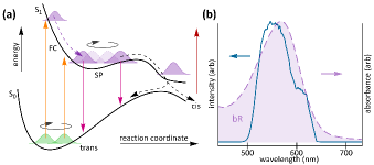
The differential absorbance map of RPSB in bR after optical excitation with a 10 fs pulse is shown in FIG. 2a. Early time-delays (100 fs) are dominated by the coherent artifact caused by the interaction of pump and probe pulses with the sample Dobryakov et al. (2005), rapidly followed by ground state bleach (570 nm) and stimulated emission bands (750 nm). Both signatures decay with the characteristic excited state lifetime of 0.5 ps concomitant with a growth of the photoproduct absorption band at 640 nm. Given the high time-resolution (20 fs) achieved in this work, the broad and slowly varying spectral and temporal electronic features are complemented by rapid oscillations caused by the impulsively excited nuclear wavepackets (see FIG. 1a).
Subtraction of the exponential electronic kinetics reveals the pure vibrational coherence, including two phase jumps, one at slightly lower energy than the S0 absorption maximum (580 nm) and another at the border between ground and excited state signatures (750 nm) (FIG. 2b). The near-infrared (NIR) coherence is strongly damped compared to the one observed in the S0 absorption window (650 nm), in agreement with the short S1 lifetime (0.5 ps). These observations suggest that the visible region is dominated by nuclear coherence on S0, while the NIR mainly exhibits signatures caused by vibrational wavepackets evolving on S1.
To conclusively remove any residual S0 coherences in the NIR, we determined the frequencies and dephasing times of all contributing oscillations using a combination of linear prediction singular value decomposition Johnson and Myers (1996) and nonlinear optimization. A sum of 24 exponentially decaying oscillations was sufficient to describe the observed coherence decay to within the measurement noise (FIG. 2c inset) and revealed the presence of some coherences with dephasing times exceeding 0.45 ps, all of which showed frequencies matching known S0 bands, such as the C=C band at 1526 cm-1.
After removal of these S0 signatures we are left with a spectrum representative of coherence activity on S1 only exhibiting a completely altered intensity and frequency distribution compared to the S0 frequency-domain Raman spectrum of bR (FIG. 2c). The hydrogen wag at 952 cm-1 dominates the spectrum, with lesser contributions from additional wagging (900 cm-1), Schiff base (1075 cm-1) and backbone torsional (177 cm-1) coordinates. Recent coherent infrared emission spectra on bR agree with our observations of strong activity in the 750-1150 cm-1 region, but an unambiguous assignment of the observed features was hampered by numerous additional modes of unknown origin Groma et al. (2011).
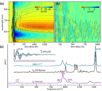
An important consequence of our detection methodology is that vibrational motion which strongly modifies the S0-S1 energy gap will also generate the most prominent oscillatory features in the transient electronic spectra (FIG. 1a). Given that the critical step during the isomerization reaction is the passage from S1 to S0 through a conical intersection, the most active modes in our experiment are likely to be the strongest contributors to excited state decay and thus the photoisomerization. Despite the complexity of the system with more than 300 nuclear degrees of freedom, the S1 vibrational coherence of RPSB in bR is dominated by the hydrogen wag at 952 cm-1, a motion essential to C=C isomerizations Kukura et al. (2005) and capable of coupling the ground and excited electronic states potential energy surfaces (PES). The results presented in FIG. 2c thus suggest a correspondence between the ability of a systems to focus energy into a few critical coordinates and the photochemical outcome of excited state decay.
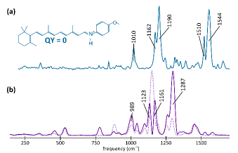
Although such an interpretation is appealing, it does not prove a correlation between the observed nuclear coherence, the shape of the PES and photochemical reactivity. By synthesizing a series of RPSBs with different Schiff bases, we found that using p-methoxyaniline (pma) as a base completely eliminated all isomerizing channels (FIG. 3a). The close resemblance of the S0 Raman spectrum of pma-RPSB to that of RPSB in bR (see FIG. 2c) suggests similar ground state geometries, excited electronic state character and initial nuclear evolution for RPSB in bR and pma-RPSB in solution. In contrast to RPSB in bR, however, the S1 coherence shows negligible activity in the hydrogen wagging (1000 cm-1) and torsional (300 cm-1) regions of the spectrum and is instead dominated by stretching coordinates with significant contributions likely originating from the aromatic Schiff base moiety (1000-1300 cm-1), as verified by comparison with fully deuterated pma (FIG. 3b).
In contrast to most current ultrafast techniques, the vibrational coherence presented in this work was generated by the photon that triggers the light induced process. Our results thus do not simply report on the structural evolution of the molecule of interest, but instead on the coherent vibrational energy flow along the S1 PES out of the FC region during the isomerization reaction. Based on the S0 Raman spectra, we would have expected strong coherent activity in backbone stretches for RPSB in bR and in solution, but observed exactly the opposite experimentally. In fact, given that S1 vibrational coherence is generated mainly in FC active modes, we would expect the power spectra of S1 coherent activity to be as similar as the respective S0 Raman spectra.
The dramatically different appearance of the S0 and S1 spectra cannot be solely attributed to vibrational structural changes caused by different bonding properties upon electronic excitation. The apparent lack of backbone stretching activity in bR is a priori surprising, given the dominant presence of C=C stretching modes in the Raman spectra of polyenic systems. Such behaviour, however, has been previously observed in the photoisomerization of stilbene Dobryakov et al. (2012). Here, totally symmetric coordinates remain dominant in the Raman spectrum of the excited electronic state for the trans-isomer but disappear almost completely for the more reactive cis-isomer likely due to significant symmetry changes caused by molecular distortion. The key question thus relates to the mechanism by which vibrational coherence can appear in initially silent and disappear from initially excited degrees of freedom. The latter can be explained by a change in equilibrium displacement between S0 and S1 as the structure of the molecule evolves on the excited electronic state. The former, however, must be related to the shape of the PES away from the FC region Takeuchi et al. (2008).
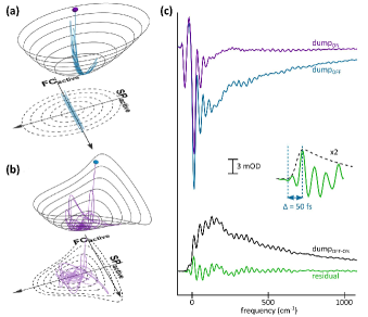
Consider a simplified, two-dimensional excited state PES consisting of two uncoupled nuclear coordinates, only one of which is displaced from its equilibrium position in the FC region. For a excitation, two such coordinates may be the symmetric C=C stretch and an asymmetric hydrogen wag. Upon population of S1, the system oscillates along the displaced stretching coordinate about its new equilibrium bond length but without gaining any momentum along the initially silent asymmetric wag (FIG. 4a). By introducing a coupling term between the two modes this behavior changes drastically. The system now gains momentum along the asymmetric coordinate after the first tens of fs, even if the initial displacement was close to zero (FIG. 4b). Such a shift, however, does not necessarily induce significant changes in the resonance Raman spectra, because only the earliest dynamics are sampled Heller (1981).
To quantify the timescale for the coherent vibrational coupling process, we used electronic population control to eliminate the otherwise dominant coherent artifact around time-zero, thus revealing the full temporal evolution of the vibrational coherence down to the earliest time-delays. Using a long (1 ps) dump pulse centered at 880 nm, we selectively removed S1 population without impulsively generating additional nuclear wavepackets or affecting the S0 population. By subtracting the transient dynamics in the absence and presence of the dump pulse we removed the coherent artifact and revealed the rapid rise of the vibrational coherence on the sub 50 fs timescale (FIG. 4c). Interestingly, this growth is comparable to the recently reported rise time of the SE signal and matches the kinetics of tentatively assigned excited state signatures at wavelengths 1 m Hasson et al. (1996); Wand et al. (2012). The major changes responsible for the observed vibrational coherence transfer does take place during the first tens of femtoseconds after photoexcitation.
In conclusion, we used ultabroadband transient absorption spectroscopy with high temporal resolution to reveal the coherent nuclear response of a molecule after absorption of a photon. In contrast to the expectation that the photon-induced structural evolution largely takes place along FC active coordinates, we observed efficient and coherent activation of initially silent vibrational modes caused by PES-induced anharmonic coupling. We further demonstrated that the nature of the coherent evolution differs dramatically with the environment of the molecule and is strongly correlated with the efficiency and outcome of the light induced process as evidenced by the comparison of RPSB in bR and pma-RPSB in solution. Interestingly, the coherent evolution takes place on the 50 fs timescale, fast enough to allow for quantum coherent effects to remain active and decisive for the eventual outcome of the process Prokhorenko et al. (2006), irrespective of the photoexcitation mechanism. Our results emphasize the importance of ultrafast energy flow in determining the outcome of light-induced processes and demonstrate the power of vibrational spectroscopy in the time-domain for studying the structural and dynamic origins of molecular relaxation phenomena.
References
- Warshel (1977) A. Warshel, Annu. Rev. Biophys. Bio. 6, 273 (1977).
- Heller (1981) E. J. Heller, Accounts Chem. Res. 14, 368 (1981).
- Takeuchi et al. (2008) S. Takeuchi, S. Ruhman, T. Tsuneda, M. Chiba, T. Taketsugu, and T. Tahara, Science 322, 1073 (2008).
- Wang et al. (1994) Q. Wang, R. W. Schoenlein, L. A. Peteanu, R. A. Mathies, and C. V. Shank, Science 266, 422 (1994).
- Engel et al. (2007) G. S. Engel, T. R. Calhoun, E. L. Read, T.-K. Ahn, T. Mancal, Y.-C. Cheng, R. E. Blankenship, and G. R. Fleming, Nature 446, 782 (2007).
- Collini et al. (2010) E. Collini, C. Y. Wong, K. E. Wilk, P. M. G. Curmi, P. Brumer, and G. D. Scholes, Nature 463, 644 (2010).
- Mathies et al. (1988) R. A. Mathies, C. H. Brito Cruz, W. T. Pollard, and C. V. Shank, Science 240, 777 (1988).
- Kandori and Sasabe (1993) H. Kandori and H. Sasabe, Chem. Phys. Lett. 216, 126 (1993).
- Tittor and Oesterhelt (1990) J. Tittor and D. Oesterhelt, FEBS Lett. 263, 269 (1990).
- Freedman and Becker (1986) K. A. Freedman and R. S. Becker, J. Am. Chem. Soc. 108, 1245 (1986).
- Smith et al. (1987) S. O. Smith, M. S. Braiman, A. B. Myers, J. A. Pardoen, J. M. L. Courtin, C. Winkel, J. Lugtenburg, and M. A. Richard, J. Am. Chem. Soc. 109, 3108 (1987).
- Levine and Martínez (2007) B. G. Levine and T. J. Martínez, Annu. Rev. Phys. Chem. 58, 613 (2007).
- Fragnito et al. (1989) H. L. Fragnito, J.-Y. Bigot, P. C. Becker, and C. V. Shank, Chem. Phys. Lett. 160, 101 (1989).
- Pollard and Mathies (1992) W. T. Pollard and R. A. Mathies, Annu. Rev. Phys. Chem. 43, 497 (1992).
- Garavelli et al. (1997) M. Garavelli, P. Celani, F. Bernardi, M. Robb, and M. Olivucci, J. Am. Chem. Soc. 119, 6891 (1997).
- Hasson et al. (1996) K. C. Hasson, F. Gai, and P. A. Anfinrud, P. Natl. Acad. Sci. USA 93, 15124 (1996).
- Ruhman et al. (2002) S. Ruhman, B. Hou, N. Friedman, M. Ottolenghi, and M. Sheves, J. Am. Chem. Soc. 124, 8854 (2002).
- Kobayashi et al. (2001) T. Kobayashi, T. Saito, and H. Ohtani, Nature 414, 531 (2001).
- Dexheimer et al. (1992) S. L. Dexheimer, Q. Wang, L. A. Peteanu, W. T. Pollard, R. A. Mathies, and C. V. Shank, Chem. Phys. Lett. 188, 61 (1992).
- Kraack et al. (2011) J. P. Kraack, T. Buckup, and M. Motzkus, Phys. Chem. Chem. Phys. 13, 21402 (2011).
- Wand et al. (2012) A. Wand, B. Loevsky, N. Friedman, M. Sheves, and S. Ruhman, J. Phys. Chem. B 117, 4670 (2012).
- Dobryakov et al. (2005) A. L. Dobryakov, S. A. Kovalenko, and N. P. Ernsting, J. Chem. Phys. 123, 044502 (2005).
- Johnson and Myers (1996) A. E. Johnson and A. B. Myers, J. Chem. Phys. 104, 2497 (1996).
- Groma et al. (2011) G. I. Groma, A. Colonna, J.-L. Martin, and M. H. Vos, Biophys. J. 100, 1578 (2011).
- Kovalenko et al. (1999) S. A. Kovalenko, A. L. Dobryakov, J. Ruthmann, and N. P. Ernsting, Phys. Rev. A 59, 2369 (1999).
- Liebel and Kukura (2013) M. Liebel and P. Kukura, J. Phys. Chem. Lett. 4, 1358 (2013).
- Kukura et al. (2005) P. Kukura, D. W. McCamant, S. Yoon, D. B. Wandschneider, and R. A. Mathies, Science 310, 1006 (2005).
- Dobryakov et al. (2012) A. L. Dobryakov, I. Ioffe, A. A. Granovsky, N. P. Ernsting, and S. A. Kovalenko, J. Chem. Phys. 137, 244505 (2012).
- Prokhorenko et al. (2006) V. I. Prokhorenko, A. M. Nagy, S. A. Waschuk, L. S. Brown, R. R. Birge, and R. J. D. Miller, Science 313, 1257 (2006).